CHAPTER 11 The tibia
Common Causes of Pain in the Anterior Aspect of the Lower Leg
Note: Knock-knee and bow-leg deformities are included with the knee joint.
Osteitis of the Tibia
Osteitis of the tibia occurs predominantly in children, with or without a history of previous trauma or sore throat. Pain is intense, tenderness is acute and initially well localised over the metaphyseal area, and there is inability to weightbear. There is systemic upset with fever and tachycardia, and often (but not always) a polymorph leukocytosis. Admission and investigation with repeated blood cultures is essential. Radiographs of the tibia are initially normal, often with a lag of 2 weeks or more before any abnormality is detectable (although MRI and CT scans may be affected somewhat earlier). The ESR and C-reactive protein are usually elevated at an early date. When this condition is suspected, it is customary to administer a broad-spectrum antibiotic effective against the penicillin-resistant Staphylococcus, and in large doses to achieve adequate bone levels, prior to the results of blood culture. Splintage of the affected area is often helpful, and in proven cases antibiotics are administered for 4 weeks. Surgical drainage is seldom necessary and is avoided unless failure of response to antibiotics, profound toxicity and spread of the infection make it essential.
Cellulitis from insect stings, small wounds and abrasions and hair follicle infections may sometimes cause difficulty in diagnosis.
Low-grade osteitis of the tibia (Brodie’s abscess) may give rise to chronic upper tibial pain.
Bone Tumours
The tibia is a common site for many primary bone tumours, so that radiographic examination of the tibia is essential in any case of undiagnosed leg pain.
Anterior Tibial Compartment Syndrome
This is a common complication of fractures of the tibial shaft, but may follow a period of intense lower limb activity: hence it is common in athletes. It gives rise to pain in the front of the leg. This is due to oedema and swelling within the confines of the anterior compartment, which lead in turn to ischaemia in the anterior tibial muscles. In severe cases where swelling is progressive there may eventually be muscle necrosis. The leg is diffusely swollen and tender, and the skin has a glossy appearance. Tibialis anterior and extensor hallucis longus are first affected, with weakness and later inability to extend the ankle and great toe. The dorsalis pedis pulse may be absent, and there may be sensory loss in the first web space due to ischaemic changes in the deep peroneal nerve. In high-risk and suspect cases compartment pressure monitoring is advisable. In severe cases immediate surgical decompression of the anterior tibial compartment is essential if muscle necrosis is to be avoided.
Stress Fracture of the Tibia
In this condition the onset of leg pain may be sudden or less acute. There is sharply localised bone tenderness and overlying oedema. Radiographic demonstration of the hairline fracture may be difficult, and with persistent pain repeated examination is essential. A radioisotope bone scan may be helpful in diagnosing a local ‘hot spot’. In many cases the diagnosis may not be firmly established until a small area of tell-tale callus is showing. The condition is also common in Paget’s disease where, of course, there is an easily identifiable radiological abnormality.
Medial Tibial Syndrome/Shin Splints
In this condition pain on the medial side of the shin in sportsmen may be severe, and there is usually tenderness along the posteromedial border of the lower part of the tibia. In a number of cases the symptoms may arise from stress fractures of the tibia, but in others the pathology is less clear. (Other causes include compartment syndromes, fascial hernias, interosseous membrane tears, periosteal avulsions, tendinitis, muscle sprains and periostitis.) Where symptoms are of a chronic nature, and fracture has been excluded, division of the attachments of the crural fascia may give relief.
Common Causes of Pain in the Posterior Aspect of the Lower Leg
Deformities of the Tibia
Alteration in the normal curvature of the tibia is not uncommon and may be a cause for complaint. The bone may curve convex laterally (tibial bowing), convex anteriorly (tibial kyphosis), or undergo a rotational deformity (tibial torsion). Deformities of these types are particularly likely to occur in infants and young children, when the immature bone may yield under the weight of a relatively heavy child. In the majority of cases no other cause is apparent, and spontaneous correction by the time the child reaches the age of 6 is the rule. Nevertheless, rickets and other osteodystrophies must be excluded and continuous observation is essential.
Pseudarthrosis of the tibia is a rare congenital abnormality which leads to progressive tibial kyphosis. The tibia becomes progressively thinner and undergoes spontaneous fracture, which proceeds to non-union. It is particularly resistant to treatment. The diagnosis is made on the radiographic findings.
In the adult, deformity of the tibia may be seen following rickets in childhood, malunited fractures, Paget’s disease and syphilis.
Guide to Commoner Causes of Leg Pain
| In children | Osteitis or other infections |
| Bone tumour | |
| Adolescents and young adults | Stress fracture tibia |
| Bone tumours (especially osteoid osteoma, osteoclastoma, osteosarcoma) | |
| Brodie’s abscess | |
| Anterior compartment syndromes | |
| Paget’s disease | |
| ‘Ruptured plantaris tendon’ | |
| Painful conditions of the foot | |
| Syphilis | |
| Bone tumours | |
| Adults | Prolapsed invertebral disc and spinal stenosis |
| Vascular insufficiency | |
| Paget’s disease | |
| ‘Ruptured plantaris tendon’ | |
| Painful conditions of the foot | |
| Syphilis | |
| Bone tumours |
11.1. Inspection (1): soft tissue swelling:
Note the site and extent of any swelling. In the case of oedema, note particularly if bilateral (suggesting a general rather than a local cause). Unilateral leg oedema in women over 40 is a common sign of intrapelvic neoplasm.
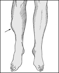
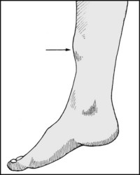
11.2. Inspection (2): localised oedema:
Localised oedema is common over inflammatory lesions and stress fractures.
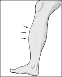
11.3. Inspection (3): local bone swelling:
This is suggestive of neoplasm (e.g. osteoid osteoma) or old fracture. Multiple or single exostoses commonly occur in the tibia in diaphyseal aclasis. Thickening of the ends of the tibia is seen in rickets and in osteoarthritis.
11.4. Inspection (4): general bone thickening:
Extensive thickening of bone is characteristic of Paget’s disease and long-standing osteitis. In the latter case there are usually other signs, such as scarring or sinuses.
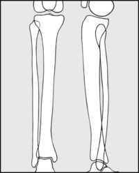
11.5. Inspection (5): tibial shape:
Note any abnormal anterior curvature (tibial kyphosis), possibly secondary to Paget’s disease, malunited fracture, syphilis or rickets. Rickets affects the distal half of both tibia and fibula, and there are associated lateral and torsional deformities.
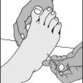
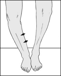
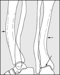
Flex the legs over the edge of the examination couch. The tibial tubercles must face directly forwards. Place the index fingers over the malleoli. The medial malleolus normally lies 20° in front of the lateral in the coronal plane).
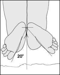
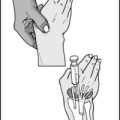
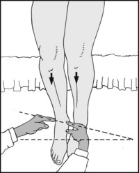
Alternatively, examine the patient in the prone position with the knees flexed to 90°. The ankle should be in the neutral position. Note the position of the medial borders of both feet in relation to the midline: this is normally in the region of 20°, but will be greater where there is a lateral tibial torsional deformity (illustrated), and less where there is a medial torsional deformity.
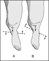
(A) Medial torsional deformity (a decrease in the angle) is associated with flat foot and intoeing. (B) Lateral torsional deformity (an increase in the angle) is seen in pes cavus. Tibial and femoral torsion may be reliably assessed using ultrasound methods.
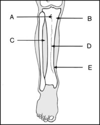
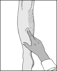
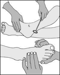
At the front of the leg, tenderness is characteristically sited in the following conditions: (A) Osgood–Schlatter’s disease, (B) Brodie’s abscess, osteitis, (C) anterior tibial compartment syndrome, (D) stress fracture, (E) shin splints.
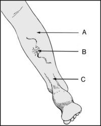
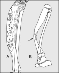
At the back of the leg, tenderness is characteristically situated in the following: (A) ‘ruptured plantaris tendon’ syndrome, (B) over varicosities in superficial thrombophlebitis, (C) over the tendocalcaneus in partial tears and complete ruptures.
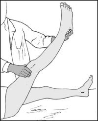
The following tests should be carried out in the investigation of any case of leg pain. The straight leg-raising test should be carried out as a first measure. In many cases pain in the leg below the knee is referred from the spine.
The lower limb reflexes should be elicited, and if indicated, test the pupils for reaction to light and accommodation. Lower leg pain is a common symptom of late syphilis. (3) The peripheral pulses should be sought. Ischaemia is an extremely common cause of leg pain.
The standard films are an anteroposterior and a lateral which include both ends of the tibia and fibula. For better visualisation of a suspect area, localised views are required. CT and MRI scans are often useful, especially in evaluating cystic defects, and radioisotope scans may be helpful in assessing local vascularity.
Begin by noting the general shape of the bones, their texture and their mineralisation. For example, in rickets during the phase of bone softening, deformity follows weightbearing. Note angulation of the plane of the ankle (which may theoretically predispose to osteoarthritis).
Deformity is common in Paget’s disease (A), where there is disturbance of form and texture, and sometimes sarcomatous change. In pseudarthrosis of the tibia (B) there is local thinning and angulation of the bone, which progresses to tibial dissolution, the fibula generally remaining relatively normal.
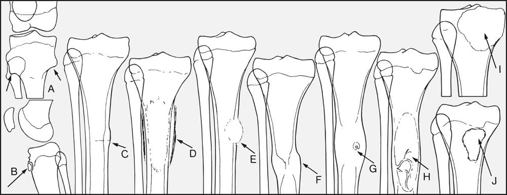
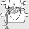
Note any localised deformity such as in (A) diaphyseal aclasis (often several bones are affected); (B) Osgood–Schlatter’s disease; (C) localised periosteal reaction in the region of a stress fracture; (D) more extensive subperiosteal new bone formation in the later stages of osteitis, or at the site of a bone tumour. Note (E) cortical bone destruction suggesting a lytic neoplasm or infection. Localised thickening of bone is seen after a healed fracture (F) or again at a tumour site (e.g. in osteoid osteoma, where there is often a central nidus (G)). Examine the cavity and ends of the bone for space-occupying lesions, such as (H) a unicameral bone cyst occurring in the shaft, (I) an osteoclastoma occurring in the epiphysis, and (J) a Brodie’s abscess in the metaphysis.
11.17. Radiographs of the tibia: examples of pathology (1):
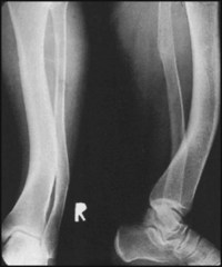
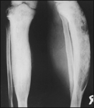
The radiograph shows curvatures of the tibia and fibula, which are convex both laterally and anteriorly. There is an additional torsional deformity which is not obvious in these films.
Diagnosis: persisting deformity of the tibia following rickets in childhood.
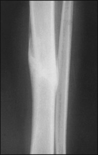
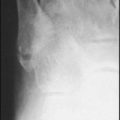
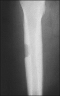
In the centre of the film the tibia is seen to be locally thickened, and just visible is an area of increased bone density running downwards from left to right.
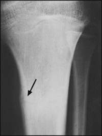
The arrow points to an area of periosteal reaction with the slight increase in bone density at the same level.
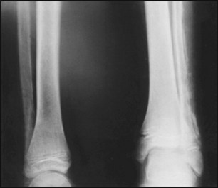
The AP view shows striking alteration in bone texture, with an increase in curvature, convex anteriorly. In both views bone thickening and increased bone density are apparent.
Diagnosis: Paget’s disease of the tibia. In this case there was no malignant change.
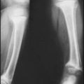
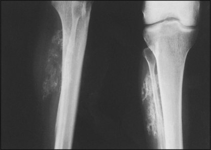
In the left fibula (on the right of the picture) there is extensive bone destruction and subperiosteal new bone formation.
Diagnosis: the appearances are characteristic of osteitis some weeks after the onset, which was heralded with fever, malaise, and severe local pain.
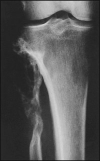
There is localised thickening of the tibia, with a large cavity which is open on its lateral aspect.
Diagnosis: Chronic osteitis of the tibia. The cavity in the bone led to a sinus. No sequestra are apparent in the radiograph.
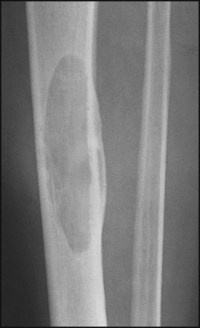
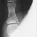
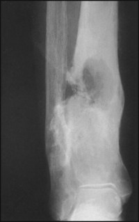
There is a large cystic space in the tibia, with well rounded margins.
Diagnosis: unicameral (simple) bone cyst involving the medullary canal of the tibia. Such abnormalities may often be brought to light by a pathological fracture.
This radiograph of the femur shows a bony lesion of the shaft with a characteristic punched-out appearance, and secondary thickening of the surrounding bone.
Diagnosis: the appearances are characteristic of a highly malignant Ewing’s tumour. Lesions in the tibia are not uncommon and have a similar appearance.
There is destruction and gross deformity of the proximal fibula.
Diagnosis: The appearances are typical of osteoclastoma. The tumour in this case was only locally malignant, but was associated with a common peroneal nerve palsy and drop foot.
There is an extensive area of new bone formation related to the shaft of the fibula. Although the lesion is more easily seen in the AP projection, its extent is best judged in this case in the lateral. There was a history of local trauma and swelling some weeks before.
Diagnosis: The appearances are typical of myositis ossificans. This was thought to have occurred in a haematoma which had formed on the lateral aspect of the leg following a small crack fracture of the fibula.