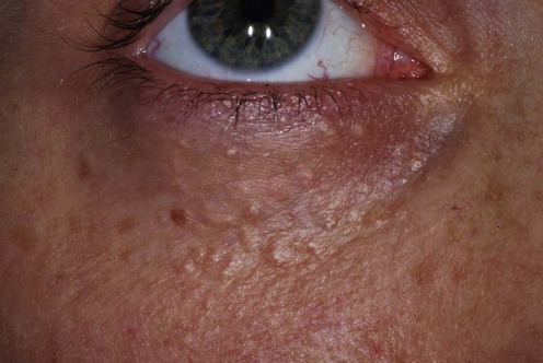
Published on 18/03/2015 by admin
Filed under Dermatology
Last modified 22/04/2025
This article have been viewed 1826 times
James A.A. Langtry
Evidence Levels: A Double-blind study B Clinical trial ≥ 20 subjects C Clinical trial < 20 subjects D Series ≥ 5 subjects E Anecdotal case reports
Syringomata are benign appendageal tumors of the intraepidermal eccrine sweat duct which have a characteristic histologic appearance. The typical clinical presentation is of individual skin- or tan-colored papules, with a rounded or flat surface, 1–5 mm in diameter. Single tumors can occur, but more commonly they are multiple and symmetric, more common in females, and from adolescence onwards. Syringomata most often involve the lower eyelids, although they may occur at other sites, including the cheeks, axillae, abdomen, and vulva. A linear distribution, familial occurrence, a variant associated with Down syndrome, as well as generalized forms have all been described. Frequent onset around puberty and reports of symptoms during pregnancy or menstruation have led to immunohistochemical studies of estrogen and progesterone receptors, with varying results and uncertain relevance.
Syringomata of the eyelids and cheeks are in a prominent site, may appear conspicuous, and treatment may be sought to improve appearance. The syringomata are situated in the upper to mid dermis. Available treatments aim to remove or flatten the papule produced by each syringoma. The majority of treatments are ablative in nature. Some patients may be troubled by only a few individual lesions, whereby excision of these is an option. Ablative modalities include: scissor excision with secondary intention healing; surgical excision of the entire cosmetic unit of the lower eyelids in patients who would also benefit from lower eyelid blepharoplasty; electrocautery; electrodesiccation; intralesional electrodesiccation; dermabrasion; cryotherapy; ablation with CO2 or erbium : YAG laser; and radiofrequency ablation.
Local anesthesia is needed prior to treatment, and this may be topical, or by local injections with or without nerve blocks. Local anesthetic injections producing a field block are most commonly employed, as good anesthesia is helpful when using ablative treatments near the eye. Patients should be warned about the possibility of postoperative bruising. Eye protection is of paramount importance, and specific precautions relevant to the use of lasers must be taken if laser treatment is used.
Ablative treatments will produce some degree of scarring and the aim is to make this imperceptible and produce an excellent cosmetic result. Possible sequelae, including scarring and hypo- or hyperpigmentation (especially with increasing skin pigmentation), should always be discussed prior to treatment.
There are no studies comparing different treatment modalities for syringomata, and a dearth of long-term follow-up data on which to base recommendations for treatment. On the basis of experience and the limited evidence available, it is not necessary to have the latest and most expensive technology to achieve good results. Expertise and good outcomes with simple, ‘low-tech’ methods are as important as with ‘high-tech’ modalities. Each has benefits as well as pitfalls for the novice or unwary. It is more important to be expert in the use and application of one or more modalities than to ‘have a go’ at them all.
The clinical features of periorbital syringomata are usually diagnostic and a skin biopsy may be undertaken for confirmation or when there is uncertainty.
Stegman SS, Tromovitch TA, Glogau RG, eds. Chicago: Year Book Medical, 1992; 32.
A commonsense approach to treatment of syringomata, advocating the use of surgical excision, electrosurgery, or laser.
Maloney ME. J Dermatol Surg Oncol 1982; 8: 973–5.
A single case is reported with a good outcome following removal of four to six lesions per session, in 12 sessions over 5 months.
A good photographic demonstration of the removal of periorbital syringomata with fine ophthalmic spring-action scissors.
Langtry JAA, Carruthers JA. J Cutan Med Surg 1997; 2: 60–3.
The technique of electrocautery is described and good results reported in a number of benign skin lesions, including syringomata.
Karma P, Benedetto AV. Dermatol Surg 1997; 23: 921–4.
Twelve patients were treated with electrodesiccation via a fine electrode into the center of the syringoma, with the aim of localizing the effect and minimizing scarring. All reported excellent results and no recurrence after a follow-up of 18–48 months. Two patients with Fitzpatrick skin type IV had focal hyperpigmentation, which cleared in 2 to 3 months.
Hong S-W, Lee H-J, Cho S-H, Soe J-K, Lee D, Sung H-S. Ann Dermatol 2010; 22: 367–9.
Favorable outcome reported in two patients treated by intralesional electrosurgery with insulated needles.
Wang JJ, Roenigk Jr HH. Dermatol Surg 1999; 25: 136–9.
A description of 10 patients treated with CO2 laser reporting excellent results. Patients with more lesions needed more treatment sessions. The median follow-up was 16 months, and one patient had new syringomata at other periorbital sites 18 months after treatment. Erythema lasted 6 to 12 weeks in all patients. One patient with Fitzpatrick type IV skin had minimal focal areas of hyperpigmentation, which cleared after 2 to 3 months.
Cho SB, Kim HJ, Lee SJ, Kim YK, Lee JH. Dermatol Surg 2011; 37: 433–8.
Thirty-five patients with periorbital syringomata were treated with two sessions of fractional CO2 laser at 10 month intervals. At 2 months follow-up clinical improvement (pre- and post-treatment clinical photographs and patient satisfaction rates) was total in 9%, marked in 43%, moderate in 34%, and minimal in 14%.
Hasson A, Farias MM, Nicklas C, Navarrete C. J Drugs Dermatol 2012; 11: 879–80.
Good cosmetic results reported in five patients with periorbital syringomata treated with the combination of radiofrequency and CO2 laser.
Stevenson TR, Swanson SA. Am Plast Surg 1985; 15: 151–4.
The technique is described and well-illustrated. Good results are reported, but there is no description of numbers of patients treated, clinical details, or follow-up data using this technique.
Dawber RPR. In: Lask GP, Moy RL, eds. Principles and techniques of cutaneous surgery. New York: McGraw-Hill, 1996; 154.
Syringoma is listed as a condition treatable by cryotherapy.
Details are not given and periorbital syringomata are not specifically mentioned.
Kang H, Kim NS, Kim YB, Shim WC. Dermatol Surg 1998; 24: 1370–4.
This study evaluates the histopathology and efficacy of combined CO2 laser and 50% trichloroacetic acid treatment in 20 Korean patients with periorbital syringomata. Results were reported as excellent (11 patients), good (six), and fair (three), without complications such as scarring, infection, or textural change, using the technique detailed.
Fulton JE. J Dermatol Surg Oncol 1978; 4: 777–9.
High-speed dermabrasion is described and good results are reported in 65 patients with acne scarring, actinic damage, adenoma sebaceum, and syringomata.
Frazier CC, Camacho AP, Cockerell CJ. Dermatol Surg 2001; 27: 489–92.
A single case report of eruptive facial syringomata in an African-American woman treated by 35% trichloroacetic acid peel, followed 2 weeks later by CO2 laser, with acceptable cosmetic results and without significant side effects.
Sanchez TS, Dauden E, Casas AP, Garcia-Diez A. J Am Acad Dermatol 2001; 44: 148–9.
A single case report of pruritic syringomata of the chest and neck improving with topical 1% atropine.
Gomez MI, Perez B, Azana JM, Nunez M, Ledo A. Dermatology 1994; 189: 105–6.
Treatment of Skin Disease Comprehensive Therapeutic Strategies 4e
WhatsApp us