Published on 18/03/2015 by admin
Filed under Dermatology
Last modified 22/04/2025
This article have been viewed 2260 times
Nicholas A. Soter
Evidence Levels: A Double-blind study B Clinical trial ≥ 20 subjects C Clinical trial < 20 subjects D Series ≥ 5 subjects E Anecdotal case reports
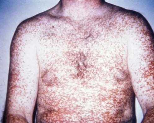
The mastocytoses are a group of disorders of mast cell proliferation that may exhibit both cutaneous and systemic features. Clinical manifestations result from mast-cell activation and from infiltration of various organs. The World Health Organization has classified mastocytosis into disease categories: cutaneous mastocytosis, indolent systemic mastocytosis, systemic mastocytosis with a non-mast cell clonal hematologic disorder, aggressive systemic mastocytosis, mast cell leukemia, mast cell sarcoma, and extracutaneous mastocytoma. The most frequent site of organ involvement in individuals with mastocytosis is the skin. Cutaneous forms include urticaria pigmentosa (shown here), mastocytoma, diffuse and erythrodermic cutaneous mastocytosis, and telangiectasia macularis eruptiva perstans. The cutaneous forms of mastocytosis may be present with or without systemic manifestations. Only the treatment of the cutaneous features will be discussed.
An important aspect of therapy of the cutaneous lesions of mastocytosis is avoidance of triggering factors, which may include temperature changes, friction, physical exertion, ingestion of alcohol, the use of non-steroidal anti-inflammatory agents or opiate analgesics, and emotional stress. Of concern is the possibility of anaphylaxis after stings by Hymenoptera spp., which may occur even in patients receiving venom immunotherapy.
A history seeking systemic features should be undertaken, as well as a physical examination to determine the types of skin lesions and to assess for lymphadenopathy and hepatosplenomegaly. The presence of specific systemic manifestations will dictate the type of specialty physician to whom a referral should be made.
Patients with systemic mastocytosis require long-term follow-up, as 10–30% may develop associated non-mast-cell clonal hematologic disorders such as myelodysplastic or myeloproliferative syndromes, leukemias, or lymphomas.
A skin biopsy should be obtained in all individuals with cutaneous lesions. A complete blood count with differential analysis, a blood chemistry profile that includes liver function tests, and a blood tryptase level should be obtained in all patients with cutaneous lesions, except those with mastocytomas. If there are abnormalities of the complete blood count, a bone marrow examination should be obtained. Plasma histamine levels are not useful to screen patients. Twenty-four hour urine levels of mast cell mediators may be obtained in patients with systemic features. Abdominal imaging studies provide a non-invasive means to assess the lymph nodes, liver, and spleen. In patients with bone pain, a 99Tc bone scan is useful. Osteoporosis may be detected by bone density analysis.
In many cases cutaneous mastocytomas may involute spontaneously; it is rarely described in adults. Childhood urticaria pigmentosa regresses spontaneously in approximately 50% of cases and urticaria pigmentosa in adults in 10%. Diffuse and erythrodermic cutaneous mastocytosis usually resolves spontaneously between the ages of 15 months and 5 years. Telangiectasia macularis eruptiva perstans tends to be a chronic condition.
Most of the therapeutic reports have been in patients with urticaria pigmentosa and, to a lesser extent, in diffuse and erythrodermic cutaneous mastocytosis. The major therapeutic measure is the administration of oral H1 antihistamines to alleviate pruritus and whealing. The use of antihistamines that interfere with the HERG K+ channel and cause cardiac arrhythmias should be avoided. Oral disodium cromoglycate has been efficacious in some individuals. The role and efficacy of topical high-potency glucocorticoid preparations with plastic-film occlusion, of narrow-band ultraviolet B (NB-UVB) phototherapy, of psoralens and ultraviolet A (PUVA) photochemotherapy, and UVA1 phototherapy have not been subjected to controlled clinical trials or remain anecdotal. Cytoreductive therapy to reduce mast-cell proliferation is not recommended in patients with only cutaneous manifestations. There is no therapy that will eradicate the mast cells in the cutaneous lesions.
Blood tryptase and IL-6 levels
Bone density scan, 24-hour urine histamine, histamine metabolites, and prostaglandin metabolites
Bone marrow examination
Schwartz LB. Immunol Allergy Clin North Am 2006; 26: 451–63.
Total and mature (β) tryptase levels are elevated in patients with systematic mastocytosis. Total tryptase levels reflect the increased burden of mast cells in patients with all forms of systemic mastocytosis. Mature tryptase levels reflect the magnitude of mast cell activation. The levels of tryptase are normal in patients with cutaneous mastocytosis.
Brockow K, Akin C, Huber M, Metcalfe DD. Clin Immunol 2005; 115: 216–23.
In patients with systemic mastocytosis, plasma IL-6 levels were elevated and there was a significant correlation between plasma IL-6 and total tryptase levels (p<0.003). In patients with indolent systemic mastocytosis, the plasma IL-6 levels correlated with the extent of urticaria pigmentosa. Plasma levels of IL-6 in patients with cutaneous mastocytosis were not significantly different from those of healthy controls.
Elevated blood tryptase levels and IL-6 appear to be useful in differentiating patients with systemic disease from those with cutaneous disease.
Akin C, Metcalfe DD. Int Arch Allergy Immunol 2002; 127: 133–6.
Measurement of the histamine metabolites N-methylhistamine and methylimidazole acetic acid in 24-hour urine specimens is the preferred method of assessing baseline levels in patients with systemic mastocytosis, and these levels correlate with the amount of the mast cell burden. The levels of the major urinary metabolite of prostaglandin D2 (9α,11β-dihydroxy-15-oxo-2,3,18,19-tetranorprost-5-ene-1,20-dioxic acid) and thromboxane B2 or its metabolites in plasma and urine are elevated in patients with systemic mastocytosis. Soluble forms of CD 25, the α-chain of the interleukin-2 receptor, and CD 117, the receptor for stem cell factor, may be elevated in the circulation in systemic mastocytosis. A c-kit mutation may be detected in lesional tissues, such as skin and bone marrow in systemic mastocytosis.
Although these surrogate disease markers of mastocytosis are valuable tools in diagnosing the extent of disease, they should not be obtained routinely.
Delling G, Ritzel H, Werner M. Pathologe 2001; 22: 132–40.
In a retrospective study of 158 untreated patients with mastocytosis, the prevalence of mastocytosis in iliac crest biopsy specimens was 1.25%, with a prevalence of 2.25% in individuals younger than 45 years. Osteopenia was present in 64% of patients with mastocytosis.
Bone marrow aspirates with biopsies in cutaneous mastocytosis should be restricted to individuals with changes in the complete blood count, organomegaly, or alterations in other organ systems. Some investigators recommend a bone marrow aspirate and biopsy in all patients with adult-onset disease.
Friedman BS, Santiago ML, Berkebile C, Metcalfe DD. J Allergy Clin Immunol 1993; 92: 520–6.
In a double-blind, randomized, crossover trial in 13 subjects with urticaria pigmentosa and systemic mastocytosis, the administration of both azelastine and chlorpheniramine for 4 weeks was associated with a reduction in pruritus.
Frieri M, Alling DW, Metcalfe DD. Am J Med 1985; 78: 9–14.
Five of six patients had less pruritus and four of six had less urticaria while receiving chlorpheniramine and cimetidine. There was no beneficial effect in those receiving disodium cromoglycate.
Gasior-Chrzan B, Falk ES. Dermatology 1992; 184: 149–52.
In a single patient with telangiectasia macularis eruptiva perstans and systemic mastocytosis, there was improvement in pruritus, erythema, and urticaria after the administration of cyproheptadine and cimetidine.
Berg MJ, Bernhard H, Schentag JJ. Drug Intell Clin Pharm 1981; 15: 180–3.
An adult with systemic disease received treatment with cyproheptadine, Lomotil (diphenoxylate-atropine), and cimetidine. Prompt recurrence of gastrointestinal and dermatological symptoms occurred after stopping the cimetidine. The drug was restarted in 3 days and the symptoms subsided.
In systemic mastocytosis H2 antagonists can play an additional role in reducing gastric hyperacidity.
Soter NA, Austen KF, Wasserman SI. N Engl J Med 1979; 301: 465–9.
In a double-blind crossover study in five patients with systemic mastocytosis and urticaria pigmentosa, in 15 of 18 trials oral disodium cromoglycate ameliorated pruritus and whealing.
Lindskov R, Wantzin GL, Knudsen L, Søndergaard I. Dermatologica 1984; 169: 49–52.
In three of four patients with urticaria pigmentosa, the oral administration of disodium cromoglycate was associated with a reduction in pruritus and whealing as assessed by a diminution in dermographism.
Barton J, Lavker RM, Schechter NM, Lazarus GS. Arch Dermatol 1985; 121: 1516–23.
In six patients with urticaria pigmentosa, topical application of betamethasone dipropionate ointment 0.05% under occlusion for 8 hours daily for 6 weeks was associated with an absence of pruritus and Darier sign. All patients remained clear of lesions at a mean follow-up of 11.5 months (range 9–15 months).
Guzzo C, Lavker R, Roberts LJ II, Fox K, Schechter N, Lazarus G. Arch Dermatol 1991; 127: 191–6.
In seven of nine adult patients with urticaria pigmentosa, the topical application of betamethasone dipropionate ointment 0.05% under occlusion overnight to one-half of the body for 6 weeks was associated with resolution of the lesions, with a maximum response within 3 to 12 weeks after the cessation of treatment. Lesions began to recur between 6 and 9 months after completing therapy. Re-treatment for 6 months followed by once-weekly application of betamethasone dipropionate ointment under occlusion kept the patients clear of lesions, with the longest follow-up time being 2.5 years.
The appropriate method for using topical glucocorticoids in patients with urticaria pigmentosa needs to be determined in controlled trials.
Verma KK, Bhat R, Singh MK. Indian J Pediatr 2004; 71: 261–3.
In a 7-month-old girl with diffuse cutaneous mastocytosis with bullae and flushing, treatment with oral betamethasone 0.1 mg/kg/day caused remission within 4 weeks.
Mateo JR. J Eur Acad Dermatol Venereol 2001; 15: 492–4.
Four infants aged between 2.5 and 6 months with mastocytomas were treated with twice-daily applications of clobetasol propionate 0.05% for 6 weeks and on alternate days for 6 weeks with tapering for a total treatment of 6 months. At the end of treatment there were residual macules with atrophy and a negative Darier sign.
Kang NG, Kim TH. J Dermatol 2002; 29: 536–8.
In a 2-month-old boy with a solitary mastocytoma treated with three intralesional injections of triamcinolone acetonide, the lesion became flat with a reduction in erythema and subjective symptoms 9 months after treatment.
Brazzelli V, Grasso V, Marina G, Barbaccia V, Merante S, Bouer E, et al. J Eur Acad Dermatol Venereol 2012; 26: 465–9.
In five adult patients with indolent systemic mastocytosis, the pruritus was controlled and urticaria pigmentosa resolved after a median of 3.5 months of therapy. At a 6-month follow-up evaluation, there was no relapse of the pruritus or urticaria pigmentosa.
Owing to its excellent side-effect profile, NB-UVB phototherapy is preferred to PUVA photochemotherapy or UVA1 phototherapy.
Vella Briffa D, Eady RAJ, James MP, Gatti S, Bleehen S. Br J Dermatol 1983; 109: 67–75.
In eight patients with urticaria pigmentosa treated with PUVA photochemotherapy twice weekly for 15–69 exposures, the amount of pruritus and whealing was reduced. The disease tended to relapse once the treatment was discontinued.
Godt O, Proksch E, Streit V, Christophers E. Dermatology 1997; 195: 35–9.
In 20 patients with urticaria pigmentosa treated with PUVA photochemotherapy, improvement in pruritus was seen in eight of 15 patients. Darier sign was suppressed in seven and reduced in eight patients. There was follow-up for 18 years; 25% showed improvement for more than 5 years. Bath PUVA photochemotherapy with 8-methoxypsoralen was without effect. The success of PUVA photochemotherapy lasted from only a few weeks to more than 10 years.
Smith ML, Orton PW, Chu H, Weston WL. Pediatr Dermatol 1990; 7: 251–5.
In three infants and one child with diffuse cutaneous mastocytosis treated with oral PUVA photochemotherapy twice weekly for 3 to 5 months, there was reduced pruritus, reduced blister formation, and a less thick skin with persistence of dermographism.
Sotiriou E, Apalla Z, Ioannides D. Photodermatol Photoimmunol Photomed 2010; 26: 46–7.
In a 58-year-old woman treated with PUVA photochemotherapy three times a week for 16 weeks then once a week for 8 weeks, there was a clearance of the skin lesions, which was maintained 6 months later.
Gobello T, Mazzanti C, Sordi D, Annessi G, Abeni D, Chinni LM, et al. J Am Acad Dermatol 2003; 49: 679–84.
In 12 patients with urticaria pigmentosa, treatment with both medium-dose (60 J/cm2) for 15 days and high-dose (130 J/cm2) for 10 days UVA1 was associated with less pruritus, with a decrease in the number of mast cells, and a change in the number of lesions over a 6-month period.
The appropriate method and schedules for the use of oral and bath PUVA photochemotherapy, UVA1 phototherapy, and even UVB phototherapy should be determined by controlled trials.
Kolde G, Sunderkötter C, Luger TA. Br J Dermatol 1995; 133: 91–4.
In six adult patients with urticaria pigmentosa, the subcutaneous administration of interferon-α 5×10⁶ U for 6 to 20 weeks then 3×10⁶ U for 6 to 38 weeks was associated with improvement of pruritus and whealing. There was no reduction in the number or appearance of the skin lesions or in the number or structural organization of the lesional mast cells with light or electron microscopic studies.
Correia O, Duarte AF, Quirino P, Azevedo R, Delgado L. Dermatol Online J 2010; 16: 8.
In two children, 14 to 26 months with a mastocytoma and in four children, 7 to 16 months with urticaria pigmentosa, pimecrolimus cream was applied twice daily, and substantial improvement was noted within 3 months.
Tolar J, Tope WD, Neglia JP. N Engl J Med 2004; 350: 735–6.
In a 2-month-old boy with systemic mastocytosis and skin lesions, wheezing, and hepatomegaly, when montelukast 0.25 mg/kg twice daily was not given, wheezing and cutaneous vesicles reappeared and subsided when the drug was re-administered.
The leukotriene-receptor inhibitors should be evaluated in mastocytosis in a controlled trial.
Kurosawa M, Amano H, Kanbe N, Igarashi Y, Nagata H, Yamashita T, et al. J Allergy Clin Immunol 1999; 103: S412–20.
In a single adult man with aggressive systemic mastocytosis with urticaria pigmentosa, the administration of cyclosporine 100 mg daily and methylprednisolone 4 mg daily was associated with control of pruritus and a reduction in the extent of urticaria pigmentosa.
This single anecdotal report does not allow assessment of the effect of cyclosporine.
Fairley JA, Pentland AP, Voorhees JJ. J Am Acad Dermatol 1984; 11: 740–3.
In a single adult woman with urticaria pigmentosa, the administration of nifedipine 10 mg three times daily was associated with a reduction in urtication of the skin lesions.
Damaj G, Bernit E, Ghez D, Claisse J-F, Schleinitz N, Harlé JR, et al. Br J Haematol 2008; 141: 249–53.
In two patients with systemic mastocytosis treated with thalidomide, the skin lesions resolved in one individual and decreased in the other, with resolution of pruritus.
Ashinoff R, Soter NA, Freedberg IM. J Dermatol Surg Oncol 1993; 19: 487–8.
In a 33-year-old woman, a solitary mastocytoma was excised.
Rose RF, Daly BM, Sheehan-Dave R. Dermatol Surg 2007; 31: 851–3.
In a 12-year-old girl with a solitary mast cell lesion with features of solitary mastocytoma and telangiectasia macularis eruptiva perstans treated with a 585 nm pulsed-dye laser, there was cosmetic improvement and reduction in the severity of wheals after six treatments.
Bedlow AJ, Gharrie S, Harland CC. J Cutan Laser Ther 2000; 2: 45–7.
In a 30-year-old woman with urticaria pigmentosa treated with a frequency-doubled Q-switched Nd : YAG laser, there was initial improvement in the clinical appearance, but the lesions recurred after 9 months.
Resh B, Jones E, Glaser DA. Cosmet Dermatol 2005; 4: 78–82.
In a 19-year-old man with urticaria pigmentosa treated with a 532 nm diode-pumped Nd : YAG laser there was a reduction in the number of lesions.
The efficacy of lasers in the treatment of various forms of cutaneous skin lesions of mastocytosis remains unknown.
Monahan TP, Petropolis AA. Cutis 2003; 71: 357–9.
In a 60-year-old man with telangiectasia macularis eruptiva perstans treated with 4000 cGy in 40 fractionated treatments, both pruritus and cutaneous lesions resolved with a 1-year follow-up.
Alvarez-Twose J, González P, Morgado JM, Jara-Acevedo M, Sánchez-Muñoz L, Matito A, et al. J Clin Oncol 2012; 30(12):e126–129.
In a 65-year-old man with well-differentiated systemic mastocytosis and urticaria pigmentosa, imatinib masylate was administered at a dose of 100 mg daily, increased to 200 mg daily after day 10, and increased to 300 mg daily at 6 months. Improvement of the skin lesions was noted at 8 months, with total remission at 18 months when imatinib mesylate was discontinued. Eight months after discontinuing the treatment, the skin lesions remained in remission.
Kluin-Nelemans HC, Oldhoff JM, van Doormaal JJ, van’t Wout JW, Verhoef G, Gerrits WBJ, et al. Blood 2003; 102: 4270–6.
In each of seven patients with systemic mastocytosis and urticaria pigmentosa treated with cladribine, there was a reduction in the number of skin lesions to near disappearance and a reduction in mast cells in skin biopsy specimens.
Siebenhaar F, Kühn W, Zuberbier T, Maurer M. J Allergy Clin Immunol 2007; 120: 213–15.
In a 56-year-old woman with cutaneous mastocytosis described as red-brown macules and papules with an increase in mast cells in a skin biopsy specimen, treatment with omalizumab 150 mg every 2 weeks for 6 weeks, then maintenance monthly, controlled wheal formation and pruritus, although the skin lesions persisted.
Interferon-α, tyrosine kinase inhibitors, cytoreductive agents, and omalizumab have been used in patients with systemic mastocytosis, with improvement of the skin symptoms, or a reduction in the skin lesions, in some patients. The use of these therapeutic agents in extensive cutaneous mastocytosis without systemic disease has not been examined.
Treatment of Skin Disease Comprehensive Therapeutic Strategies 4e
WhatsApp us


 H1 antihistamines
 H2 antihistamines
 Disodium cromoglycate
 Topical glucocorticoid with plastic-film occlusion
 Oral glucocorticoids
 Intralesional glucocorticoids
 NB-UVB phototherapy
 Oral PUVA photochemotherapy
 Psoralen baths plus UVA photochemotherapy
 UVA1 phototherapy
 Interferon-α
 Topical calcineurin inhibitors
 Leukotriene receptor inhibitor
 Cyclosporine
 Nifedipine
 Thalidomide
 Surgical excision
 Flashlamp-pumped pulsed dye and Nd : YAG lasers
 Electron beam radiation
 Imatinib mesylate
 Cladribine
 Omalizumab