Published on 18/03/2015 by admin
Filed under Dermatology
Last modified 22/04/2025
This article have been viewed 2746 times
Mouhammad Aouthmany, Amy E. Flischel, Stephen E. Helms and Robert T. Brodell
Evidence Levels: A Double-blind study B Clinical trial ≥ 20 subjects C Clinical trial < 20 subjects D Series ≥ 5 subjects E Anecdotal case reports
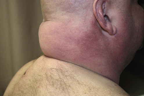
Scleredema (scleredema adultorum or scleredema of Buschke) is a connective tissue disorder characterized by progressive, symmetric induration and thickening of the skin secondary to increased amounts of collagen and glycosaminoglycans. Clinically, scleredema most commonly involves the posterior neck, shoulders, trunk, face, and arms. The three clinical forms are: (1) scleredema following acute viral or bacterial infection, which usually resolves spontaneously in 6 months to 2 years; (2) scleredema associated with diabetes mellitus, which persists indefinitely; and (3) scleredema associated with malignancy (monoclonal gammopathy, insulinoma, and carcinoma of the gallbladder).
Treatment of scleredema is difficult and usually unsatisfactory (or frequently inadequate) however, there are case-based data to support the effectiveness of several therapies. In many cases, a candid discussion with the patient regarding limitations of treatment, cost, and side effects will lead to a decision to withhold treatment. This is particularly appropriate in patients with the post-infectious form, which can resolve spontaneously without any specific therapy. Of course, identification of a specific etiology, such as streptococcal pharyngitis, should lead to appropriate antibiotic treatment, even in the absence of evidence that antibiotics would alter the rate of clearing in this self-limited form of scleredema. In the forms associated with diabetes mellitus and monoclonal gammopathy, progressive involvement can lead to discomfort, unsightly thickening, and even systemic complications such as restrictive pulmonary function, dysphagia secondary to tongue swelling, and cardiac arrhythmias. In these cases, patients will demand treatment.
Bath or cream PUVA is recommended as initial therapy in moderately severe disease. More recently, narrowband UVB and UVA1 have shown to be moderately effective. Electron beam therapy is the primary recommendation for patients with severe disease, especially cases with restrictive pulmonary function. Alternative therapies include cyclosporine and high-dose penicillin. Anti-diabetic therapy has no effect on the evolution of scleredema in diabetics, as the progression of scleredema has been found to be unrelated to control of serum glucose levels.
Venencie PY, Powell FC, Su D, Perry HO. J Am Acad Dermatol 1984; 11: 128–34.
Rongioletti F, Rebora A. In: Bolognia J, Jorizzo J, Schaffer JV, eds. Dermatology, 3rd ed. Edinburgh: Elsevier, 2012; 687–98.
Fasting blood sugar, glucose tolerance test, hemoglobin A1c (glycosylated hemoglobin)
Serum protein electrophoresis, immunoelectrophoresis
Antistreptolysin O, bacterial culture, other efforts to identify an acute infectious agent, erythrocyte sedimentation rate
Alp H, Orbak Z, Aktas A. Pediatr Int 2003; 45: 101–3.
In 65–95% of cases, scleredema occurs within a few days to 6 weeks after an acute febrile illness. Of these infections, 58% are streptococcal. These infections may present as tonsillitis, pharyngitis, scarlet fever, erysipelas, cervical adenitis, pneumonia, otitis media, pyoderma, impetigo, or rheumatic fever. Appropriate studies will rapidly determine if an underlying infection is associated with scleredema.
Since 50% of cases of scleredema occur in childhood, the adultorum appellation is a misnomer.
Kovary PM, Vakilzadeh F, Macher E, Zaun H, Merk H, Goerz G. Arch Dermatol 1981; 117: 536–9.
Immunoelectrophoresis studies in six patients with severe persistent scleredema revealed that three patients also had a monoclonal gammopathy, and many subsequent reports have confirmed this association. The skin manifestations often precede the development of the gammopathy, and, therefore, immunoelectrophoresis should be performed at regular intervals in all cases of widespread scleredema.
Manchanda Y, Das S, Sharma VK, Srivastava DN. Br J Dermatol 2005; 152: 1373–4.
Scleredema has been associated with internal malignancies including one report in a female with carcinoma of the gallbladder.
When clinically indicated, an evaluation for internal malignancies may be appropriate.
Fleischmajer R. Arch Dermatol 1970; 101: 21–6.
Scleredema in patients with diabetes mellitus generally follows a progressive course that is unrelated to control of serum glucose levels.
A review of 33 cases of scleredema, in which systemic corticosteroids, methotrexate, and D-penicillamine demonstrated no effect on the disease course. Both diabetic and non-diabetic patients were studied, and, although some patients in both groups suffered from mild complications such as dysphagia and ECG changes, the authors emphasize that scleredema is generally a mild disease that poses no threat to overall health.
Curtis AC, Shulak BM. Arch Dermatol 1965; 92: 526–41.
A review of 223 scleredema patients revealed that 25% have had symptoms for more than 2 years. The following treatments demonstrated no therapeutic benefit: calcium gluconate, estradiol, fever, hot baths, hyaluronidase, nicotinic acid, ovarian extract, p-aminobenzoate, penicillin, pituitary extract, corticosteroids, thyroid hormone, and vitamin D.
Because of the expense and potential side effects of the second-line therapies discussed below, as well as the recognition that many patients with mild scleredema either improve without treatment or do not respond to the therapy, a policy of ‘do no harm’ and withholding of treatment is recommended in patients with mild disease.
Xiao T, Yang Z, He C, Chen H. J Dermatol 2007; 34: 270–2.
A 56-year-old Chinese man with scleredema was treated with NB-UVB at an initial dose of 0.2 J/cm2. This was administered three times weekly and increased by 25% per treatment. Significant improvement occurred after 10 treatments and the patient received a total of 54 exposures (33.8 J/cm2 dose). Beneficial effects were still evident 14 months after treatment was discontinued. NB-UVB phototherapy has several advantages over PUVA. It has shorter irradiation times, no need for protective glasses following treatment, and reduced risk of photocarcinogenesis. Further large-scale studies are needed to define the role of NB-UVB in scleredema.
Tuchinda C, Kerr H, Taylor C, Jacobe H, Bergamo B, Elmets C, et al. Photodermatol Photoimmunol Photomed 2006; 22: 247–53.
In this retrospective study, six patients with scleredema adultorum were treated with UVA1 phototherapy (five received low-dose regimens and one a medium-dose regimen). One patient developed polymorphous light eruption and therapy was discontinued. Moderate to good responses were seen in four out of five patients treated. In recalcitrant cases UVA1 phototherapy should be considered as a therapeutic option.
Kroft EB, de Jong EM. Arch Dermatol 2008; 144(7): 947–8.
Three cases of scleredema diabeticorum with substantial clinical improvement after a course of UVA1 radiation therapy. Two patients were treated with 35 J/cm2 per session and the third patient with 60 J/cm2 per session. Clinical improvement and durometer measurements indicated skin softening and increased mobility in all three patients.
Thumpimukvatana N, Wongpraparut C, Lim HW. J Dermatol 2010; 37: 1036–9.
UVA1 phototherapy at 60 J/cm2 produced substantial clinical improvement in two patients who also had type 2 diabetes mellitus. After 40 treatments one patient continued to have more than a 75% clinical improvement 2 years post treatment. The second patient demonstrated clinical improvement for 10 months but subsequently required additional treatments.
Yüksek J, Sezer E, Köseoğlu D, Markoç F, Yıldız H. Photodermatol Photoimmunol Photomed 2010; 26: 257–60.
A 23-year-old female with biopsy-confirmed scleredema was treated with low-dose broadband UVA phototherapy 15 J/cm2, three times weekly for 40 sessions (total cumulative dose of 600 J/cm2) and colchicine 1800 mg/day for 1 year. Histopathologic and clinical improvement was noted. The authors conclude that more long-term follow-up studies in a larger series are needed.
Tamburin LM, Pena JR, Meredith R. Arch Dermatol 1998; 134: 419–22.
A patient with scleredema and insulin-dependent diabetes mellitus experienced complete resolution of skin induration after receiving electron beam therapy twice weekly for 36 days. Prior treatment with topical, intralesional and systemic corticosteroids had failed. This patient also suffered significant restrictive pulmonary disease thought to be secondary to scleredema, and pulmonary function tests following electron beam therapy revealed marked improvement in lung function. This treatment should be considered in patients with severe persistent disease and systemic complications.
Angeli-Besson C, Koeppel M, Jacquet P, Andrac L, Sayag J. Br J Dermatol 1994; 130: 394–7.
Ten sessions of electron beam therapy produced significant clinical improvement in the skin lesions of a patient with scleredema associated with IgAκ monoclonal gammopathy. A trial of factor XIII and cyclofenil had proved unsuccessful. Although the patient had initially responded to systemic corticosteroids, she subsequently became resistant and the condition progressed. At that point, electron beam therapy was begun.
Hager CM, Sobhi HA, Humzelmann N, Wickenhauser C, Scharenberg R, Krieg T,et al. J Am Acad Dermatol 1998; 38: 240–2.
Bath PUVA therapy (median of 59 treatments) produced substantial clinical improvement in three patients with scleredema. Although one patient did not have a history of diabetes mellitus or preceding infection, the remaining two patients had diabetes. The successful use of bath PUVA in these cases may be significant, since diabetic patients with scleredema tend to have a long unremitting course without treatment.
Grundmann-Kollmann M, Ochsendorf F, Zollner TM, Spieth K, Kaufmann R, Podda M. Br J Dermatol 2000; 142: 1058–9.
Cream PUVA therapy (35 irradiations) produced marked clinical improvement and softening of the skin in a patient with a 10-year history of scleredema who also suffered non-insulin-dependent diabetes mellitus. Bath PUVA was contraindicated in this patient due to coronary artery disease, and prior treatment with intravenous penicillin was unsuccessful. Cream PUVA therapy is easier to administer than bath PUVA and may become an important treatment option for scleredema.
Bray SM, Varghese S, English JC 3rd. Arch Dermatol 2010; 146: 453–4.
A 42-year-old man with diabetes-associated scleredema was treated with hydroxychloroquine and psoralen UVA without clinical improvement. Physical therapy with ultrasonic massage, active range-of-motion, and flexibility exercises three times a week was initiated. Ultrasonic massage therapy was performed using a Dynatron 709 series machine with the following parameters: 100%, 1 MHz, and 1.5 W/cm2 for 10 minutes per session. The patient showed objective improvement in nine of 10 range-of-motion parameters measured after 1 year of physical therapy. Ultrasonic massage and physical therapy may improve the quality of life for patients with scleredema.
Alsaeedi SH, Lee P. J Rheumatol 2010; 37: 2636–7.
A 61-year-old woman with diabetic scleredema was treated with tamoxifen 20 mg twice daily after failing to improve after treatment with methotrexate and D-penicillamine. After 2 months, her symptoms improved and after 48 months there was clinical evidence of softening of the skin on her back as well as improvement of shoulder flexibility. A 54-year-old female with diabetic scleredema was also treated with tamoxifen 20 mg twice daily and demonstrated similar improvement. The authors conclude that tamoxifen’s antifibrotic effect may be an effective treatment for scleredema.
Krasagakis K, Hettmannsperger U, Trautmann C, Tebbe B, Garbe C. Br J Dermatol 1996; 134: 597–8.
High-dose (3 × 10⁶ IU daily) intravenous penicillin for 7 days led to a reduction in the degree of scleredema in a patient with comorbid severe diabetes mellitus. The antifibrotic action of penicillin, causing a decreased dermal thickness, is postulated to be the mechanism of action in this case. The observation that treatment with erythromycin was not effective in this patient and that antibiotics in general have not been successful in treating scleredema supports the role of an antifibrotic rather than an antibiotic action of penicillin at work in this case.
Mattheou-Vakali G, Ioannides D, Thomas T, Lazaridou E, Tsogas P, Minas A. J Am Acad Dermatol 1996; 35: 990–1.
Cyclosporine at a dose of 5 mg/kg daily for 5 weeks was effective in completely clearing scleredema in two patients, neither of whom had coexistent monoclonal gammopathy or diabetes mellitus. Both patients presented with the post-infectious form of scleredema and thus would have likely experienced resolution of their symptoms even without treatment.
Stables GI, Taylor PC, Highet AS. Br J Dermatol 2000; 142: 781–3.
A patient with scleredema associated with paraproteinemia demonstrated a significant improvement in skin lesions following treatment with extracorporeal photopheresis. When the treatment sessions were reduced from two per month to one, the patient’s condition deteriorated, suggesting that the improvement was due to therapy rather than spontaneous resolution. Since scleredema associated with paraproteinemia usually has a progressive course, this treatment should be considered in patients with a similar presentation.
Koga N. Ther Apher 2001; 5: 506–12.
A 59-year-old woman with diabetic scleredema and familial hypercholesterolemia, treated with weekly LDL apheresis over a period of 3 years, had significant improvement in her scleredema when her lipid levels were reduced to normal. Histopathologic as well as clinical improvement was noted. The author concluded that LDL apheresis therapy is an effective second-line treatment for resistant diabetic scleredema.
Santos-Juanes J, Osuna CG, Iglesias JR, De Quiros JF, del Rio JS. Int J Dermatol 2001; 40: 720–1.
A 70-year-old woman with IgA myeloma and scleredema was treated with oral melphalan and prednisone over a 6-month period. During the 18 months she was followed, clinical evidence of softening of the indurated and taut skin was observed. Similar cases were cited in which chemotherapy for myeloma resulted in significant improvement of associated scleredema.
Treatment of Skin Disease Comprehensive Therapeutic Strategies 4e
WhatsApp us


 Identify and treat any underlying disease (diabetes mellitus, monoclonal gammopathy, acute infection)
 Conservative management using the principle ‘do no harm’
 Narrowband UVB phototherapy
 UVA1 phototherapy
 Electron beam therapy
 Bath PUVA and cream PUVA
 Physical therapy
 Tamoxifen
 High-dose penicillin
 Cyclosporine
 Extracorporeal photephoresis
 Chemotherapy (melphalan)