Xanthomas

Published on 18/03/2015 by admin

Filed under Dermatology

Last modified 18/03/2015

Print this page

rate 1 star rate 2 star rate 3 star rate 4 star rate 5 star
Your rating: none, Average: 0 (0 votes)

This article have been viewed 1277 times

Xanthomas

Lucile E. White, Marcelo G. Horenstein and Christopher R. Shea

Evidence Levels:  A Double-blind study  B Clinical trial ≥ 20 subjects  C Clinical trial < 20 subjects  D Series ≥ 5 subjects  E Anecdotal case reports

image

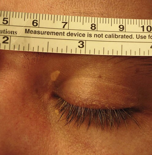
(Courtesy of Arlene Ruiz de Luzuriaga, MD, MPH)

Xanthomas are flat, yellow plaques or nodules consisting of abnormal lipid deposits. Clinically, xanthomas can be classified as eruptive, tuberoeruptive, tuberous, tendinous, verruciform, or plane. Plane xanthomas are the most common and include xanthelasma palpebrarum, xanthoma striatum palmare, and intertriginous xanthomas. Verruciform xanthomas typically affect genital skin, and may be a consequence of lymphedema. Necrobiotic xanthogranulomas are scarring, commonly ulcerated nodules with a predilection for the periorbital areas, and associated with paraproteinemia.

Management strategy

Xanthomas may be idiopathic or a sign of underlying hyperlipidemia. Diagnosing and treating the underlying disease is necessary not only to decrease the size of the xanthomas, but also to reduce the risk of atherosclerosis associated with lipoprotein disorders. Treatment of the hyperlipidemia initially consists of diet and lipid-lowering agents such as statins, fibrates, bile acid-binding resins, probucol, or nicotinic acid. The lipid-lowering effects of these agents have been well documented, but few studies document the efficacy of these drugs at resolving xanthomas, limiting an evidence-based evaluation of what would appear to be a rational management approach. Anecdotally, eruptive xanthomas typically appear to resolve within weeks of initiating systemic treatment, and tuberous xanthomas after some months, but tendinous xanthomas may take years to resolve or even persist indefinitely. Surgery or locally destructive modalities can be used for idiopathic or unresponsive xanthomas.

Specific investigations

Excluding an underlying condition is essential in the management of most clinical forms of xanthomas. Eruptive xanthomas typically occur in the setting of hypertriglyceridemia. Hypertriglyceridemia can be the result of lipoprotein lipase deficiency, familial hyperlipoproteinemia, or secondary causes such as diabetes mellitus, alcohol ingestion, or exogenous estrogens. Tuberoeruptive and tuberous xanthomas represent parts of a spectrum and are seen most commonly in the setting of familial dysbetalipoproteinemia. Tuberous xanthomas may also be a presentation of homozygous familial hypercholesterolemia, cerebrotendinous xanthomatosis (associated with neuropsychiatric symptoms), or sitosterolemia. Patients with sitosterolemia and cerebrotendinous xanthomatosis may have normal serum lipid panels; therefore diagnosis may require liquid chromatography for plant sterols or urinary gas chromatography. Tendinous xanthomas may also occur with cerebrotendinous xanthomatosis, sitosterolemia or, more commonly, heterozygous familial hypercholesterolemia. Certain xanthomas are diagnostic for an inherited hyperlipidemia: xanthoma striatum palmare for familial dysbetalipoproteinemia, and intertriginous xanthomas for homozygous familial hypercholesterolemia. Even in the presence of these classic presentations, a serum lipid panel is still indicated to confirm the diagnosis.

Regarding xanthelasma palpebrarum, a type of plane xanthoma, levels of total cholesterol are elevated in only about half of patients; however, patients with xanthelasma may have elevated apolipoprotein B and decreased apolipoprotein A1 levels, as well as subclinical increases in carotid intima-media thickness. Moreover, xanthelasma is a risk factor for myocardial infarction, ischemic heart disease, severe atherosclerosis, and death in the general population, independently of plasma cholesterol and triglyceride concentrations. Less commonly, plane xanthomas can signal a monoclonal gammopathy. In this situation, the differential diagnosis of necrobiotic xanthogranuloma with paraproteinemia, a condition usually associated with lymphoproliferative disorders, should be considered.

First-line therapies

imageLow-fat diet and systemic lipid-lowering therapy: statins, bile acid-binding resins, fibrates, and/or nicotinic acid B