Published on 18/03/2015 by admin
Filed under Dermatology
Last modified 22/04/2025
This article have been viewed 1585 times
David P. D’Cruz
Evidence Levels: A Double-blind study B Clinical trial ≥ 20 subjects C Clinical trial < 20 subjects D Series ≥ 5 subjects E Anecdotal case reports
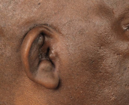
Relapsing polychondritis is an autoimmune rheumatic disorder characterized by cartilage inflammation. Characteristic features include nasal bridge, auricular and ocular inflammation, and major airway disease. It is rare with around 800 patients described in the literature and there is often delay in establishing a diagnosis.
The diagnosis is established by the presence of chondritis in two of three characteristic anatomical sites: auricular, nasal, laryngotracheal, or one of these sites and two other features, including ocular inflammation, audiovestibular damage or seronegative inflammatory arthritis. In most patients, histological confirmation is not necessary for diagnosis.
Auricular chondritis manifests as ear pain, redness, and swelling with sparing of the non-cartilaginous lobule. After repeated relapses, the pinnae may be floppy and distorted or have a cauliflower like appearance. The ear may become rigid following extensive calcification. Recurrent nasal chondritis results in a saddle-nose deformity.
Skin manifestations include oral ulceration, sometimes with genital ulceration, nodules, purpura, papules, sterile pustules, superficial phlebitis, livedo reticularis, skin ulcers, and distal necrosis.
Laryngeal chondritis presents with hoarseness, tracheal ring tenderness, cough, breathlessness, and stridor. All patients with suspected pulmonary disease should undergo computed tomography (CT) imaging including end-inspiratory and dynamic expiratory volumetric imaging. End-inspiratory scanning may reveal tracheal and bronchial stenosis, wall thickening, and calcification. Expiratory scans may demonstrate tracheobronchial malacia with airway collapse and air trapping.
Cardiovascular complications eventually occur in half of all patients and are the second most frequent cause of mortality. Aortic valve inflammation, the most common cardiac manifestation (10% of patients), may occur in asymptomatic patients and can silently progress during seemingly effective systemic corticosteroid therapy. Atrioventricular block, mitral regurgitation, and acute pericarditis may also occur.
Central and peripheral nerve involvement is rare. Cranial nerve lesions are the most common, but other complications include seizures, cerebral dysfunction, confusion, headaches, cerebral aneurysm, and rhomboencephalitis.
All patients should be monitored for the development of renal disease with routine urine dip testing for blood and protein.
Disease activity is assessed clinically by standard methods. There is now an objective scoring system, yet to be validated in clinical practice, that may provide objective disease assessment, for use in clinical studies.
Pearson CM, Kline HM, Newcomer VD. N Engl J Med 1960; 263: 51–8.
In this seminal paper, Pearson and colleagues coined the term ‘relapsing polychondritis’. They recognized the episodic inflammatory nature and the significant morbidity and mortality of relapsing polychondritis. Excellent clinical responses to prednisone were reported with relapses occurring on tapering the corticosteroid dosage.
Michet CJ Jr, McKenna CH, Luthra HS, O’Fallon WM. Ann Intern Med 1986; 104: 74–8.
Any major airway involvement in relapsing polychondritis is associated with a poorer prognosis than isolated head and neck disease affecting the ears, eyes or nose. This report describes the poor long-term prognostic factors: 5- and 10-year survival rates were 74% and 55%, respectively. The most frequent causes of death were infection, systemic vasculitis, and malignancy, and only 10% of the deaths were attributed to airway involvement by chondritis. Anemia at diagnosis was a marker for decreased survival. Corticosteroid therapy did not influence survival and is not a disease-modifying therapy.
Bachor E, Blevins NH, Karmody C, Kühnel T. Auris Nasus Larynx 2006; 33: 135–41.
A good description of the broad spectrum of ear manifestations. No patients died, suggesting patients with limited disease have a good prognosis.
Up to 46% of patients have impaired hearing, and inadequately treated patients can suffer permanent hearing loss. Screening audiometry is highlighted.
Yoo JH, Chodosh J, Dana R. Semin Ophthalmol 2011; 26: 261–9.
A thorough description of the ocular manifestations of relapsing polychondritis and an up-to-date review of the current therapeutic approaches to systemic disease manifestations.
Lee KS, Ernst A, Trentham DE, Lunn W, Feller-Kopman DJ, Boiselle PM. Radiology 2006; 240: 565–73.
Dynamic expiratory CT scans demonstrated abnormalities such as tracheomalacia and air trapping in 94% of relapsing polychondritis patients who had pulmonary symptoms, yet only half the patients demonstrated abnormalities on routine inspiratory CT scans. The most common findings were air trapping (94%), malacia (72%), and calcification (39%).
Expiratory CT scans show clinically relevant bronchopulmonary abnormalities earlier than standard inspiratory CT scans, allowing earlier institution of aggressive therapy to prevent disease progression.
Airway manifestations are ultimately present in over 50% of patients, and are the leading cause of death. Airway obstruction may be asymptomatic in the earlier stages, detected only on pulmonary function testing. Other sources stress the importance of plain radiography, CT, and MRI for early detection of tracheal narrowing and upper airways disease.
Francès C, el Rassi R, Laporte JL, Rybojad M, Papo T, Piette JC. Medicine (Baltimore) 2001; 80: 173–9.
The largest series in the literature of 200 patients. Oral ulceration was the most common finding in patients with relapsing polychondritis who did not have any other disease. Nodules on the limbs were the most common skin lesions and were described as erythema nodosum-like lesions with septal panniculitis. Histologic findings included vasculitis in 19 patients (leukocytoclastic in 17 and lymphocytic in two), neutrophilic infiltrates in six, thrombosis of skin vessels in four, septal panniculitis in three, and minor non-specific changes in two patients. Myelodysplastic syndrome was described in 22 patients who were more likely to have dermatological manifestations, including limb nodules, purpura, papules, and livedo reticularis. The authors recommend that elderly patients with relapsing polychondritis and skin lesions should be investigated for an underlying myelodysplastic syndrome. This confirmed previous data from Hebbar et al. 1995 (see below).
Hebbar M, Brouillard M, Wattel E, Decoulx M, Hatron PY, Devulder B, et al. Leukemia 1995; 9: 731–3.
Twenty-eight percent (five of 18) of relapsing polychondritis patients over a period of 13 years were found to have myelodysplastic syndromes.
Anemia is a poor prognostic sign in patients with relapsing polychondritis; those with concurrent myelodysplasia often develop refractory anemia requiring transfusions. Several patients have progressed to leukemia.
Del Rosso A, Rosa Petrix N, Pratesi M, Bini A. Semin Arthritis Rheum 1997; 26: 840–4.
The most frequent cardiovascular abnormalities are aortic regurgitation and aortic aneurysm and several cases of atrioventricular block, mitral regurgitation, and acute pericarditis have been reported.
For early diagnosis and treatment of these severe complications, periodic cardiovascular examination is mandatory.
Nadeau SE. Neurol Clin 2002; 20: 151–78.
This review covers the main neurological complications of relapsing polychondritis, amongst other conditions.
Chang-Miller A, Okamura M, Torres VE, Michet CJ, Wagoner RD, Donadio JV Jr, Offord KP, Holley KE. Medicine (Baltimore) 1987; 66: 202–17.
A description of renal involvement in 29 of 129 patients. Renal patients tended to be older and had a significantly worse survival than those without renal involvement.
Arnaud L, Devilliers H, Peng SL, Mathian A, et al. for the RPDAI study group. Autoimmun Rev 2012; 12: 204–9.
A novel disease activity scoring system is proposed which is simple to use and, once validated, may be useful in clinical studies.
Treatment of relapsing polychondritis is aimed at reducing inflammation, which may progressively destroy the ears, nose, eyes, joints, respiratory tract, and cardiovascular system. The most common cause of mortality is laryngotracheal involvement. A multidisciplinary approach, including referral to specialist centers, is crucial to evaluate and treat multiple organ involvement. There are no controlled trials: treatment is empirical and should be tailored to the severity of disease.
Mildly affected patients may be managed using non-steroidal anti-inflammatory agents. They should be used sparingly and for short periods given the long-term risks of gastrointestinal, renal, and cardiovascular complications. Colchicine 0.5 mg twice daily and dapsone 50–100 mg daily have also provided benefit but toxicity may occur, especially with dapsone and G6PD (glucose-6-phosphate dehydrogenase) deficiency.
Corticosteroids are widely used for more severe disease. Life-threatening airway obstruction may be treated with pulsed intravenous methylprednisolone (500 mg to 1 g/day for 3 days). The traditional dose of 1 mg/kg/day is completely non-evidence-based, is excessive, and should be abandoned. High-dose corticosteroids contribute significantly to long-term damage which is associated with premature mortality. Most patients with moderate disease will benefit from doses of prednisolone between 10 and 20 mg daily in a tapering dose irrespective of body weight. The lowest possible maintenance dose should be used and some patients may be able to use intermittent short courses of prednisolone 20 mg daily for 1 to 2 weeks. Bone prophylaxis with calcium and vitamin D supplementation should be considered in corticosteroid-dependent patients.
Second-line therapies should be considered for patients unable to taper corticosteroid doses. The most commonly used agents are methotrexate (considered first-line by most experts) 5–25 mg weekly, azathioprine (1.5–2 mg/kg/day), cyclosporine 5 mg/kg/day, mycophenolate mofetil 2–3 g/day, leflunomide, and chlorambucil. Intravenous cyclophosphamide has been used for severe rapidly progressive and life-threatening disease, especially for aortitis or glomerulonephritis. Plasma exchange and intravenous immuneglobulin (IVIG) therapy are usually not successful but isolated case reports have described clinical benefit.
Biologic agents are increasingly being used off label for treatment resistant patients with some success. Infliximab, adalimumab, etanercept, tocilizumab, and anakinra have all been reported to improve disease control in isolated case reports.
Topical steroids may be used to treat skin manifestations and are standard therapy for ocular inflammation. Inhaled steroids may be useful in mild airway inflammation and nebulized ephedrine has been used occasionally.
Respiratory support with continuous positive airway pressure and other non-invasive ventilation devices may improve quality of life for patients where surgical intervention or stenting is not feasible.
Surgical intervention may be needed acutely when tracheostomy is needed for tracheal stenosis. Other surgical interventions to manage fibrotic or stenotic complications should only be considered electively when the disease is in remission. Tracheal surgery, including reconstruction procedures, may be needed for localized stenosis. Balloon dilatation may be used either alone or in combination with other surgical procedures. Large airway stenting has been successfully used, although metallic stents, usually used for malignant disease, may erode the airways over time. Silicon stents are more likely to migrate or be expectorated. Endobronchial ultrasound has been used to identify localized stenotic lesions and to measure airway size prior to positioning of stents.
Cardiac valve replacement and aortic surgery are associated with significant surgical morbidity and mortality but several case reports describe successful outcomes. Any general anesthetic which requires intubation and ventilation prior to surgery requires careful preoperative anesthetic review.
Otolaryngological evaluation
Audiometry
Pulmonary evaluation
Chest radiograph and/or CT/MRI
Dynamic expiratory CT examination
Pulmonary function tests (especially flow/volume loops)
Endobronchial ultrasonography
Cardiovascular examination
Electrocardiogram and/or echocardiogram (CT/MRI may also be useful)
Ophthalmologic examination
Urinalysis
Barranco VP, Monor DB, Solomon H. Arch Dermatol 1976; 112: 1268–88.
Three patients were successfully treated with dapsone (100–200 mg daily), each showing complete resolution of an acute attack of relapsing polychondritis within 2 weeks of starting therapy.
Although not all patients respond to dapsone, those that do may improve dramatically within 1 to 2 weeks. The effectiveness of dapsone seems to be dose dependent, with 200 mg daily being the most commonly reported effective dose. G6PD levels should be measured prior to treatment to avoid the risk of dapsone-induced hemolytic anemia.
Kent PD, Michet CJ Jr, Luthra HS. Curr Opin Rheumatol 2004; 16: 56–61.
A well-written and comprehensive review of the clinical features, assessment and management of relapsing polychondritis. Recommendations for corticosteroid doses are given ranging from 10–20 mg daily of prednisolone for mild to moderate disease up to high-dose prednisolone and intravenous methylprednisolone 500 mg to 1 g/day for 3 days.
Trentham DE, Le CH. Ann Intern Med 1998; 129: 114–22.
In this review, 23 of 31 patients were able to reduce their prednisone dose from an average of 19 mg daily to 5 mg daily by adding methotrexate (average weekly methotrexate dose 15.5 mg).
A good review article.
Lahmer T, Treiber M, von Werder A, Foerger F, Knopf A, Heemann U, Thuermel K. Autoimmun Rev 2010; 9: 540–6.
An excellent clinical review of relapsing polychondritis and its treatment, covering the use of corticosteroids, immunosuppressive agents, and biologics in some depth.
Goldenberg G, Sangueza OP, Jorizzo JL. J Dermatol Treat 2006; 17: 158–9.
A 50-year-old man with bilateral ear pain was successfully treated with a prednisone taper and mycophenolate 3 g/day (increased from an initial dose of 2 g/day). At 17 months’ follow-up the patient maintained his improvement on prednisone 5 mg daily and mycophenolate 3 g/day.
Richez C, Dumoulin C, Coutoly X, Schaeverbeke T. Clin Exp Rheumatol 2004; 22: 629–31.
One patient had marked improvement in symptoms 4 days after an infusion of 4 mg/kg infliximab. Control was maintained with repeat treatments every 6 to 8 weeks.
Infliximab seems to be an effective therapy for relapsing polychondritis unresponsive to conventional therapy, as well as a steroid-sparing agent.
Subrahmanyam P, Balakrishnan C, Dasgupta B. Scand J Rheumatol 2008; 37: 239–40.
A 54-year-old woman with relapsing polychondritis complicated by tracheomalacia experienced a dramatic improvement in her symptoms and an 18-month sustained response after etanercept was substituted for infliximab.
Seymour MW, Home DM, Williams RO, Allard SA. Rheumatology 2007; 46: 1739–41.
A 43-year-old woman who had undergone aortic valve replacement continued to experience aortitis despite a variety of immunosuppressive drugs and cytotoxic agents. Despite an initial good response to infliximab, after 10 months her symptoms returned. Switching her antitumor necrosis factor (TNF) agent to adalimumab and continuing methotrexate and corticosteroids resulted in improvement; she has subsequently been maintained on adalimumab and low-dose corticosteroids.
These two reports suggest that switching anti-TNF agents may benefit patients with recalcitrant relapsing polychondritis.
Terrier B, Aouba A, Bienvenu B, Bloch-Queyrat C, Delair E, Mallet J, et al. Clin Exp Rheumatol 2007; 25: 136–8.
A young woman with relapsing episodes of nasal chondritis and severe scleritis with scleromalacia received IVIG every 3 then every 4 weeks (2 g/kg on 2 days) in association with 25 mg/day of prednisone after treatment with corticosteroids, infliximab, methotrexate, mycophenolate, and cyclophosphamide failed to control her symptoms. Dramatic improvement of symptoms was sustained for 11 months, but when IVIG treatments were spaced every 6 weeks hearing loss and episcleritis recurred. Re-institution treatment every fourth week brought her symptoms under control.
Tsuburai T, Suzuki M, Tsurikisawa N, Ono E, Oshikata C, Taniguchi M, et al. Respirology 2009; 14: 299–301.
A 19-year-old patient with relapsing polychondritis used high-dose fluticasone propionate which reduced oral corticosteroid requirements and dramatically decreased the patient’s obstructive airway impairment.
Adliff M, Ngato D, Keshavjee S, Brenaman S, Granton JT. Chest 1997; 112: 1701–4.
A patient with relapsing polychondritis of the trachea and bronchi was treated with nasal continuous positive airway pressure. This approach is now widely used.
Sarodia SD, Dasgupta A, Mehta AC. Chest 1999; 116: 1669–75.
Five patients with severe respiratory involvement (three of whom required continuous mechanical ventilation due to airway collapse) benefited from placement of self-expandable metallic tracheobronchial stents. A total of 17 stents of varying sizes were placed in these patients over a period of 3 years, with favorable outcomes in four patients.
When severe, progressive disease leads to extensive destruction of the tracheobronchial tree despite maximal medical therapy, tracheostomy or tracheobronchial stent placement may preserve or improve respiratory function and reduce patients’ reliance on mechanical ventilation.
Miyazu Y, Miyazawa T, Kurimoto N, Iwamoto Y, Ishida A, Kanoh K, et al. Chest 2003; 124: 2393–5.
Endobronchial ultrasonography revealed poorly defined bronchial wall structure with two patterns of cartilaginous damage: fragmentation, and edema. Successful treatment was achieved by the implantation of nitinol stents, the sizes of which were determined by endobronchial ultrasonography.
Dib C, Moustafa SE, Mookadam M, Zehr KJ, Michet CJ Jr, Mookadam F. Mayo Clin Proc 2006; 81: 772–6.
Clinically important aortic or mitral regurgitation occurs in about 10% of relapsing polychondritis patients, aortic regurgitation being the more common and more urgent. In this retrospective series and literature review the mean time between initial onset of relapsing polychondritis and surgery was about 5 years. In contrast to previous reports of 70% 1-year mortality after valve replacement, in this analysis 50% of patients were alive 1 year after surgery.
Aortic regurgitation is a serious complication of relapsing polychondritis. Baseline chest CT, MRI, or transesophageal echocardiography should be performed upon diagnosis and repeated every 6 months. All aortic segments should be regularly evaluated because involvement of multiple thoracic and abdominal aneurysms has been reported in several patients.
Biro P, Rohling R, Schmid S, Matter C, Lang M. J Clin Anaesth 1994; 6: 59–62.
This case report highlights the need for careful preoperative evaluation of vital organ functions, with particular reference to airway management, so that the anesthetic approach can be tailored to the individual needs of the patient.
Treatment of Skin Disease Comprehensive Therapeutic Strategies 4e
WhatsApp us