Published on 18/03/2015 by admin
Filed under Dermatology
Last modified 22/04/2025
This article have been viewed 2631 times
Danielle M. DeHoratius
Evidence Levels: A Double-blind study B Clinical trial ≥ 20 subjects C Clinical trial < 20 subjects D Series ≥ 5 subjects E Anecdotal case reports
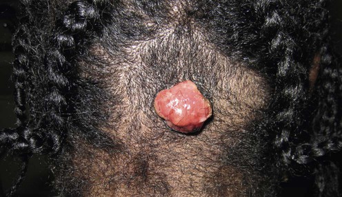
Pyogenic granuloma, also known as a lobular capillary hemangioma, is a common benign vascular growth. It can rapidly appear and is a solitary, erythematous papule. Pyogenic granulomas are often friable and can frequently ulcerate. They most commonly occur in children and young adults. The etiology is unclear, although reactive neovascularization is suspected because of their occurrence at sites of previous trauma. There is no gender or racial predominance. The most common locations are the head and neck region (including the oral mucosa, especially in pregnant women – granuloma gravidarum) and digits. Occasionally, pyogenic granulomas have been found in subcutaneous or intravascular locations. The term pyogenic granuloma, however, is a misnomer, as there is not an infectious or a granulomatous component to these lesions. Over time they can resolve on their own. Dermoscopy of these lesions can be useful but should not substitute histology. The most sensitive and specific pattern is a reddish homogeneous area, white collarette, and white rail lines.
Pyogenic granulomas are most commonly managed by destruction. This can be completed through shave excision with electrocautery to the base, curettage with electrodesiccation, or cryotherapy. Histologic confirmation is beneficial as other disorders may clinically mimic pyogenic granulomas, examples being amelanotic melanoma, Kaposi’s sarcoma, and bacillary angiomatosis. There is a possibility of recurrence and/or the development of satellite lesions, but these options are less invasive and do not result in significant scarring. Complete excision requiring sutures may lower the recurrence rate and reduce the possibility of bleeding; however, a linear scar will be present. Hemostasis can be obtained by either electrocautery, silver nitrate, or argon laser photocoagulation, as all are shown to be effective. As this is a benign growth, it is important to consider the cosmetic outcome of the therapeutic intervention.
Cryotherapy is also effective. With this modality, patients should be seen within 1 to 2 weeks to assess the response and need for additional treatments. Because the pyogenic granuloma is not completely removed, there is a possibility of recurrence; additionally, no tissue is obtained for sampling.
Vascular lasers also destroy these lesions using selective photothermolysis. Usually multiple treatments are required, and there is no histologic confirmation. Pulsed dye laser has proved to be more successful with smaller lesions. For larger lesions the Nd:YAG laser has been efficacious. Sclerotherapy destroys these vascular lesions and has been reported to have a very high cure rate in experienced hands. Various application schedules of imiquimod 5% have resolved these lesions, presumably owing to its anti-angiogenic properties. Recently, photodynamic therapy has been a modality shown to be effective in the destruction of these lesions with very few adverse events.
Histology
History to assess previous trauma, medication, and topical exposures
In general, clinical suspicion is very useful in diagnosing pyogenic granulomas, although histologic confirmation is important. Pyogenic granulomas should be differentiated from other vascular lesions, especially bacillary angiomatosis. Amelanotic melanomas may mimic pyogenic granulomas. The remainder of the differential includes angiosarcoma, basal cell carcinoma, and Kaposi’s sarcoma. Because many of the methods described below result in removal, this tissue can be sent to confirm the clinical diagnosis.
Some drugs have been reported to cause pyogenic granulomas. These include oral contraceptives, isotretinoin, acitretin, reverse transcriptase inhibitors, epidermal growth factor inhibitors, systemic 5-fluorouracil, capecitabine, mTOR inhibitors, monoclonal anti-CD20 antibodies, and topical tretinoin. Recently, there have been reports of these lesions arising in both port-wine stains and cherry angiomas when treated with the pulsed dye laser. Rarely eruptive lesions have been reported in response to a drug hypersensitivity reaction.
Pagliai KA, Cohen BA. Pediatr Dermatol 2004; 21: 10–3.
A retrospective study of 128 children with pyogenic granulomas and a follow-up phone interview of 76 patients. Of these, 72.3% underwent a shave excision with electrocautery. The second most common treatment was laser therapy (16.9%). Fifty-five percent of the children in the first group reported a subtle scar, and 33% of the CO2 laser group and 44% of the pulsed dye laser group reported a similar scar. All patients were pleased with the cosmetic result.
Ghodsi SZ, Raziel M, Taheri A, Karami M, Mansoori P, Farnaghi F. Br J Dermatol 2006; 154: 671–5.
Eighty-nine patients were randomized for treatment with either liquid nitrogen cryotherapy or curettage followed by electrodesiccation. Of the 86 patients who completed the study, all had complete resolution of the lesions after one to three sessions (mean 1.42) in the cryotherapy group, and after one to two sessions (mean 1.03) in the curettage group. No scar or residual pigmentation was reported in 57% of the cryotherapy group or in 69% of the curettage group. The authors concluded that although both treatments were safe and effective, curettage should be first-line as fewer treatment sessions were necessary and cosmesis was better.
Kirschner R, Low D. Plast Reconstruct Surg 1999; 104: 1346–9.
The shave excision technique was performed on the exophytic portion and photocoagulation of the base through a glass slide. The lasers used were an argon, argon-pumped tunable yellow dye, or KTP laser. All treatments were performed until complete hemostasis was achieved. Complete resolution was seen in 18 of the 19 patients after a single treatment. Of note, the preoperative diagnosis was erroneous in 18.8% of the cases.
Although the final diagnoses were benign in nature, this technique allows the clinical diagnosis to be confirmed and minimizes scar formation.
Quitkin HM, Rosenwasser MP, Strauch RJ. J Hand Surg [Am] 2003; 28: 435–8.
Thirteen lesions were treated with simple removal and cautery of the base using silver nitrate. Postoperative care was to keep the area completely dry for 2 weeks, as the authors previously observed that recurrence was more common in moist environments. Eighty-five percent of the lesions had complete resolution with an average of 1.6 treatments. The average time to complete healing was 3.5 weeks. There was no need for expensive equipment, and this was simple and cost-effective.
Giblin AV, Clover AJ, Athanassopoulos A, Budny PG. Plast Reconstruct Aesthet Surg 2007; 60: 1030–5.
A retrospective study of 408 cases analyzed between 1994 and 2004 assessing the most successful form of treatment on the basis of multiple parameters. The excision and direct closure group had the fewest recurrences, although all techniques investigated showed an acceptably low recurrence rate.
Whatever technique is used, it must yield material for histologic analysis to ensure the exclusion of other diagnoses.
Mirshams M, Daneshpazhooh M, Mirshekari A, Taheri A, Mansoori P, Hekmat S. J Eur Acad Dermatol Venereol 2006; 20: 788–90.
A prospective observational study of 135 patients treated with liquid nitrogen cryotherapy using a cotton-tipped applicator. Patients required anywhere from one to four treatments (mean 1.58). All patients had complete resolution, with 96.2% occurring after three treatments. Minor scars were reported in 11.8%, and 5.1% had hypopigmentation.
These authors report that cryotherapy should be considered first-line as it is an easy and inexpensive technique; however, the main limitation is that no tissue is obtained for histology.
Tay YK, Weston WL, Morelli JG. Pediatrics 1997; 99: 368–70.
Twenty-two children with solitary lesions were treated with a vascular-specific (585 nm), pulsed (450 ms) dye laser using a 5 nm spot size with a laser energy of 6–7 J/cm2 without anesthesia. This treatment was successful in 91% and all healed without scarring. Fifteen patients required from two to six treatments at 2-week intervals, and seven required three or more. The two patients who did not respond had larger lesions (0.5–1 cm). No recurrences were reported during the follow-up period (6 months to 3 years). The limitation of this laser modality was that the depth of penetration was only 1 mm.
This can be a useful modality in children, as no anesthesia is required and there is minimal scarring. The drawback is that no tissue is obtained for histology.
Raulin C, Greve B, Hammes S. Arch Dermatol 2002; 138: 33–7.
One hundred patients were selected from a population-based sample and underwent treatment with the CO2 laser. The laser was first used in the continuous mode (power 15 W) and then in the pulsed mode (pulse length 0.6–0.9 ms; energy fluence 500 mJ/pulse). Follow-up was 6 weeks and 6 months, and 98 of 100 patients had complete resolution after one treatment. In 88 patients there were no visible scars, and 10 had only slight textural changes. No erythema or pigmentary changes were observed.
Hammes S, Kaiser K, Pohl L, Metelmann HR, Enk A, Raulin C. Dermatol Surg 2012; 38: 918–23.
Twenty patients were treated using this modality and required one to four treatment sessions (settings used: fluences 60–180 J/cm2, spot size of 7 mm, and a pulse duration of 40 ms). Nineteen of 20 patients had recurrence-free healing (follow-up duration 6 to 22 months) with good cosmetic results. Only slight textural changes were noted. This can be a satisfactory laser modality especially when the diameter of the lesion is larger.
Holbe HC, Frosch PJ, Herbst RA. J Am Acad Dermatol 2003; 49: 509–10.
This demonstrated a pedunculated pyogenic granuloma that was ligated using an absorbable suture tightly tied at the base. Over a few days the lesion became necrotic and fell off.
This was atraumatic and required no anesthesia, but there was also no histologic confirmation.
Tritton SM, Smith A, Wong LC, Zagarella S, Fischer G. Pediatr Dermatol 2009; 26: 269–72.
Ten healthy children with a mean age of 2.5 years were instructed to apply imiquimod 5% either once a day, twice a day, or three times per week depending on the clinical response. They were assessed either weekly or bi-weekly. All lesions were located on the face (3–6 mm) and local erythema was unanimously observed. Three had no residual disease while five had either small hypopigmented or erythematous lesions which were continuing to improve at the completion of the study. No systemic side effects were reported.
The authors suggest a trial of three times per week initially, increasing to daily if tolerated for up to 2 months. Treatment should be discontinued 1 week after complete disappearance of the lesion. This appears a reasonable alternative to surgical excision.
Moon SE, Hwang EJ, Cho KH. Arch Dermatol 2005; 141: 644–6.
Fifteen pyogenic granulomas were injected with 0.5% sodium tetradecyl sulfate until blanching appeared. Follow-up was every 1 to 2 weeks, and at 6 weeks 80% showed complete resolution. In two patients a small shallow vascular area remained which responded to the CO2 laser.
The authors felt that this treatment can be an alternative to excision because of its simplicity and lack of scarring; however, multiple treatments may be necessary to achieve resolution. There is no tissue for histologic sampling. This treatment can be considered for larger lesions or those in challenging locations.
Lee DJ, Kim EH, Jang YH, Kim YC. Arch Dermatol 2012; 148: 126–8.
Fourteen pyogenic granulomas were injected (26-gauge needle) with 0.3 mL/cm3 of 5-aminolevulinic acid, 20% solution followed by occlusion with polyurethane film. The lesions were then illuminated with red light (600–720 nm, light dose 100 J/cm2 and fluence 100 mW/cm2). Eleven patients showed a marked response and had no recurrence at 1-year follow-up. One patient showed moderate response (lesion was on the lip) and two did not respond (lesions large >1 cm). Only three patients complained of perilesional swelling.
The authors felt that this treatment can be an alternative to standard therapy especially in patients with small lesions who refuse surgery. It is important to consider the location and size of the lesion. In addition, intralesional was suggested to be more effective than topical application of the photosensitizer.
Niiyama S, Amoh Y, Katsuoka K. J Plast Reconstr Aesthet Surg 2009; 62: 153–4.
A case report involving two lesions, one on the finger and the other on the toe. They were injected with triamcinolone acetonide at the dose of 2 mg weekly for a total of seven to eight times. The lesions became smaller and were subsequently excised.
This can be used when the lesion is in an unfavorable location for simple excision.
Daya M. J Plast Reconstr Aesthet Surg 2010; 63: e331–3.
A 63-year-old woman presented with a recurrent giant pyogenic granuloma (5 cm × 6 cm). Previous treatments included surgical excision and intralesional steroids. The lesion was injected with a total of 8 mL of 0.5 mg/mL of bleomycin given under general anesthesia with tourniquet control. The lesion regressed after 2 months and did not recur. The patient reported only mild hypersensitivity within the scar.
Losa Iglesias ME, Becerro de Bengoa Vallejo R. Dermatol Surg 2010; 36: 675–8.
A report of 18 patients treated with a 98% phenol solution after a thorough cleansing of the area. The phenol was applied to the lesion in three applications of 1 minute each, consecutively. The entire tumor and a small surrounding area turned white. The areas were then treated with 10% silver sulfadiazine and 10% povidone iodine and wrapped in sterile gauze. The frequency of additional treatments varied based on size. At the 14-week endpoint, all lesions had resolved. The treatments were well tolerated with no scarring or adverse events.
This approach is simple to perform, fairly inexpensive, and relatively pain free; however, recurrence is possible and treatment may necessitate frequent office visits.
Treatment of Skin Disease Comprehensive Therapeutic Strategies 4e
WhatsApp us


 Simple shave excision/curettage with electrocautery of the base
 Full-thickness skin excision
 Cryotherapy
 Silver nitrate cautery
 Pulsed dye
 CO2 laser
 Nd:YAG laser
 Ligation
 Imiquimod 5%
 Sclerotherapy
 Photodynamic therapy
 Intralesional corticosteroids
 Intralesional bleomycin
 Topical phenol