Published on 18/03/2015 by admin
Filed under Dermatology
Last modified 22/04/2025
This article have been viewed 2554 times
Gabriele Weichert
Evidence Levels: A Double-blind study B Clinical trial ≥ 20 subjects C Clinical trial < 20 subjects D Series ≥ 5 subjects E Anecdotal case reports
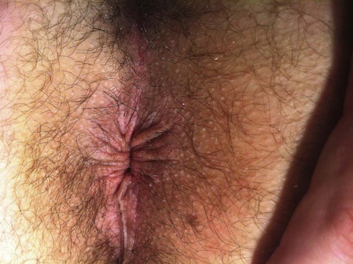
Pruritus ani represents a chronic, idiopathic intensely pruritic sensation of perianal skin. Long-standing cases are associated with significant discomfort, embarrassment, and sleep disturbance. Chronic primary pruritus ani shows lichenification and excoriations of the perianal area in the absence of primary skin disorders, infections, or neoplasms.
When evaluating a patient, any contributing primary skin disorders must be identified. These may include atopic dermatitis, psoriasis, or lichen sclerosis. Neoplasms, hemorrhoids and anal fissures, infectious etiologies such as genital warts, tinea, candidiasis, infestations such as pinworms and scabies, and bacterial infections (e.g., β-hemolytic streptococcus) must be ruled out by clinical examination and appropriate investigations. With this approach, most causes of acute pruritus ani can be identified and treated. It is the patient in whom none of the above factors are identified who suffers from chronic or idiopathic pruritus ani. History may reveal atopy or sensitive skin, leading to an increased itch sensation from all causes.
A history of the management, including cleansing habits, may reveal a sensitizer (such as a wet wipe) causing a contact dermatitis. Over-cleansing is not uncommon and may be irritating. Many of these patients suffer from low-grade fecal incontinence. This is often evident on examination of the underclothes or of the perianal skin. A history of over-zealous cleansing routines or sleep disturbance is common. Potential topical sensitizers should be stopped. A biopsy should be performed if the diagnosis is in doubt. Avoidance of toilet paper to cleanse the area after bowel movements can be helpful, as this may be abrasive.
Advise patients to cleanse the perianal skin twice a day and after bowel movements using cotton balls or cotton squares (make-up removal pads) moistened with warm water or a liquid cleanser. Patients with low-grade fecal incontinence should perform this routine several times a day. Cleansing has been shown to be as effective as topical steroids. A short (few weeks) course of a low- to mid-potency topical steroid is recommended. Caution must be taken with prolonged high-potency steroid use, as this area is prone to atrophy. Potency should be reduced as symptoms improve. Topical tacrolimus may be used in clearance or maintenance treatment. Topical zinc paste can limit the degree of irritant dermatitis in patients with fecal incontinence. An evening sedating antihistamine may provide welcome sleep in the early weeks of treatment.
If a patient fails to improve, lower gastrointestinal investigations should be considered to rule out neoplastic disease. Patch testing should be performed in patients who fail to improve. Avoidance of caffeine and increased dietary fiber may be helpful. For second-line therapy, topical capsaicin 0.006% three times a day for 4 weeks could be used. Capsaicin will cause perianal irritation in most patients during the therapy. Finally, intralesional steroid injections to the perianal skin may be considered. Intradermal methylene blue injections are reported to damage dermal nerve endings and provide relief. Injection with phenol in almond oil has been reported to be beneficial.
Bacterial swab
Fungal microscopy and culture
Biopsy
Skin scrapings for scabies
Patch testing
Gastrointestinal investigations
Bauer A, Geier J, Elsner P. J Reprod Med 2000; 45: 649–54.
A 5-year collection of patients patch tested for consideration of allergic anogenital contact dermatitis demonstrated an increased incidence of benzocaine and (chloro-) methyl-isothiazolinone allergy. A diagnosis of allergic contact dermatitis was made in 34.8%. The authors recommend using the standard tray plus dibucaine, propolis, bufexamac, and other ingredients gained from the patient’s history.
Silvestri D, Barmettler S. Dermatitis 2011; 22: 50–5.
A patient with recalcitrant pruritus ani was found to be highly nickel allergic on patch testing. A low-nickel diet led to resolution of his symptoms and recurrence appeared with re-challenge.
Dasan S, Neill SM, Donaldson DR, Scott HJ. Br J Surg 1999; 86: 1337–40.
In a series of 40 patients with pruritus ani, 34 had a recognizable dermatosis and 18 had a positive patch test result. The authors suggest early assessment by a dermatologist may be appropriate.
Farouk R, Duthie GS, Pryde A, Bartolo DC. Br J Surg 1994; 81: 603–6.
Abnormal rectal sphincter tone was demonstrated in patients with pruritus ani. This may well be the contributing factor in patients who have occult fecal leakage as a component of their itch.
Daniel GL, Longo WE, Vernava AM. Dis Colon Rectum 1994; 37: 670–4.
In this series of 104 patients presenting with pruritus ani as their primary symptom, 52% had anorectal disease (including hemorrhoids, anal fissures, genital warts, and fistulas) and 23% had an existing colorectal neoplasm found with investigations. In those without neoplasia, there was a direct correlation between increased caffeine intake and severity of irritation. Patients with primary pruritus ani improved with dietary restrictions (not defined), dietary fiber, steroid creams, and drying agents.
Al-Ghnaniem R, Short K, Pullen A, Fuller LC, Rennie JA, Leather AJ. Int J Colorectal Dis 2007; 22: 1463–7.
Nineteen patients were randomized to 1% hydrocortisone in paraffin twice daily or paraffin base for 2 weeks followed by crossover treatment for 2 weeks. Blinded analysis of visual analog score (VAS) showed a 68% reduction in VAS with steroid treatment.
Suys E. J Am Acad Dermatol 2012; 66: 327–8.
Twenty-one patients were randomized to 0.1% tacrolimus versus petrolatum bid for 4 weeks with a further 4-week crossover phase. There was improvement in itch intensity and frequency on tacrolimus but DLQI was not significantly different compared to placebo. Only one patient had temporary burning sensation.
Oztas MO, Oztas P, Onder M. Postgrad Med J 2004; 80: 295–7.
In this study, 28 patients applied topical methylprednisolone cream twice daily for 2 weeks. In a separate group, 32 patients were instructed to cleanse the perianal area twice daily with a liquid cleanser. Effectiveness was similar at 92.3% in the first and 90.6% in the second group (p>0.05).
Lysy J, Sistiery-Ittah M, Israelit Y, Shmueli A, Strauss-Liviatan N, Mindrul V, et al. Gut 2003; 52: 1323–6.
In this double-blind placebo-controlled crossover study, capsaicin 0.006% cream was applied three times a day for 1 month, followed by 1 month of control treatment using 1% menthol cream. Of 44 patients, 31 experienced partial relief of symptoms while using the capsaicin cream (p<0.0001). All patients experienced some degree of perianal burning with use of the capsaicin cream. The menthol cream was not effective.
Tomi N, Weiser R, Strohal R, et al. Br J Nurs 2012; 21: 98–102.
Twenty-eight patients applied an acrylate protection cream (Cavilon Durable Barrier Cream, 3M) daily for 3 weeks. There was an 80% reduction in VAS.
Minvielle L, Hernandez VL. Dis Colon Rectum 1969; 12: 340–3.
Nineteen patients were treated weekly for 4 weeks with intralesional triamcinolone at doses of 5–20 mg/week. At the end of the treatment period, nine of 19 (73.6%) patients had excellent progress, two of 19 reported fair improvement, and three failed to improve. No perianal atrophy was noted at the end of 4 weeks.
Sutherland AD, Faragher IG, Frizelle FA. Colorectal Dis 2009; 11: 282–7.
Forty-nine patients received perianal inection with a solution of 1% methylene blue (10 mL), 0.5% marcaine (20 mL), and methylprednisolone 40 mg/mL (1 mL) under general anesthesia; 96% had improved symptoms and 57% were symptom free. There was transient incontinence in seven patients.
Methylene blue has been shown by electron microscopy to cause damage to dermal nerve endings. This is the proposed mechanism of action for this modality.
Shafik A. Int Surg 1990; 75: 43–6.
In this series, 67 patients were treated with an injection of 5% phenol in almond oil: 62/67 (92.4%) experienced complete relief. Five patients relapsed after a period of remission. Repeat injection led to cure.
Treatment of Skin Disease Comprehensive Therapeutic Strategies 4e
WhatsApp us


 Topical corticosteroids
 Topical tacrolimus
 Hygiene
 Topical capsaicin
 Topical acrylate barrier
 Intralesional corticosteroids
 Intradermal methylene blue
 Subcutaneous phenol injection