Perforating dermatoses
 Reactive perforating collagenosis, which is characterized by the transepidermal elimination of collagen
 Elastosis perforans serpiginosa (EPS), characterized by transepidermal extrusion of elastic material
 Perforating folliculitis, in which epidermal perforation involves hair follicles
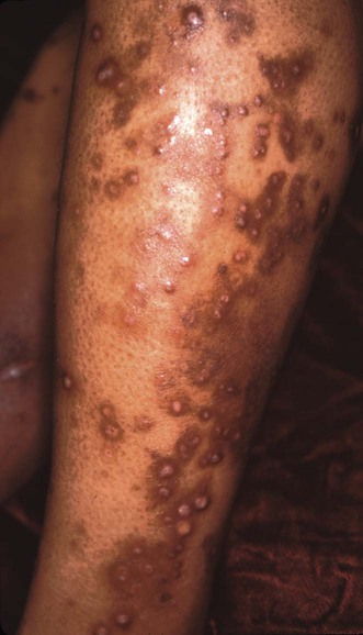
 Kyrle disease, in which dermal connective tissue perforates through the epidermis.
Specific investigations


First-line therapies
 Tretinoin 0.1%
 Tazarotene gel 0.1%
 UVB
UVB Narrowband UVB
Second-line therapies
 Allopurinol
 Isotretinoin
 PUVA
PUVA Acitretin
Third-line therapies
 Doxycycline
 Oral metronidazole
 Oral clindamycin
 Oral hydroxychloroquine
 Imiquimod
 Surgical debridement
 Cryotherapy
 Cantharidin
 Ultrapulse laser
 CO2 laser
 Photodynamic therapy
 Transcutaneous electrical nerve stimulation (TENS)
