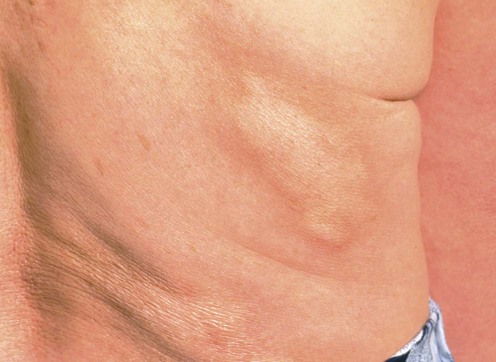
Morphea

Management strategy
Initial evaluation
The initial evaluation of patients with morphea should involve:
 Questions regarding recent development of new lesions/extension of existing lesions to determine level of activity
 Symptoms (pain/itch) associated with activity, musculoskeletal complaints, eye complaints (especially en coup de sabre/hemifacial atrophy)
 Total body examination of the skin (patients frequently do not appreciate the extent of lesions) looking for active/inactive lesions
 Palpation of affected areas for depth of sclerosis
 Examination for limited range of motion, contracture, limb length discrepancy, and evidence of musculoskeletal involvement
 Imaging with MRI if musculoskeletal involvement is suspected.
Active morphea lesions should be treated as the lesions are frequently symptomatic and can produce permanent cosmetic and functional sequelae. Initial evaluation should be focused on determining the extent, severity, and activity of morphea lesions laying the groundwork for rational therapeutic choices.
Specific investigations
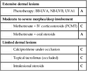
First-line therapies
Second-line therapies


 Phototherapy: BB-UVA, NB-UVB, UVA1
 Methotrexate + IV corticosteroids (PCMT)
 Methotrexate + oral steroids
 Calciprotriene under occlusion
 Topical tacrolimus (occluded)
 Intralesional steroids
 PUVA (bath or cream)
 Mycophenolate mofetil
 Abatacept
 Imiquimod
 Calciprotriol–betamethasone
 Cyclosporine
 Combination of oral medications
 Combination of oral medications with light


