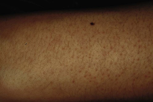
Keratosis pilaris and variants

Published on 18/03/2015 by admin

Filed under Dermatology

Last modified 18/03/2015

Print this page

rate 1 star rate 2 star rate 3 star rate 4 star rate 5 star
Your rating: none, Average: 0 (0 votes)

This article have been viewed 2963 times

Keratosis pilaris and variants

Nisha C. Desai and Robert A. Silverman

Evidence Levels:  A Double-blind study  B Clinical trial ≥ 20 subjects  C Clinical trial < 20 subjects  D Series ≥ 5 subjects  E Anecdotal case reports

image

Keratosis pilaris (KP) is a common inherited disorder of unknown etiology characterized by tenacious keratin plugging of follicular orifices in specific locations on the body. It typically becomes apparent during childhood on the extensor aspect of the upper arms, anterior surface of the thighs, and lateral aspects of the cheeks. In severe cases, it may extend onto the distal extremities, shoulders, and buttocks. Perifollicular erythema and a background of erythema on the cheeks is not uncommon (keratosis pilaris rubra, KPR). Variants of KP include keratosis pilaris atrophicans (ulerythema ophryogenes), keratosis follicularis spinulosa decalvans (KFSD), folliculitis spinulosa decalvans (FSD), and atrophoderma vermiculatum; these are also addressed in this chapter. KP is a common, though minor < feature of patients with atopic dermatitis. It may be exacerbated during pregnancy and is a feature of infants with malnutrition. Extensive or persistent disease may be observed in patients with Down syndrome, and cardiofaciocutaneous syndrome among others. KP has also been observed in patients treated with vemurafenib.

Management strategy

KP is usually asymptomatic and often does not require treatment. Those with extensive or symptomatic involvement often desire treatment, though, even without therapy, the condition often becomes less prominent with increasing age.

Initial treatment involves measures to decrease excessive skin roughness and follicular accentuation that often waxes and wanes over a period of months. Harsh soaps are to be avoided, but cleansers containing 2% salicylic acid or glycolic acid may be helpful. Bathing should be followed by application of an emollient to damp skin. Keratolytic agents such as glycolic acid, ammonium lactate, salicylic acid, and urea containing humectants are the mainstays of treatment. Salicylic acid 2% in 20% urea cream or salicylic acid 6% in propylene glycol combines the properties of an emollient with a keratolytic agent. Twice daily application of one of these compounds for at least a 3-week trial is recommended. The addition of gentle massage with a polyester sponge such as a Buf-Puf® during a shower or bath is particularly helpful. Vigorous scowering can lead to irritation. Once adequate relief of symptoms has been achieved, maintenance therapy of weekly or twice-weekly application of 20% urea cream (Carmol 20®) is recommended.

Topical retinoids may be tried in some cases but can be irritating and expensive if large areas are treated. Lower concentrations of topical tretinoin such as 0.025% cream can be used at first. If there is no significant irritation, higher-concentration preparations can be applied. Tazarotene 0.05% cream has also been shown to decrease the pruritus, erythema, and roughness of KP in a recent placebo-controlled trial.

If a significant inflammatory component is present, the inflammation can be treated for defined, short periods with a medium potency topical corticosteroid in an emollient base. Once inflammation has abated, corticosteroids are discontinued and keratolytics are introduced. With the presence of pustules, suggestive of a bacterial infection, topical antibiotics, such as clindamycin lotion, can be effective. A 3-month course of oral vitamin A, 50 000 units three times a day, has been advocated for some patients. Oral isotretinoin has been helpful in some patients with ulerythema ophryogenes and atrophoderma vermiculatum. In the author’s experience, calcipotriol and calcineurin inhibitors have not been helpful. In one small double blind study, Aquaphor™ improved KP as much as topical tacrolimus.

Cutaneous laser therapy has been utilized for several variants of KP. Pulsed-dye laser (PDL) treatment has recently been demonstrated to be a safe treatment for the erythema associated with keratosis pilaris atrophicans and KPR. Long pulsed 1064 nm Nd:YAG laser also significantly improved KP in a single blinded observational study. Potassium titanyl phosphate (KTP) laser has also cleared the keratotic papules and reduced the erythema of KPR lesions. However, reports involved only a few cases and made no mention of sustained long-term effectiveness. KPSD has been treated with laser-assisted hair removal using a normal mode, non-Q-switched, high-energy, pulsed ruby laser. This mode of treatment has been effective in producing persistent reductions of inflammation in this KP variant, albeit at the expense of permanent hair loss in treated areas.

Specific investigations

The prevalence of cutaneous manifestations in young patients with type 1 diabetes.

Pavlović MD, Milenković T, Dinić M, Misović M, Daković D, Todorović S, et al. Diabetes Care 2007; 30: 1964–7.

The presence and frequency of skin manifestations were examined and compared in 212 unselected type 1 diabetic patients and 196 healthy sex- and age-matched control subjects. KP affected 12% of the diabetic patients compared with 1.5% of control subjects. Significant association was also found between acquired ichthyosis and KP.

Other conditions associated with KP include high body mass index, dry scaly legs, atopic conditions, pregnancy, cardiofaciocutaneous syndrome, and hyperandrogenism in obese females. KP-like lesions have been observed in patients treated with vemurafenib for metastatic melanoma.