Juvenile xanthogranuloma

Published on 18/03/2015 by admin

Filed under Dermatology

Last modified 22/04/2025

Print this page

rate 1 star rate 2 star rate 3 star rate 4 star rate 5 star
Your rating: none, Average: 1 (1 votes)

This article have been viewed 2866 times

Juvenile xanthogranuloma

Megan Mowbray and Olivia M.V. Schofield

Evidence Levels:  A Double-blind study  B Clinical trial ≥ 20 subjects  C Clinical trial < 20 subjects  D Series ≥ 5 subjects  E Anecdotal case reports

image

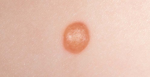
Courtesy of Mary Glover, Great Ormond Street Hospital, London, UK

Juvenile xanthogranuloma (JXG) is a benign disorder characterized by solitary or multiple, yellow-red nodules in the skin, and, occasionally, in other organs. The lesions spontaneously regress by age 6 months to 3 years. It occurs most commonly in infancy and early childhood, but adults may be affected. Treatment is only recommended for systemic lesions if their location interferes with organ function.

Management strategy

The diagnosis of JXG is usually made clinically, but a biopsy is required for atypical clinical variants (giant, plaque-like, paired, clustered, infiltrative, lichenoid, linear, subcutaneous, and intramuscular) and if multiple lesions are present. JXG is classified as a ‘non-Langerhans cell histiocytosis’ and characterized histologically by an accumulation of foamy histiocytes lacking Birbeck granules (non-Langerhans cells) and multinucleated Touton-type giant cells. Immunohistochemical staining of JXG lesions shows expression of factor XIIIa, CD68, CD163, fascin, HLA-DR, and CD14, but not S100 or CD1a. This pattern of expression suggests that the cell of origin of JXG is the dermal dendrocyte. The histogenesis of this group of conditions is unclear but the relationship to LCH is thought to be a divergent differentiation from a common precursor CD34+ myeloid cell. Reported cases of the two conditions occurring in the same individual suggest that this divergent maturation is not irreversible.

The ‘JXG family’ can be divided into three groups:

In the most common solitary type of JXG (60–89% of all cases), there is a male to female preponderance of 1.5 : 1. The usually affected sites are the head, neck, and upper trunk. In young infants with multiple lesions the male : female ratio increases to 12 : 1. In the majority of cases no further investigation is required. The lesions will resolve spontaneously within months or years and no follow-up is necessary. As involvement of other organs can occasionally occur in the cutaneous group, a review of systems and general examination is recommended to look for involvement of other sites and to examine for café-au-lait macules (see below).

Specific investigations

First-line therapies

imageNone E
imageSurgical resection for symptomatic cutaneous and extracutaneous lesions D

Second-line therapies

imageOcular lesions treated with topical, intralesional corticosteroids D
imageOral corticosteroids E
imageOcular surgery E
imageCutaneous lesions treated with CO2 laser E

Third-line therapies

imageChemotherapy including oral corticosteroids D
imageRadiotherapy D
imagePulsed methylprednisolone E

Treatment of JXG.

Stover DG, Alapati S, Regueira O, Turner C, Whitlock JA. Pediatr Blood Cancer 2008; 51: 130–3.

Two case reports of multisystem JXG treated with LCH-based chemotherapy regimens which resulted in prompt resolution of symptoms. This paper includes a literature review of all reported multisystem JXG cases treated with chemotherapy. Ten prior studies describe 15 cases (29 chemotherapeutic regimens of multisystem JXG). Twelve of the 15 patients received some form of corticosteroid, nine of the 12 receiving corticosteroid had stable disease (SD), partial response (PR), or complete resolution (CR). Regimens that included corticosteroids and a vinca alkaloid had the highest rates of CR, PR, or SD. Regimens that did not include corticosteroids had the lowest rates of CR, PR, or SD.

Share this: