Hereditary hemorrhagic telangiectasia

Published on 18/03/2015 by admin

Filed under Dermatology

Last modified 22/04/2025

Print this page

rate 1 star rate 2 star rate 3 star rate 4 star rate 5 star
Your rating: none, Average: 0 (0 votes)

This article have been viewed 1657 times

Hereditary hemorrhagic telangiectasia

Mitchell S. Cappell and Oscar Lebwohl

Evidence Levels:  A Double-blind study  B Clinical trial ≥ 20 subjects  C Clinical trial < 20 subjects  D Series ≥ 5 subjects  E Anecdotal case reports

image

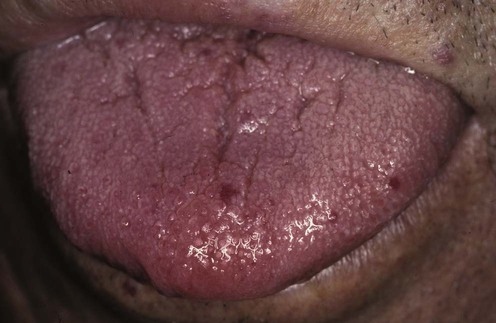
Hereditary hemorrhagic telangiectasia (HHT, or Osler–Weber–Rendu syndrome), an autosomal dominant disease, produces a syndrome of multiple orocutaneous telangiectasias, especially on the face, lips, tongue, oral mucosa, and fingertips, together with multiple internal telangiectasias, especially on the mucosa in the nose and gastrointestinal tract. Cutaneous or mucosal lesions typically are 3–10 mm wide, macular, bright red, non-pulsatile, and spidery or punctate in shape with a fine reticular internal structure. They tend to blanch under pressure (with diascopy), and to slowly increase in size and number with age. Although the dermatologic lesions are usually only a minor, cosmetic problem, the nasal and gastrointestinal telangiectasias often bleed significantly and repeatedly. Chronic blood loss may result in iron-deficiency anemia, whereas acute blood loss may cause hypovolemia and systemic hypotension. Internal shunts created by direct arteriovenous communications in HHT infrequently cause clinical manifestations, including hypoxia from pulmonary arteriovenous shunts; cerebral ischemia, hemorrhage, or abscesses from intracerebral shunts; and portal hypertension, biliary tract disease, or high-output cardiac failure from intrahepatic shunts.

The prevalence of HHT is estimated at 1 per 6000 individuals. The basic lesion in HHT is a defect in the wall of small vessels that leads to direct arteriovenous communications without intervening capillaries. Most cases of HHT are due to underlying genetic mutations.

Management strategy

HHT is reliably diagnosed by the presence of three or more findings among the following clinical tetrad: recurrent, spontaneous epistaxis; mucocutaneous telangiectasias; gastrointestinal telangiectasias or visceral arteriovenous malformations; and a first-degree relative with HHT (Curacao criteria). HHT is highly unlikely in adult patients satisfying only one or none of these criteria. The diagnosis may be confirmed by genetic tests for the individual underlying mutations, but such tests are generally not required for diagnosis or clinical management. Patients may have telangiectasias solely on the face with rosacea or lupus erythematosus, but lack other manifestations of HHT. Patients with sporadic gastrointestinal angiodysplasia are differentiated from patients with HHT by clinical presentation in old age, a paucity of the characteristic lesions, a negative family history, and by exclusively gastrointestinal involvement without epistaxis or intranasal telangiectasia.

For epistaxis, the bleeding severity is determined by inspection, vital signs, and laboratory tests. The bleeding site is precisely localized by nasal examination using bright, shadow-free illumination, with a headlight or head mirror, and spot suction, using a fine-caliber rigid tube. For gastrointestinal bleeding, the bleeding severity is indicated by patient history, vital signs, physical findings, rectal examination, nasogastric aspiration, and transfusion requirements. The bleeding site and source are conclusively diagnosed by gastrointestinal endoscopy.

Choice of therapy depends on the bleeding site, severity, and chronicity as well as individual physician expertise. Significant acute blood loss is treated with intravenous fluid resuscitation and transfusion of packed erythrocytes as needed. Chronic blood loss is treated with iron supplementation as needed. Epistaxis is initially treated by non-specific local therapy such as nasal packing to tamponade the bleeding, and topical vasoconstrictors to reduce local blood flow. Estrogen with progesterone is used to prevent or arrest chronic epistaxis by promoting vascular integrity. Significant, refractory, chronic epistaxis is definitively treated by septal dermoplasty. Actively bleeding gastrointestinal telangiectasias are ablated at endoscopy by argon plasma coagulation, thermocoagulation, electrocoagulation, or photocoagulation. Chronically bleeding gastrointestinal telangiectasias may be treated with estrogen and progesterone. Surgical resection is reserved for well-localized, active bleeding from gastrointestinal telangiectasias refractory to other therapy, because patients tend to subsequently rebleed from telangiectasias at other gastrointestinal sites. Where available, angiographic embolization can obviate the need for gastrointestinal surgery.

Specific investigations

Serial hematocrit determinations are important, particularly for acute bleeding, to determine the need for transfusions of packed erythrocytes. Serum levels of iron, total iron-binding capacity (TIBC), and ferritin are important, particularly for chronic bleeding, to determine the need for iron replacement therapy. Upper gastrointestinal bleeding, usually manifested by hematemesis or melena, should be investigated by esophagogastroduodenoscopy. Lower gastrointestinal bleeding, usually manifested by hematochezia or fecal occult blood, and occasionally manifested by melena, should be investigated by colonoscopy. Barium contrast radiography or virtual colonoscopy should not be performed to evaluate lower gastrointestinal bleeding in patients with HHT because the telangiectasias are mucosal and macular, and not visualized with barium contrast. Small intestinal bleeding, usually manifested by hematochezia, melena, or fecal occult blood, can be investigated by capsule endoscopy after exclusion of upper and lower gastrointestinal bleeding by the aforementioned endoscopic tests. Double-balloon enteroscopy is being increasingly used instead of capsule endoscopy for small intestinal bleeding because of its greater diagnostic sensitivity and therapeutic capabilities despite its greater costs.

At endoscopy, the gastrointestinal lesions resemble the cutaneous telangiectasias in size and shape. Angiography is indicated for active gastrointestinal bleeding if colonoscopy and esophagogastroduodenoscopy failed to diagnose the source of bleeding. Telangiectasias bleed only intermittently, and extravasation of contrast material from telangiectasias is infrequently detected at angiography.

First-line therapies

For epistaxis or gastrointestinal bleeding  
image Non-specific therapy: avoid aspirin, other anti-platelet drugs, and anticoagulation; transfuse packed erythrocytes for significant, acute bleeding; administer iron therapy for significant iron-deficiency anemia from chronic blood loss A
image Oral estrogen with progesterone B
For epistaxis  
image Non-specific therapy: room humidification and nasal moisteners A
image Nasal packing A
image Septal dermoplasty A
For gastrointestinal bleeding  
image Endoscopic therapy: argon plasma coagulation, thermocoagulation, electrocoagulation, or photocoagulation B
image Angiographic embolization B
image Segmental bowel resection A
For cutaneous lesions  
image Cosmetic therapy: laser or other focal ablation techniques for cutaneous lesions A

Outcome of septal dermoplasty in patients with hereditary hemorrhagic telangiectasia.

Fiorella ML, Ross D, Henderson KJ, White Jr, RI. Laryngoscope 2005; 115: 301–5.

Retrospective study reporting outcome of 32 consecutive patients undergoing septal dermoplasty in whom accurate records of transfusion requirements were available. Mean blood transfusion requirements significantly declined from a mean of 21 units of packed erythrocytes in the year before surgery to a mean of 1 unit in the year after surgery (p < 0.001). This study illustrates the fairly large literature supporting the efficacy of septal dermoplasty for treating recurrent, relatively severe epistaxis from HHT.

Nasal moisteners and room humidification promote mucosal integrity and reduce nasal mucosal injury and epistaxis. Nasal packing often temporarily stems epistaxis by vascular tamponade. Septal dermoplasty is indicated for severe, recurrent epistaxis, especially from the anterior nasal mucosa. In this procedure, skin, removed from the upper thigh, is grafted on to the anterior nasal septum and floor to cover and protect the fragile mucosal telangiectasias from local trauma. The procedure is well tolerated using only local anesthesia, and complications, other than recurrent epistaxis, are rare. Septal dermoplasty is effective in more than 75% of cases. Failure results from inadequate graft coverage with bleeding from lesions at the border or beyond the grafted area. Focal laser therapy has been used to ablate discrete bleeding intranasal lesions.

Long-term outcome of argon plasma ablation therapy for bleeding in 100 consecutive patients with colonic angiodysplasia.

Olmos JA, Marcolongo M, Pogorelsky V, Herrera L, Tobal F, Davalos JR. Dis Colon Rectum 2006; 49: 1507–16.

In this prospective trial, 85 of 100 patients with moderate to severe bleeding from colonic angiodysplasia treated at colonoscopy with argon plasma coagulation (mean 3.9 angiodyplasias treated per patient) had no further overt bleeding and had a stable hemoglobin level without further blood transfusions or iron therapy during a mean follow-up of 20 years. Only two minor procedure complications occurred. Although not specifically addressing argon plasma coagulation for telangiectasias in HHT, this large, prospective study demonstrated high efficacy of this therapy for preventing rebleeding from the similar lesions of sporadic colonic angiodysplasia.

Mucosal vascular malformations of the gastrointestinal tract: clinical observations and results of neodymium: yttrium-aluminum-garnet laser therapy.

Gostout CJ, Bowyer BA, Ahlquist DA, Viggiano TR, Balm RK. Mayo Clin Proc 1988; 63: 993–1003.

Gastrointestinal bleeding from telangiectasias was successfully controlled in nine of 10 patients with HHT and in 72 of 83 patients with sporadic telangiectasias by endoscopic photocoagulation using the Nd : YAG laser. Three gastrointestinal perforations occurred in 243 treatment sessions.

Non-specific therapy for bleeding from HHT includes avoidance of aspirin, NSAIDs, other antiplatelet drugs, and anticoagulants; and administration of iron for patients with significant iron-deficiency anemia. At endoscopy, isolated actively bleeding gastrointestinal telangiectasias are treated with endoscopic argon plasma coagulation, thermocoagulation, electrocoagulation, or photocoagulation. Endoscopists increasingly prefer argon plasma coagulation because of the ease of application and the potentially greater safety due to more superficial tissue ablation. Nonetheless, all these endoscopic therapies are relatively safe and highly successful at achieving hemostasis when performed by an experienced endoscopist. However, patients with HHT often subsequently rebleed from other, untreated gastrointestinal telangiectasias, and therefore require multiple sessions of endoscopic therapy to treat these other lesions. Angiographic embolization or segmental bowel resection are reserved for severe, active bleeding, localized to a single region and refractory to medical and endoscopic therapy. These therapies often provide only intermediate-term relief owing to the recurrence of telangiectasias at other gastrointestinal sites.

Use of estrogen in treatment of familial hemorrhagic telangiectasia.

Harrison DFN. Laryngoscope 1982; 92: 314–20.

Report of successful control of epistaxis with estrogen therapy in 67 patients with HHT, with few complications.

Several studies have reported a markedly reduced incidence of chronic gastrointestinal or nasal bleeding from telangiectasias after instituting estrogen therapy, either alone or with progesterone, owing to the promotion of endothelial integrity. Estrogen therapy can be combined with other therapies, such as local endoscopic therapy, to increase efficacy. Despite controversy concerning efficacy, this therapy should be considered before performing gastrointestinal or nasal surgery for chronic bleeding, because of its low risk and the risk of recurrent bleeding after surgery. Estrogen therapy is less desirable in males than in females because it can cause feminization.

Second-line or temporizing therapies

For epistaxis  
image Arterial ligation B
image Topical vasoconstrictors B
image Cryosurgery/electrical cautery/argon plasma coagulation B
image Unilateral or bilateral surgical closure of the nostrils B
image Arterial embolization B
image Bevacizumab B
image Tamoxifen B
image Submucosal resection C
image Topical aminocaproic acid D
For gastrointestinal bleeding  
image Oral aminocaproic acid therapy D
image Bevacizumab C
image α-Interferon D

Treatment of hereditary hemorrhagic telangiectasia with submucosal and topical bevacizumab therapy.

Karnezis TT, Davidson TM. Larygoscope 2012; 122: 495–7.

In a prospective trial of 19 patients, the epistaxis severity score significantly decreased to 2.00, 2 months after intranasal, submucosal injection of bevacizumab compared to the baseline, pretreatment, severity score of 8.12 (p<0.0001). Bevacizumab therapy statistically significantly protected against epistaxis for all 12 months of the clinical trial.

Recent data suggest that patients with severe bleeding from HHT may respond dramatically to therapy with bevacizumab, a vascular endothelial factor inhibitor. Some recent data support use of tamoxifen, an estrogen antagonist, to prevent bleeding from telangiectasia in HHT. A few patients have responded to therapy with α-interferon, which has known anti-angiogenic properties, but this therapy is currently experimental as there are insufficient data.

Local non-specific therapy of arterial ligation, cryosurgery, electrical cautery, argon plasma coagulation, or submucosal resection provide temporary relief of epistaxis from HHT. However, these treatments can cause mucosal scarring, which diminishes the efficacy of subsequent septal dermoplasty. Topical therapy with vasoconstrictors or aminocaproic acid rarely produces scarring. Non-specific therapies are indicated only for temporary relief of acute bleeding. Life-threatening epistaxis refractory to septal dermoplasty is treated by surgical closure of one or both nostrils, depending on the bleeding source.

Aminocaproic acid promotes thrombosis and retards bleeding by inhibiting fibrinolysis. Oral aminocaproic acid therapy has had mixed success with HHT. This therapy rarely causes hypotension or rhabdomyolysis. Aminocaproic acid is a temporary, second-line therapy.

Share this: