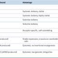
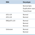
FLT1, Fms-like tyrosine kinase 1, VEGFR1; RGD, Arg-Gly-Asp; RRM2, ribonucleotide reductase subunit M2.
Gene Knockout
Antisense
Activation of proto-oncogenes such as
Ras, B-Raf, Myc, or
EGFR through various mechanisms is a key factor in carcinogenesis. For example, mutations of the
Ras oncogene occur in many types, including pancreatic cancer, where about 90% of cases carry a mutation of the K-ras gene.
13,14 Knockout of oncogenes driving tumor development in transgenic mouse models of cancer can lead to complete regression, confirming
their potential value as drug targets.
15 Antisense technology has been studied as a strategy to knock down expression of oncogenes. This approach is based on the possibility of inhibiting transcription of a particular mRNA by transfecting short double-stranded DNA oligonucleotides into target cells that bind and inactivate the target mRNA in a sequence-specific manner.
16 The development of oligonucleotides with modified DNA backbone that increases the stability of the molecule in vivo allowed for the development of clinical protocols involving intravenous application of antisense molecules targeting, for example, mutant Kras or the anti-apoptotic genes
BCL-2 and
Survivin. 17 Clinical trials with oblimersen sodium (G3139), an antisense oligonucleotide directed against BCL-2, have demonstrated increased survival of patients with advanced melanoma in combination with chemotherapy compared to chemotherapy alone.
18 An interesting variant of the antisense approach that is currently being tested clinically is GRN163L, a lipid-modified 13-mer DNA-oligonucleotide that acts as a telomerase RNA template antagonist.
19
Table 54-2
Cancer-Selective Viruses in Clinical Trials
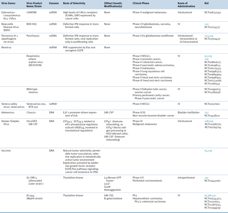
Phase I/II, Phase II, or Phase III clinical trials. June 2012. Clinicaltrials.gov.
EGFR, epidermal growth factor receptor; GFP, green fluorescent protein; GusA, β-glucuronidase; ICAM1, intercellular adhesion molecule 1; IFN, interferon; IV, intravenous; DAF, decay accelerating factor; GM-CSF, granulocyte macrophage colony-stimulating factor; LacZ, β-galactosidase; NSCLC, non–small-cell lung cancer; PKR, protein kinase R.
RNAi
The discovery of the possibility of silencing gene expression in mammalian cells with high efficiency using RNA interference (RNAi) has spurred renewed interest in the gene knock-down as a therapeutic strategy. This approach, first described in the worm
Caenorhabditis elegans, takes advantage of a cellular gene silencing machinery that involves the RNA-induced silencing complex (RISC) and degrades double-stranded RNAs with high efficacy.
20 The introduction of short RNAs with complementary sequence to any cellular transcript activates this mechanism. RNAi can silence target genes with high efficacy. However, off-target effects that result in sequence-specific though unpredictable knock-down of additional genes represent a potential problem that has not been fully resolved at this point.
21 Nevertheless, novel targeting and delivery mechanisms have been developed that hold the promise of therapeutic application of RNAi-based agents in cancer.
20 For instance, a recently reported nanoparticle-based delivery system shows great promise for systemic delivery of siRNA. These synthetic nanoparticles consist of a cyclodextrin-based polymer, a human transferrin protein (TF) targeting ligand displayed on the exterior of the nanoparticle to engage TF receptors (TFR) on the surface of the cancer cells, a hydrophilic polymer (polyethylene glycol [PEG] used to promote nanoparticle stability in biological fluids), and siRNA designed to reduce the expression of the Ribonucleotide Reductase subunit M2 (RRM2). Tumor-specific targeting is achieved via the interaction of the TF targeting ligand with TFR, which is known to be upregulated in malignant cells.
22 Moreover, the size of these nanoparticles, about 70 nm in diameter, favors their exit from the bloodstream in leaky tumor vasculature and accumulation in the tumor bed via the enhanced permeability and retention (EPR) effect. RRM2, an enzyme catalyzing a rate-limiting step in DNA synthesis, is an established anticancer target.
23 These particles (CALAA-01), currently in Phase I clinical studies, have been administered systemically to humans and have shown specific gene inhibition—a reduction in both mRNA and protein.
24
Another well-established anticancer target is the KRAS oncogene. Most pancreatic adenocarcinomas are caused by a somatic mutation in KRAS, most commonly KRAS G12D. In vitro and animal studies suggest that selective inhibition of the mutant, but not the WT KRAS expression, leads to apoptosis of targeted cancer cells. An siRNA specifically targeting mRNA encoding KRAS G12D, coated by a biodegradable polymeric matrix, siG12D LODER (Local Drug EluteR), is currently in Phase 0/I to evaluate its safety and tolerability. Apart from these two particular RNAi therapeutics, at least another six are currently undergoing clinical testing, thanks to recent advancements in the understanding of RNAi biology and in the areas of RNAi specificity, stability, and delivery. Most are delivered employing synthetic carriers, such as cationic liposomes, anionic liposomes, and polymeric particles. As more attention is focused on safe and effective methods for delivering siRNA to tumors, the clinical value of this approach will likely increase dramatically. siRNA-based therapy has the potential of addressing undruggable oncogene targets, combinatorial approaches to cancer cell killing, and drug resistance, but this potential will not be realized until the challenges of delivery and uptake have been fully addressed.
Killing Cancer Cells by Delivering Prodrug Converting Enzymes and Other Enzymes
For more than 60 years chemotherapeutic agents have been used for the treatment of cancer. However, their use is often limited by damage to normal cells, drug resistance, and low chemical stability. One strategy to overcome limitations of classical chemotherapeutic agents is the use of prodrugs. A prodrug is a fairly nontoxic compound that needs to be transformed before acting as a pharmacon. Such a transformation can be catalyzed by endogenous enzymes, in which case tissue distribution of such endogenous enzymes dictates where the active pharmacon is produced. Alternatively, gene-directed enzyme prodrug therapy (GDEPT) can specifically deliver such enzymes to diseased cells where they can activate nontoxic prodrugs into toxic agents. Progress in this field has been reviewed recently by Duarte and colleagues.
25
Thymidine Kinase
The first GDEPT system described was the thymidine kinase gene of the herpes simplex virus (HSV-TK) in combination with the prodrug nucleoside analog ganciclovir. The enzyme thymidine kinase (TK) is naturally present in bacteria, viruses, and mammals, where it is involved in the salvage pathway of nucleotide biosynthesis. Thus, high TK activity is found in proliferating cells such as cancer cells. TK also converts ganciclovir to ganciclovir monophosphate, which is subsequently converted by cellular kinases into the toxic ganciclovir triphosphate nucleotide. The HSV-TK is three orders of magnitude more efficient than any human kinase catalyzing this first activation step. Hence, several gene therapy approaches combining HSV-TK and ganciclovir have been developed.
For instance, retroviral vectors were used to deliver HSV-TK in a brain tumor model. Because retroviruses integrate only in proliferating cells, gene delivery and expression would be tumor selective in the context of normal, nonproliferating brain cells.
26 A further advantage of this approach was thought to result from the “bystander effect,” the killing of uninfected neighboring cells that occurs when HSV-TK–expressing cells are exposed to ganciclovir, which can be observed in vitro and in vivo.
27 To increase transduction of target cells with retroviruses, virus-producing cells (VPCs) were used to inoculate target tumors instead of virus suspension. Preclinical studies in a rat glioma model demonstrated that this delivery of a retrovirus expressing HSV-TK resulted in high transduction levels and frequent tumor regressions following ganciclovir administration.
28 Initial clinical studies were promising and demonstrated in responses of small glioblastomas following VPC injection.
29 However, when standard therapy (surgical resection and radiotherapy) was compared to standard therapy plus injection of retrovirus-producing cells in a Phase III trial, no differences in progression-free and overall survival were observed between the two groups.
30 Smaller, preoperative studies suggested that lack of transduction of tumor cells is the dominant reason for the failure of this approach.
31 Despite the disappointing results of this trial, it represents an early and innovative effort to use gene therapy to kill cancer cells selectively.
Cytosine Deaminase
In contrast to thymidine kinase, the enzyme cytosine deaminase (CD) is not present in mammalian cells, but in several bacteria and fungi. It catalyzes the amidine hydrolysis of cytosine to uracil and ammonia, and several cytosine analogs such as halogenated cytosines are substrates as well. One such substrate is the prodrug 5′-fluorocytosine (5′-FC) which is activated by CD to 5′-fluorouracil (5′-FU). 5′-FU kills cells mainly by inhibition of thymidilate synthetase and by incorporation into DNA. The safety and efficacy of a CD and 5′-FC prodrug therapy are currently being assessed in Phase I/II trials in which 5′-FC and a recombinant Bifidobacterium longum (a live bacterium normally found in the digestive tract) that has been modified to produce CD are given orally to patients with various solid tumors.
Carboxylesterase
Another well-studied prodrug-converting enzyme is carboxylesterase (CE). CE expression is widely distributed in human tissues; nonetheless, targeted gene delivery to enhance CE expression specifically in tumor cells is a promising approach to increase the local availability of cytostatic drugs. CE is involved in the activating metabolism of various commonly used chemotherapeutics: It activates paclitaxel-2-ethylcarbonate to the microtubule-stabilizing agent paclitaxel/taxol and is involved in the multistep conversion of capecitabine to the pyrimidine analog 5′-fluorocytidine (5′-FC). Moreover, CE converts the relatively nontoxic camptothecin analog CPT-11/irinotecan to SN38, a potent topoisomerase I inhibitor. Because the human CE is comparatively inefficient in catalyzing this conversion, several gene therapeutic strategies using the more effective rabbit CE were developed and shown to be successful in tissue culture and animal models. Also, secreted mutants of this enzyme are being studied to enhance the spread of the toxic metabolite and thereby the bystander effect. For instance, Oosterhoff and colleagues show increased killing of colon carcinoma cells on infection with adenovirus expressing secreted CE (Ad5-Δ24-sCE) combined with CPT-11 treatment.
32 Even though CPT-11 also reduced viral replication, the overall cytotoxicity of virus combined with CPT-11 was still higher than cell killing achieved by the virus alone. Nonetheless, this exemplifies the complications of using chemotherapeutic agents in combination with oncolytic viruses; it is crucial to know the kinetics of both viral replication and drug metabolism to design a suitably timed treatment regimen. This is not only essential for oncolytic viruses as vectors for GDEPT, but for the combination of any oncolytic virus with chemotherapeutic agents.
33
Sodium-Iodide Symporter
Not only prodrug-converting enzymes are being pursued as gene therapeutics. Several vectors have been developed
to deliver genes for various other enzymes, including the one for the sodium iodide symporter (
NIS). The endogenously expressed NIS is an integral plasma membrane glycoprotein that mediates active I
− transport into thyroid follicular cells. The rationale for its use in gene therapy approaches is twofold: targeted expression of
NIS in cancer cells renders these cells susceptible to uptake of radioactive iodine, which, depending on the isotope used, can be harnessed to kill and/or to image targeted cells. For instance, measles virus (MV) strains that have been engineered to express the human NIS (MV-NIS) have shown significant antitumor activity against various cancer lines and orthotopic xenografts. Expression of NIS in infected glioma cells resulted in effective concentration of radioactive iodine, which allowed for in vivo monitoring of localization of MV-NIS infection by measuring uptake of
123I and led to a significant increase of MV-NIS antitumor activity.
34 A Phase I study is ongoing to establish the safety of intrapleural administration of this MV-NIS in patients with malignant pleural mesothelioma.
Another interesting approach is the retroviral delivery of a multidrug resistance pump (
MDR1) gene into peripheral blood progenitor cells (PBPCs). The rationale behind this is not to kill cancer cells, but to prevent some of the toxicities of an intensive chemotherapy regimen in patients. PBPCs are isolated from the patient, genetically modified ex vivo, and transferred back into the patient. The hope is that introduction of the
MDR1 gene into the patients’ PBPCs renders these cells and their offspring resistant to the toxic effects of certain chemotherapeutic agents, by pumping out chemotherapeutic agents before they can exert a cytotoxic effect.
35–39
Many other strategies have been devised to express prodrug-converting enzymes, or other potentially toxic genes, in cancer cells selectively. Most of these depend on tumor-selective gene expression to drive the gene of interest, such as the promoters for the androgen receptor or prostate-specific antigen (PSA),
40–42 for tyrosinase (selective for melanoma),
43 or for alpha-lactalbumin (selective for breast cancer).
44 More generic tumor-selective–specific promoters include E2F-1, which is upregulated through loss of the RB checkpoint, and telomerase reverse transcriptase (TERT), which is also upregulated in most cancers.
45,46
Genes That Boost the Immune System
Although oncolytic viruses can kill cancer cells directly via viral replication and by delivery of cytotoxic genes or prodrug-converting enzymes, it is becoming more and more evident that the great potential of viruses as gene therapeutic vectors lies in their immune-modulatory capability. This feature can be further enhanced by the addition of immune-modulatory genes into the viral genome. For virus-based therapies to be successful, both host immune system avoidance and potent immune stimulation need to be combined. One approach to boosting antitumor immune responses involves the administration of cytokines to increase the activity of immune effector cells systemically or to enhance the presentation of antigens in tumor cells themselves. A wide range of cytokine genes have been engineered into an equally wide range of vectors and viruses. IL-2, IL-4, and IL-12 have been studied extensively, as well as interferons, members of the tumor necrosis factor (TNF) family, and granulocyte-macrophage colony-stimulating factor (GM-CSF).
TNF
TNFerade is an exciting example of this approach. This replication-incompetent adenovirus expresses TNF under the control of the Early Growth Response Factor 1 (EGRr-1) promoter, which is strongly activated in response to cellular stress such as iodizing radiation or chemotherapy.
47 TNFerade was injected in Phase I studies to up to 4 × 10
11 pu (particle units) without dose-limiting toxicities occurring. In agreement with this favorable safety profile, serum TNF levels remained consistently low. However, in esophageal cancer, relatively high rates of thromboembolic complications occurred that were potentially induced by the study medication. This problem was not observed in trials of this agent in pancreatic cancer. In this disease the maximum tolerated dose (MTD) of TNFerade in combination with chemoradiation treatment was determined to be 4 × 10
11 pu. At that dose level, clinical responses and evidence for prolonged median survival were seen in an interim analysis of a Phase III study in locally advanced pancreatic cancer in combination with chemoradiotherapy.
48
GM-CSF
Of cytokines tested in oncolytic virus and vaccine models so far, GM-CSF seems to be the most potent at generating antitumor immune responses. HSV-, adenovirus-, and vaccinia virus–based vectors encoding GM-CSF have shown great promise in early clinical trials, and studies are still ongoing (see
Table 54-2). Arming viruses with GM-CSF aims to activate the immune system primarily by attracting and activating dendritic cells (DCs). GM-CSF has been clinically tested in peptide and whole-cell vaccine strategies as well as oncolytic virus approaches. Notably, only virus-based
GM-CSF expression seems to generate a potent and sustained antitumor immune response, suggesting that the context in which GM-CSF is expressed is crucial. Virus-based GM-CSF expression and DC recruitment are tightly coupled to tumor cell death and subsequent release of a vast array of tumor antigens. Remarkably, for both HSV and vaccinia expressing GM-CSF, not only injected but also noninjected tumors responded. The studies mentioned earlier are among the first to combine oncolysis with immune system activation using armed, replication-competent viruses. Further exploitation of the immune system is a focus of current gene therapy and vaccine studies.
49
Naturally Occurring Viruses That Replicate Selectively in Cancer Cells
Cancer cells provide an environment that is permissive for replication of a number of naturally occurring viruses. This is because checkpoints and defense mechanisms are disabled in cancer cells, allowing them to grow and survive and to evade detection by the immune system. In some cases these mechanisms are also used to defend normal cells against virus replication. Cancer cells may therefore be vulnerable to virus infection while normal cells are protected. Because infection usually leads to cell death, this vulnerability could potentially be exploited for cancer therapy. Indeed, several naturally occurring viruses are under clinical evaluation in a variety of cancer indications (see
Table 54-2).
Reovirus
Reoviruses replicate selectively in many cancer cells. During infection of normal cells, their double-stranded RNA genomes activate a cellular protein kinase (PKR) that restricts viral replication by blocking translation of viral mRNA. For reasons that are unclear, this kinase activity is suppressed in cancer cells in which the Ras pathway is hyperactivated, allowing productive viral replication.
50 Based on this selectivity, and the capacity of reoviruses to replicate quickly and kill infected cells, reoviruses are undergoing clinical evaluation. One of these, Reolysin, is being tested in Canada, the United Kingdom, and the United States using clinical protocols that include local or systemic delivery of Reolysin as a monotherapy, and local delivery in combination with radiation therapy for patients with advanced cancers. Intravenous administration of this virus was well tolerated in a Phase I study. Although no objective tumor responses were observed, disease stabilization of up to 6 months was observed in a subset of patients.
51
Vesicular Stomatitis Virus
The rhabdovirus vesicular stomatitis virus (VSV) has a single-stranded RNA genome. Selectivity for cancer cells is thought to be the result of their failure to elicit a protective interferon response, thus allowing lytic replication. VSV variants with mutations in the matrix (M) protein enhance VSV’s effectiveness, at least in animal models.
52 Systemic delivery of VSV has been shown to be effective and safe against laboratory models of multifocal and invasive malignant gliomas.
53 M protein mutant viruses have also shown efficacy against prostate cancer cell lines and others.
52
Measles
Measles viruses, like VSV, contain negative-stranded RNA genomes but are members of the paramyxovirus family. Replication-competent attenuated Edmonston B measles vaccine strain (MV-Edm) is nonpathogenic and has potent antitumor activity against several human tumors. The virus is selectively oncolytic, eliciting extensive cell-to-cell fusion and ultimately leading to cell death. An attenuated strain of MV has been genetically engineered to produce carcinoembryonic antigen, which can be used as a serum marker of virus replication.
12 This virus had potent antitumor activity against gliomas in vitro, as well as in animal models.
54 This virus is undergoing clinical evaluation in patients with glioblastoma multiforme and multiple myeloma.
Newcastle Disease Virus
Lytic strains of the avian paramyxovirus Newcastle disease virus (NDV) selectively kill cancer cells in culture and in mouse models.
55 The molecular basis of selectivity is not fully understood, but appears to be facilitated by high levels of N-myc, at least in neuroblastoma cells. Cytotoxicity is due to multiple caspase-dependent pathways of apoptosis independent of interferon signaling competence.
55,56 Several Phase I studies of intravenously infused NDV have been performed using various doses and administration schedules.
57,58 Main toxicities included moderate flu-like symptoms and mild gastrointestinal symptoms. Interestingly, a two-step intrapatient dose-escalation of the NDV strain PV701 aiming at desensitizing resulted in significant reduction of the intensity of adverse events. Patients developed only moderate levels of neutralizing antibodies, and the serum clearing of virus was not significantly different during the course of treatment.
59 Disease stabilization as well as objective tumor responses were observed in Phase I studies, in particular in patients
who had received higher doses. A complete remission was observed in a patient with glioblastoma multiforme treated with the NDV strain NDV-HUJ.
57
Viruses Engineered to Replicate Selectively
In addition to naturally occurring viruses, many efforts have been made to engineer viruses to replicate in tumor cells selectively. Such agents kill cells through lytic mechanisms and potentially spread from one infected cell to another, amplifying the dose of the selective killing agent. Selectivity for cancer cells can be achieved by several strategies. The first uses DNA synthetic enzymes produced by proliferating tumor cells to support replication of DNA viruses that are otherwise defective. The second takes advantage of genetic defects in cancer cells that supply functions that have been specifically deleted from the oncolytic agent, and the third uses tumor-selective promoters to drive replication of conditionally replicating viruses.
Herpes Simplex Viruses
One of the first viruses designed to replicate in cancer selectively were HSVs that had been engineered so that they were unable to express viral genes necessary for DNA replication, such as thymidine kinase or ribonucleotide reductase. Proliferating cells would provide these essential functions, whereas resting normal cells would not. HSV G207 is an example of such an oncolytic virus. In addition to inactivation of a subunit of the viral ribonucleotide reductase gene, both copies of the neurovirulence gene, the gamma(1)34.5 gene, are deleted to further reduce replication in normal tissues.
60 Although direct tumor cell killing represents a major mechanism of action of these viruses, evidence from experiments in immunocompetent mouse models suggests that also a vaccination effect mediated by activated T lymphocytes contributes to the effect.
61 Phase I clinical trials of G207 and a related virus, HSV1716, have been completed and demonstrated the safety of these viruses.
62 Another related HSV mutant, NV1020, has been tested in a Phase I study in patients with hepatic metastases from colorectal cancer. The virus was administered into the hepatic artery in a Phase I study. Only mild toxicity was observed, and a decline in CEA levels was suggestive of some antitumor activity.
63 Many innovative approaches have been employed to improve the clinical value of HSV viruses, including expression of prodrug-converting enzymes to elicit bystander cells and the addition of genes encoding cytokines to boost immune recognition of tumor cells. The latter approach has been taken into the clinic in the form of the HSV mutant OncoVex(GM-CSF), a conditionally replicating HSV-1 mutant that expresses the cytokine GM-CSF. A Phase I/II trial with intratumorally injected Oncovex GM-CSF demonstrated that this agent is well tolerated.
64 Side effects included mainly fever, flu-like symptoms, and inflammation at the injection site. Clinical evidence for tumor necrosis was found; however, no detailed objective response assessments have been published at this point.
Vaccinia
Using a similar strategy, attenuated strains of another large DNA virus, vaccinia, have been created and shown to replicate selectively in cancer cells.
65 Vaccinia virus (VV) strains have several attributes that render them ideally suited for their use as oncolytic agents. First, VV vectors are derived from vaccine strains that have been used safely in millions of children worldwide for immunization against smallpox. Thus, their safety profile, genetic stability, and other pharmacological parameters are extremely well documented. Second, these viruses have several suitable biological properties: vaccinia has evolved mechanisms for stability in the bloodstream and spread to distant sites, including resistance to antibody and complement-mediated neutralization. Moreover, because of their relatively large size (about 200 nm), vaccinia virions preferentially accumulate in tumor tissues, where neovasculature has increased permeability (“leaky tumor vasculature”). The relatively large size also results in a large transgene capacity (25 to 50 kb), which allows for the expression of several therapeutic and monitoring genes. Last but not least, VV replication is promoted by EGFR/Ras pathway signaling,
66,67 which is frequently hyperactivated in cancer cells. A genetically engineered vaccinia that has shown great promise in recent clinical trials is JX-594.
68 This virus is a derivative of a smallpox-vaccine strain carrying an inactivated
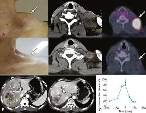
TK gene to increase tumor specificity, and two transgenes: one encoding GM-CSF to stimulate antitumor immune responses and the other β-galactosidase, as a surrogate marker for viral gene expression. JX-594 has demonstrated great success on local-regional and intravenous delivery to patients in terms of safety, cancer selectivity, oncolysis, chemosensitization, and induction of immune and antitumor responses (
Figure 54-1 ,
Table 54-3 ). Notably, in a Phase I clinical trial, intravenously delivered JX-594 was capable of replicating selectively in metastases from a variety of tumor types, representing a milestone in the development of oncolytic agents for systemic administration. Moreover, intravenous JX-594 therapy led to a reduction in the outgrowth of new metastases in patients over time. Seven Phase I and II clinical trials are currently ongoing to test this agent’s safety and efficacy further in various solid tumors.
69,70
Figure 54-1 Representative tumor response to JX-594 treatment (A) Patient 201 with a primary hepatocellular carcinoma received direct intratumoral injections into the liver tumor, which led to a RECIST PR. Direct injection of a previously noninjected tumor in the neck, after four previous cycles of JX-594 treatment in the liver, led to RECIST and Choi response at this site as well, despite the presence of high concentrations of neutralizing antibodies to JX-594. Physical examination and CT and PET-CT scans of the metastatic tumor in the neck region, before and after induction of high-titer antibodies, illustrate this response. (B, C) Patient 103 with metastatic squamous cell carcinoma of the lung received six rounds of JX-594 intratumoral injections. (B) CT scan and (C) tumor cross-section areas before and after treatment. Arrow indicates initiation of JX-594 treatment. CT, Computed tomography; PET, positron emission tomography; PR, partial response; RECIST, Response Evaluation Criteria in Solid Tumors. (Adapted from Park BH, Hwang T, Liu TC, et al. Use of a targeted oncolytic poxvirus, JX-594, in patients with refractory primary or metastatic liver cancer: a phase I trial. Lancet Oncol. 2008;9:533-542.)
Table 54-3
Representative Patient Response to JX-594
∗ RECIST: Response Evaluation Criteria in Solid Tumors: Partial response (PR) is a maximum diameter decrease of ≥30%, progressive disease (PD) is an increase of ≥20%, and stable disease (SD) is a change in diameter between these two cutoffs.
∗∗ Choi criteria: response (+) is maximum diameter decrease of ≥10% or density decrease of ≥15%.
† HU: Hounsfield units, also CT units: related to the composition and nature of the tissue imaged, represents the density of tissue.
Adapted from Park BH, Hwang T, Liu TC, et al. Use of a targeted oncolytic poxvirus, JX-594, in patients with refractory primary or metastatic liver cancer: a phase I trial. Lancet Oncol. 2008;9:533-542.
Adenovirus
Whereas G207 and related viruses were engineered to take advantage of permissive conditions in cancer cells relative to quiescent cells, ONYX-015 was designed to exploit lack of functional p53 in tumor cells. ONYX-015 is an adenovirus that lacks the E1B 55K gene. This gene encodes a protein that binds p53 and targets it for degradation. In the absence of E1B 55K, ONYX-015 was expected to replicate poorly in normal cells, in which functional p53 could abort lytic,
productive replication. In contrast, cancer cells should be permissive for ONYX-015 because E1B 55k should be unnecessary in cells that lack p53.
71
Extensive analysis of the molecular mechanisms underlying ONYX-015 replication revealed that the virus does indeed replicate selectively in tumor cells. Whereas replication of this virus is mediated through the p53 pathway, tumor selectivity is mostly based on the ability of tumor cells to complement other functions of E1B 55K unrelated to p53.
72–74 These functions relate to the ability of E1B 55K to facilitate the export of viral mRNAs and shut down host protein synthesis. Nonetheless, ONYX-015 advanced to Phase III trials in the United States, after encouraging signs of safety and efficacy. The analysis of clinical tumor specimens obtained following injection of ONYX-015 provided unambiguous evidence for its tumor selectivity. Replicating virus was found (1) in head and neck cancer following intratumoral injection, (2) in liver metastases from gastrointestinal cancer after intra-arterial injection, and (3) in lung tumors following intravenous injection.
75–77 Furthermore, objective tumor responses were observed in several clinical Phase II studies. Single-agent treatment with ONYX-015 induced objective responses in 14% of patients with recurrent head and neck cancer.
77 In a subsequent study, the virus was combined with standard chemotherapy. Only a single tumor was injected with ONYX-015, leaving a subgroup of patients with additional uninjected control lesions. Interestingly, the response rate of tumors injected with the virus was significantly greater than those of noninjected lesions, and the time to progression was significantly longer for injected tumors.
75 Further evidence of antitumor activity of ONYX-015 was found in three patients with 5-FU/leucovorin-refractory liver metastases from colorectal cancer that experienced minor responses (30% to 48% shrinkage) following intra-arterial infusion of ONYX-015 into the hepatic artery.
78
Meanwhile, a closely related adenovirus, H101, has recently been developed by Shanghai Sunway Biotech and approved for treatment of head and neck cancer in China
3 after a clinical Phase III study demonstrated a dramatically higher tumor response rate in patients who had received H101 in combination with cisplatin and 5-fluorouracil (78.8% versus 39.6%, respectively).
79
Delta-24 (also known as
dl922-947 or ONYX-838) is another adenovirus mutant that targets cancer cells selectively.
80 The E1a region contains a small deletion that prevents binding to RB. As a result, this virus cannot replicate efficiently in normal cells, because RB represses E2F activity that is essential for replication (in addition to transcribing genes involved in DNA synthesis, E2F activates transcription of the viral E2 region; this is how E2F was first named and identified). In cancer cells, E2F activity is not repressed by RB, because RB itself is mutated or inactivated indirectly through loss of p16INK4a, amplification of cyclin D1, or by other means. Tumor cells therefore provide a permissive environment for replication of Delta-24. A modified version of Delta-24 has entered clinical trials for the treatment of glioblastoma.
81,82 This virus, Delta-24 RGD, has been engineered to increase infectivity by the addition of an RGD sequence in its fiber gene.
83 Likewise, adenoviruses designated KD1 and KD3 contain two small deletions in E1A that abolish its binding to pRB but leave the ability of E1A to transactivate viral genes intact. These have been shown to replicate with great efficiency in tumor cells, but fail to replicate efficiently in normal cells.
84
A second-generation version of Delta-24 was engineered to express human p53.
85,86 Ad24-p53 more effectively killed most human cancer cell lines tested in vitro than did its parent Ad24 and had significant activity against xenografts in vivo
. 85 To further improve potency of this virus, the p53 transgene was engineered so that it is resistant to degradation by Mdm2.
85 Adenoviruses have also been engineered to take advantage of unregulated TCF transcriptional activity, a characteristic of colorectal cancer cells that lack the APC tumor suppressor or contain activating mutations in beta-catenin.
87,88
An array of creative approaches has been employed to make viruses replicate selectively based on abnormal transcription activity in cancer cells. These include viruses that use unregulated E2F activity resulting from loss of the RB tumor suppressor pathway to drive the E2F-1 promoter, the telomerase promoter, prostate-specific promoters and regulatory elements to drive proliferation in prostate cancer cells, and many others. These approaches have been reviewed recently by Fukazawa and colleagues.
89
Challenges and Future Perspective
Preclinical Development
Gene therapeutic agents and oncolytic viruses have highly diverse physical and biological properties and act through complex molecular mechanisms, making predictions about their pharmacological and pharmacodynamic behavior in humans difficult. Preclinical model systems are therefore particularly important elements in the selection process for further clinical development.
Mouse xenograft tumor models of human cancer cell lines in nude mice allow for efficient assessment of antitumor activities of novel gene delivery and oncolytic agents in a broad variety of human tumor types. However, these models have significant limitations due to the lack of a functional immune system and differences in structure and composition
of the tumor stroma. To address these limitations, immunocompetent tumor models have been developed. Such models have been instrumental for assessing the impact of immune-modulatory genes in the genome of oncolytic adenoviruses on virus replication and antitumor effect.
90 In addition, orthotopic implantation of allografts in immunocompetent models allows for testing novel vectors in tumors growing an organotypic microenvironment that more closely resembles the situation in humans.
91–93 Despite these improved murine models, limitations remain. For example, normal and malignant mouse tissues only poorly support replication of human adenoviruses. Another example is the difference between the sequences of human and mouse cytokines, which makes the generation of mouse-specific variants of immune-modulatory agents necessary. Other species offer potentially advantageous features. For example, normal and malignant Syrian hamster cells support adenovirus replication. Using this model, intratumoral injection of an oncolytic adenovirus resulted in suppression not only of the primary tumor but also of distant metastases following virus entry into the bloodstream.
94 Similar results were obtained in cotton rats.
95 In addition, replication-competent viruses for use in canine models have been developed.
96
Biodistribution and systemic effects of novel gene therapy and oncolytic viral agents are fundamentally different from small-molecule or antibody-based anticancer therapies and therefore difficult to predict. To address this, novel in vivo imaging strategies allowing for real-time monitoring of the effects of such agents in animals as well as in humans have been developed and will play a major role in the further development of this therapeutic approach. Distribution of nonviral and viral particles can be directly assessed by radioactive or fluorescent labeling. The biodistribution of liposomes, for example, can be followed after labeling with radioactive isotopes (e.g.,
99mTc) or gadolinium by scintigraphy or magnetic resonance imaging, respectively.
97,98 A variety of strategies have been pursued to monitor viral agents. Green-fluorescent protein and firefly luciferase are transgenes that allow for the detection of cells infected with viruses carrying an expression cassette for either of these genes through the detection of fluorescent light or bioluminescence, respectively—in animal models, at least.
99,100 The use of prodrug-converting enzymes opens the possibility of using the enzyme activity for imaging purposes. The most developed approach is expression of the prodrug converting enzyme HSV-TK, which not only converts ganciclovir into cytotoxic phosphorylated derivatives but also phosphorylates uracil derivatives labeled with radioactive iodine and acylguanosines labeled with radioactive fluorine, which are then also retained within the cell and detectable by positron emission tomography (PET). This approach has been successfully used in a variety of vectors in small animals. Recently, it has also been demonstrated to be an effective imaging strategy in a pilot study in patients with liver cancer.
101 A similar approach involves the vector-mediated delivery of receptors with only limited physiologic expression, such as the dopamine D2 receptor or the somatostatin receptor subtype 2. PET imaging probes for both receptors are already clinically available for imaging of neuroendocrine tumors. A third technology has been developed using the sodium-iodide symporter, which is a transmembrane transporter protein that is physiologically predominantly expressed in the thyroid gland.
102 Ectopic expression of this protein leads to accumulation of radioactive iodine, which can be detected by radionuclide imaging using a gamma camera or PET. The potential advantage of this approach is that cytotoxic radionuclides such as
131 I can be used therapeutically.
103
The complexity of the host-vector interaction and the resulting dynamics of virus and tumor cell replication are theoretically amenable to in silico modeling of virus and tumor cell population dynamics. Such mathematical models created important insights into the kinetics of human immunodeficiency virus (HIV) infection and treatment
104,105 and have been, at a theoretical level, developed for oncolytic viruses and their interaction with tumor cells and the host immune system.
106,107 Experimental validation of these models is currently being actively pursued by several laboratories.
Safety and Toxicity
The use of viruses either as vectors for the delivery of therapeutic genes or, in mutant form, as therapeutics themselves raised significant concerns not only in regard to the safety of individuals treated with such agents but also because of the potential risks for others. In particular, the occurrence of recombinant viruses that regain wild-type properties or demonstrate even greater toxicity was feared. For this reason, extensive safety studies were performed. For example, the biohazard potential of AdCMV-p53 was investigated in France in the context of clinical trials of this virus in head and neck cancer. No evidence for any environmental risk from intratumoral injection of the virus was found.
108
Immune and Cytokine Response
A major concern with respect to the use of viral particles as therapeutic agents is the induction of neutralizing antibodies that could limit the efficacy of such agents. In the case of adenovirus this is of particular relevance, as at least 50%
of patients present with preexisting antibodies against adenovirus type 5, resulting from earlier infections.
75–78,109,110 It is therefore not surprising that almost all of the patients treated with viral agents developed high titers of neutralizing antibodies following the first administration of the virus. Preclinical studies suggest that the presence of such antibodies might reduce the efficacy of adenoviral treatments.
111 At this point, this has not been clearly demonstrated clinically. In contrast, following intra-arterial infusion of ONYX-015 into the hepatic artery or intravenous administration, the virus was cleared rapidly from the bloodstream.
Improving Tumor Killing by Improving Tumor Cell Access
Viral vectors and oncolytic agents have been engineered to improve their ability to infect cancer cells, either to increase selectivity or to increase potency. Adenoviruses have been the focus of many of these efforts. This is because it is believed that the utility of adenovirus vectors is limited because of the low expression of CAR, the high-affinity receptor that is necessary for efficient attachment to the cell membrane.
112 This is of particular concern in many advanced cancers, in which CAR levels are often low relative to normal cells or well-differentiated cancers.
113,114 Many attempts have been made to address this problem. Wickham and co-workers
115 modified the C terminus of the adenoviral fiber protein by the addition of either an RGD-containing peptide or seven lysine residues. Dmitriev and colleagues
116,117 have also shown that the incorporation of an RGD-containing peptide in the H1 loop of the fiber knob domain results in the ability of the virus to utilize an alternative receptor during the cell entry process. The modified virus was able to infect primary tumor cells and tumor cell lines more efficiently than unmodified virus.
116,118 The RGD/fiber modification was subsequently introduced into the Delta-24 virus, described above. Gu and associates have successfully redirected cell binding and uptake of an adenovirus through fibroblast growth factor receptors (FGFRs), suggesting that redirecting the native tropism of adenovirus may offer therapeutic benefit.
119
Conclusion
Virotherapy and other gene-therapeutic approaches represent a novel class of targeted anticancer agents that is distinct from traditional treatment modalities.
The initial focus of this field was to invent and test new agents that kill cancer cells selectively, based on the genetic lesions that cause this disease. This goal has been achieved using a rich variety of viruses, vectors, genes, and approaches, in laboratory and animal model settings. Some of these approaches have been translated into clinical research projects; however, until very recently, clinical results have been disappointing in terms of increased survival or meaningful patient benefit.
120 Although most treatment regimens have shown great safety, some early gene therapeutic trials raised serious concerns in this regard—the death of a patient with ornithine transcarbamylase deficiency, and the leukemia caused by gene therapy in four severe combined immunodeficiency (SCID) patients.
121 These rare adverse effects and the initial lack of compelling clinical activity, together with the potential risk and cost of manufacturing these agents, has dampened the initial enthusiasm in this field, as well as interfered with significant investments. However, substantial advances in our understanding of cancer biology and validation of targets, as well as the promise shown by some of the very recent gene therapeutic clinical trials, have led to a paradigm shift: anticancer gene therapy is now on an upswing again.
Nonetheless, significant hurdles still exist and need to be addressed. One of the biggest remaining challenges is delivery, including delivery to the tumor site, spread within the tumor, undesired clearance by the liver, neutralizing antibodies, and complement. Another significant difficulty for many of the approaches currently in development is the lack of appropriate preclinical models.
For gene therapy strategies to be clinically successful, they likely have to be included in multimodality treatment approaches and go far beyond killing cancer cells: they need to be anti-angiogenic and immune-modulatory, and they must prevent or overcome resistance to therapy. Intelligent vector design and clever combination regimens will, it is hoped, bring us closer to achieving this goal. Recent studies have demonstrated synergistic effects of combination therapies consisting of conventional chemo- or radio- and virotherapy. Moreover, combinatorial therapies with more “modern” approaches, such as the use of nanotechnology and cellular carriers, hold great promise. For instance, polymer-coated adenoviruses have shown a significantly enhanced half-life when compared to naked viruses, and the use of appropriate cellular carriers has been demonstrated to improve delivery and induction of desirable immune responses against the tumor.
Furthermore, clinical studies will likely yield better patient benefits and increase the chance for approval if factors can be pinpointed that predict efficacy—such as tumor type, histology, and molecular signatures.
Taken together, the recent advances in cancer biology, virology, biotechnology, and nanotechnology, as well as interdisciplinary collaborations, will expedite the development of this therapeutic platform and, we can hope, improve patients’ lives in the very near future.
1. Clevers H. Wnt breakers in colon cancer . Cancer Cell . 2004 ; 5 : 5 – 6 .
2. Kaye F.J. RB and cyclin dependent kinase pathways: defining a distinction between RB and p16 loss in lung cancer . Oncogene . 2002 ; 21 : 6908 – 6914 .
3. Garber K. China approves world’s first oncolytic virus therapy for cancer treatment . J Natl Cancer Inst . 2006 ; 98 : 298 – 300 .
4. Zhang W.W. , Alemany R. , Wang J. , Koch P.E. , Ordonez N.G. , Roth J.A. Safety evaluation of Ad5CMV-p53 in vitro and in vivo . Hum Gene Ther . 1995 ; 6 : 155 – 164 .
5. Barak Y. , Juven T. , Haffner R. , Oren M. mdm2 expression is induced by wild type p53 activity . EMBO J . 1993 ; 12 : 461 – 468 .
6. Xu M. , Kumar D. , Srinivas S. et al. Parenteral gene therapy with p53 inhibits human breast tumors in vivo through a bystander mechanism without evidence of toxicity . Hum Gene Ther . 1997 ; 8 : 177 – 185 .
7. Reed J.C. , Miyashita T. , Krajewski S. et al. Bcl-2 family proteins and the regulation of programmed cell death in leukemia and lymphoma . Cancer Treat Res . 1996 ; 84 : 31 – 72 .
8. Molinier-Frenkel V. , Le Boulaire C. , Le Gal F.A. et al. Longitudinal follow-up of cellular and humoral immunity induced by recombinant adenovirus-mediated gene therapy in cancer patients . Hum Gene Ther . 2000 ; 11 : 1911 – 1920 .
9.
Buy Membership for Hematology, Oncology and Palliative Medicine Category to continue reading.
Learn more here
Related
The Molecular Basis of Cancer Expert Consult - Online