Epigenetics and Cancer

Published on 09/04/2015 by admin

Filed under Hematology, Oncology and Palliative Medicine

Last modified 09/04/2015

Print this page

rate 1 star rate 2 star rate 3 star rate 4 star rate 5 star
Your rating: none, Average: 0 (0 votes)

This article have been viewed 2756 times

Figure 5-1 Chromatin surrounding an actively expressed gene in a normal mature cell versus surrounding that same gene when it is DNA hypermethylated and aberrantly, heritably, silenced in a tumor cell. On the left, the chromatin is composed of histone modifications associated with active transcription (H3K4me) and (H3K9acetyl), and the DNA is largely unmethylated at CpG sites (green circles) with only occasional methylation (red circles). The nucleosomes (large blue ovals) are linearly arranged as associated with the areas of active transcription defined in Figure 5-2. The gene on the right is fully transcriptionally repressed (large red X); the DNA is methylated and DNA methylating enzymes are present (DNMT1 and -3b); HDACs are present to catalyze histone deacetylation; the machinery of transcriptional repression is present, including the PcG proteins (PRC) with EZH2, which catalyzes the H3K27me3 mark (red hexagons); and the key silencing marks of H3K9me2 and me3 are also present (red hexagons). The nucleosomes are more tightly compacted, as is representative of the repressive chromatin shown in Figure 5-2. (From Ting AH, McGarvey KM, Baylin SB. The cancer epigenome: components and functional correlates. Genes Dev 2006;20:3215-3231, with permission.)
image
Figure 5-2 The normal versus cancer epigenome Top: In normal mammalian cells, CpG islands in proximal gene promoter regions (a three-exon gene is shown, with each exon marked in blue and numbered) are largely protected from DNA methylation (cytosines, open lollipops) and reside in restricted regions of open chromatin (inset, upstream of transcription start shows three nucleosomes with wide spacing), or euchromatic states, favorable for gene transcription (large gray arrow). In contrast, for most regions of the genome, such as in the bodies of many genes and areas outside genes, particularly including repeat elements and pericentromeric regions, the cytosines in CpG dinucleotides are methylated (black lollipops). This DNA methylation is characteristic of the bulk of the human genome, which is packaged as closed chromatin (the inset above methylated CpGs shows multiple nucleosomes with higher-order, tight compaction) unfavorable for transcription. Bottom: In cancer cells, there tends to be a reversal of this pattern. Proximal promoter CpG islands for many abnormally silenced genes (as represented by the same gene as shown in the top panel, which is depicted as representing the tumor suppressor genes listed) become DNA hypermethylated and reside in a closed chromatin, or more heterochromatic-type state, which is not favorable for transcription (red X). In contrast, cytosines in CpG dinucleotides in other regions of the genome display hypomethylation and are associated with states of aberrantly loosened chromatin. The overall result is abnormal chromatin packaging with the potential for underpinning an abnormal cellular memory for gene expression and for conveying abnormal structural function for chromosomes. (From Ting AH, McGarvey KM, Baylin SB. The cancer epigenome: components and functional correlates. Genes Dev 2006;20:3215-3231, with permission.)
In contradistinction to the depletion of CpGs throughout most of the genome, approximately half of the genes in the genome have regions in their promoters, termed CpG islands where the expected frequency of the nucleotide has been preserved (see Figure 5-2). For most such genes, these islands are protected from DNA methylation, and this methylation-free state is associated with active transcription of these genes, or preservation of their being in a transcription-ready state. 6,7,17 These CpG islands are the target of key epigenetic abnormalities in cancer cells, as discussed in detail in subsequent sections of this chapter.
In addition to the previously described role of DNA methylation in global DNA packaging, it is also linked to regulation of expression for specific genes in normal cells. In this regard, when localized to gene promoter regions, it may act to provide a tightening of heritable states for gene silencing. Examples include the imposition of DNA methylation in the promoters of genes shortly after other processes initiate their silencing in regions on the inactive X-chromosome of females. 18 A similar role is apparent in genes that are imprinted in mammals wherein DNA methylation of promoter regions is seen on the silenced allele of such genes. 19,20 DNA methylation also may participate in regulating expression of certain genes in normal cells that are expressed in a tissue-specific manner, such as the silencing of globin genes in all but cells actively engaged in erythropoiesis. 21,22
In the gene-silencing roles, there is a tight interplay between the modification of key histone amino acid residues and DNA methylation. Thus, at least in lower organisms such as Neurospora and Arabidopsis, methylation of lysine 9 of histone H3 (H3K9me) may help determine positions where cytosine methylation is placed in the genome. 23,24 Increased levels of the active histone modification H3K4 methylation can be inhibitory to the recruitment of the DNA methylating enzymes. 25 In turn, DNA methylation recruits a series of proteins, methylcytosine binding proteins (MBPs), which are complexed, in turn, with HDACs, which help maintain the deacetylation of H3K9 and other key histone lysines in regions of silenced genes. 6,7

Abnormalities of DNA Methylation and Chromatin Organization in Cancer: The Cancer “Epigenome”

Overall Characterization
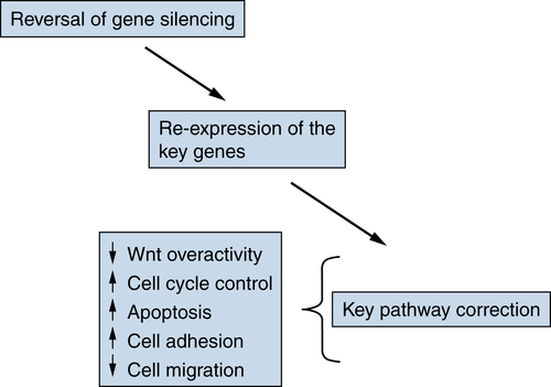
The organization of the genome, as mediated by chromatin and DNA methylation, appears to be quite abnormal in cancer cells of all types when compared with the corresponding cells in normal renewing adult tissues. 2628 In many cancers, total levels of DNA methylation are decreased with losses apparent within repeat sequences, the bodies and promoters of selected genes, and in the pericentromeric regions of chromosomes. 2628 The full ramifications of these losses are still being explored, but the changes have the potential for associating with unwanted gene expression and especially, in terms of the pericentromeric abnormalities, with chromosomal instability. 2630

Table 5-1

Examples of Pathways Affected by Aberrant Gene Silencing in Cancer

Pathway Genes
Cell cycle control p16, p15
Apoptosis DAP-kinase, ASC/TMS1, HIC1
Increased stem/developmental pathway activity (Wnt, etc.) SFRPs
DNA damage repair MLH 1 , O 6 -MGM, GST Pi
Cell adhesion E-cadherin
Cell migration TIMPs
Differentiation GATA-4, GATA-5, TGF-β receptor
Chromosomal stability CHFR
Recently, extensive analyses of DNA methylation patterns in cancer have revealed that, in addition to the regions mentioned previously, there are large megabase regions for loss of DNA methylation scattered over many chromosomes. 31 Within these regions, more localized gains of DNA methylation simultaneously reside in the normally unmethylated CpG islands of promoters of many genes. These methylation gains are, to date, the most studied of the epigenetic abnormalities in cancer 3133 and are associated with repressive chromatin changes and potentials for aberrant loss of gene expression and function. 2628,3234 In fact, it is increasingly apparent that potential disruption of gene function as a consequence of promoter DNA hypermethylation is as frequent, or more frequent, in cancers than mutations as a potential mechanism for loss of tumor suppressor gene function. 26 Individual tumors may actually contain hundreds of such affected genes, which include many of the best characterized tumor suppressor genes 2628,35 and genes involved with virtually every cellular pathway for which alterations are thought to drive the initiation and progression of cancer 36 (Table 5-1 ), 2628,35,37 including those for cell cycle events, apoptosis, developmental biology signal transduction for stem cell function, differentiation, cell-cell adhesion, cell-cell recognition, cell migration and invasion, and others. 3335,37 The list of involved genes, as identified by study of candidate genes and techniques for randomly screening the cancer epigenome, 3335,38,39 is steadily growing for virtually all major cancer types.

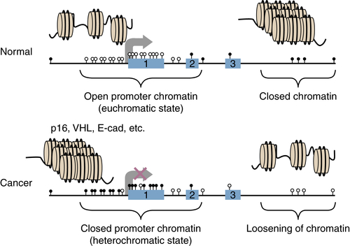
Chromatin Abnormalities in Cancer and Interplay with DNA Methylation Changes

In addition to abnormalities in DNA methylation in cancer, chromatin alterations are also frequent, and there can be interplay between the two. Indeed, one of the most active areas of cancer epigenetics research at present, and one of utmost importance to the translational impact for cancer prevention, diagnosis, and treatment, concerns delineation of the molecular underpinnings of how the cancer epigenome evolves. 32 This investigation has benefited from, and contributed to, the explosion of knowledge over the past 5 to 10 years in understanding how chromatin functions for packaging of the genome and for modulation of gene expression. 40 Although many remain to be elucidated, important findings are emerging that provide clues to the origins of epigenetic abnormalities in cancer.
The initiation of DNA methylation, its maintenance, and its role in transcriptional repression are all dependent on its interaction with chromatin organization (see Figure 5-1). As previously alluded to, the sites of DNA methylation themselves may be dependent, initially, on histone modifications. Thus, H3K9 methylation, and the histone methyltransferases that catalyze this mark, appears required for DNA methylation in lower organisms such as Arabidopsis and Neurospora. 23,24 In addition, the polycomb group of proteins, 4143 discussed in more detail later, which target another key gene repression mark to nucleosomes, H3K27me, have been implicated in the targeting and maintenance of DNA methylation. Also, a series of proteins called methylcytosine binding proteins (MBPs), and the protein complexes in which they reside, can bind to methylated CpG sites to help relay, and/or maintain, a silencing signal. 6,7 These complexes contain the previously mentioned enzymes, histone deacetylases (HDACs), which catalyze the deacetylation of key amino acid residues, such as H3K9, that are highly characteristic of transcriptionally silent regions of DNA. 6,7,44 The DNMTs themselves also interact with HDACs to help target these enzymes to sites of DNA methylation. 4547
The alterations in the levels or ratios of factors that mediate epigenetic abnormalities in cancer cells are first manifest by certain global abnormalities. Thus, increases in the levels and activities of the DNA methylation catalyzing enzymes 48 ; of the proteins in complexes that modulate the enzymes that catalyze transcriptional repression histone modifications 4951 ; and altered levels of the repressive histone marks themselves, including loss of acetylation at H4K16 and increased levels of H4K20 acetylation, 52 are all reported as common hallmarks of human cancer. Locally, at gene promoters affected by promoter DNA methylation and aberrant gene silencing (see Figure 5-1), there are decreases in histone modifications associated with active gene transcription, such as acetylation of H3K9 and H4K16; increases in modifications associated with transcriptionally repressive chromatin, including H3K9me2 and me3 and H3K27me3; and increases in the enzymes that catalyze these latter repressive marks. 48,53
The precise manner in which all of these chromatin components interact to initiate and/or maintain abnormal gene promoter DNA methylation and the attendant silencing of involved genes is not yet known. As noted earlier, in cancer, these gains of DNA methylation can occur as focal changes within large regions of loss of normal DNA methylation. 31 These data suggest that molecular maintenance of chromatin and DNA methylation boundaries “break down” during tumor progression. Factors such as “insulator” proteins, which maintain separation between transcriptionally repressive and active chromatin states, are altered and/or chromatin states that associate with such transcription states and are also shifted. 32
How might such chromatin alterations come about? One potential mechanism concerns recent exciting findings stemming from deep sequencing analyses of most solid and liquid tumor types that are revealing many frequent mutations in genes encoding for proteins that normally ensure formation of chromatin in normal cell epigenomes 32,54 (see Figure 5-2 and Table 5-1). The high frequency of these changes suggests that they are fundamentally important to the initiation and progression of cancer 32,54 and may justify considering their roles as “driver” mutations. As such, they would contribute important steps in the initiation and/or progression of cancer. A major challenge, however, is to understand the exact consequences of these mutations for key steps in tumorigenesis and for their precise contribution to cancer-specific alterations of chromatin and DNA methylation. One mutation particularly, in the IDH1 and 2 genes in glioblastomas and leukemias, 5557 appears to cause a metabolic balance that inhibits enzymes that remove key gene expression silencing histone marks and/or that can remove DNA methylation. 58,59 These mutations, when studied experimentally, appear to disrupt normal stem/progenitor cell function and commitment of cells to proper lineages. Moreover, the tumors that harbor them have an increased frequency of abnormal promoter region CpG island DNA methylation. 60,61
The studies just referred to concerning regional changes in epigenetic patterns in cancer suggest that many genes with CpG island–containing promoters are particularly vulnerable to adopting abnormal DNA methylation during the abnormal cellular expansion that underlies the earliest phases of tumor progression 48,6264 (Figure 5-3 ). A growing number of studies reveal that these genes are enriched for those that are important for developmental functions. 65 Importantly, in normal embryonic and adult stem cells, these genes are maintained in a low, poised transcription state in which their promoter regions are marked, not by DNA methylation, but by a “bivalent” chromatin pattern characterized by a broad distribution of the PcG-mediated H3K27me3, repressive histone modification accompanied by a more narrow zone of the active mark, H3K4 methylation surrounding the transcription start site. 48,6264 This bivalency may allow regulatory flexibility by keeping these genes in a low, poised quiescent transcription state to maintain stem cell pluripotency and/or prevent their undergoing premature lineage or differentiation commitment. 65 Interestingly, these genes in ESC typically reside in the large regions mentioned earlier that broadly lose DNA methylation in cancer but gain CpG island methylation focally around promoter regions. 31 Thus, it appears that for these vulnerable genes, during tumorigenesis, there is imposition of abnormal, promoter CpG island DNA methylation as opposed to the control of expression of these genes in normal cell renewal by a balance involving polycomb proteins and active gene histone marks. The more stabilizing silencing process in association with DNA methylation may, then, provide a mechanism by which inability to induce at least some of the foregoing genes could help foster maintenance of stemlike cells and/or a block in their ability to differentiate. 65,66
image
Figure 5-3 A model for the potential contribution of stem cell chromatin to the initiation and maintenance of aberrant epigenetic gene silencing in cancers During normal ES cell formation, a bivalent chromatin is recruited to the promoters of a subset of genes that need to be held in a low-expression state to prevent lineage commitment. The involvement of small interfering RNA (siRNA) species could be a trigger to this process, and the chromatin is composed of histone modifications associated with active transcription (H3K4me) and inactive transcription (H3K27me). The PcG proteins (PRC) are responsible for the H3K27me3 mark through the HMT, EZH2, and deacetylation of key histone lysine residues is catalyzed by HDACs that are recruited by multiple transcriptional repressive complexes. At such genes, DNA is largely unmethylated (green circles), and histones may be maintained in a mixture of acetylated (green hexagons) and deacetylated (red hexagons) states. Bottom left: With normal cell differentiation and lineage commitment, the genes become transcriptionally active, and the silencing marks are reduced while active histone marks are retained. DNA remains unmethylated. However, as shown at bottom right, during cancer-predisposing events, abnormal pressure for stem/progenitor cell proliferation with retained bivalent chromatin may allow polycomb proteins and/or marks to recruit other silencing marks such as H3K9me2 and H3K9me3 and DNA methyltransferases. The promoter evolves abnormal DNA methylation (red circles) and a tight heritable gene silencing (large red X), which results in loss of function for genes. Tumors may arise in such clones with subsequent progression steps. Experimentally, the potential underlying bivalent chromatin for such tumor genes, plus retained H3K9me3, can be revealed by induced DNA demethylation (large green arrow) and resultant gene re-expression. (From Ting AH, McGarvey KM, Baylin SB. The cancer epigenome: components and functional correlates. Genes Dev 2006;20:3215-3231, with permission.)

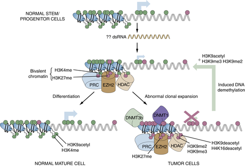
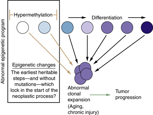
Relationships of Epigenetic Changes in General, and Aberrant Gene Silencing in Particular, to the Progression of Cancer

Although losses and gains of DNA methylation in cancer may arise at any point during tumor progression, it has become apparent that many of the changes arise early, before frank carcinomas. 34,37,48,67 In fact, it is possible that some of the events, such as silencing of key genes, could even initiate the abnormal clonal expansion that creates early preinvasive lesions, which are then at risk for subsequent genetic and epigenetic events that further tumor progression and lead to invasive and metastatic cancer (Figure 5-4 ). 34,37,48,67 The genes silenced, or groups of such genes, may provide loss of tumor suppressor function that allows cells to abnormally survive the hostile environments that are risk factors for cancer development, such as chronic inflammation, and expose cells to DNA-damaging agents such as reactive oxygen species (ROS). In fact, experimental data now indicate that increases in ROS can rapidly trigger localization of protein complexes, including DNA methyltransferases, to CpG islands in gene promoters. 68 During this period, for the types of low-transcription genes that, as discussed in a previous section, seem vulnerable to adopting abnormal promoter DNA methylation, abnormal DNA methylation begins to appear. 68 This change in key groups of genes during injury repair may enable cells that would normally undergo apoptosis from DNA damage to survive and expand. They may, then, more easily select for mutations and/or chromatin damage that may favor subsequent tumor progression (see Figure 5-4).
image
Figure 5-4 The potential that epigenetic gene-silencing events have for participation in the earliest stages of tumor progression As discussed in the text and Figure 5-3, suppression of gene transcription can be a normal event for a group of key genes in stem cells and progenitor cells as adult epithelial-cell renewal takes place (left large box). This low-level gene expression is accomplished by a balance of chromatin modifications that associate with active and repressed transcription (bivalent chromatin; see Figure 5-3), but transcription can increase in maturing cells during normal cell renewal. This balance of control for gene expression allows stem and progenitor cells to progress along a normal differentiation pathway (moving with arrow from left to right across the top of the figure). During chronic and abnormal pressures on stem-cell and progenitor-cell pools for tissue repair, there is a tendency for the gene chromatin constituents in these cells (see Figure 5-3) to recruit promoter DNA hypermethylation (top of large box), and this becomes associated with heritable silencing of the genes (abnormal epigenetic program, large box). This inability of the genes to increase with maturation cues facilitates abnormal clonal expansion of stem/progenitor cells (heavier arrows), at the expense of differentiation. Such expansion may occur in stroma, leading to an abnormal environment that helps support epithelial tumor growth. This process renders the abnormal clones at risk for further tumor progression (bottom arrow) driven by subsequent genetic or epigenetic events.
There are now several key examples of this proposed early role for DNA hypermethylation and gene silencing in tumor progression. One of the major tumor suppressor genes in cancer, where loss of function leads to cell cycle abnormalities and uncontrolled growth, is p16. 69 A role for this loss of function in early tumorigenesis, via early expansion of stem cells, would be predicted from data in p16 knockout mice revealing that germline loss of this gene can increase stem-cell lifespan. 7072 The rate of point mutations in p16 in most cancer types is low, but the gene is a frequent target for early methylation in these same tumors, such as breast cancer and non–small-cell lung cancer (NSCLC). 73,74 This methylation occurs early in tumor progression, before invasive cancer. 73,75 In fact, histologically normal mammary epithelium from some healthy women without malignancy can harbor focal p16 promoter hypermethylation. 76 Experimentally, early loss of p16 in mammary epithelial cells precedes genomic and epigenetic instability. 7779 A recent study in the Cancer Genome Atlas project (TCGA) further emphasizes how, in the squamous form of NSCLC, abnormal methylation of p16 is mutually exclusive to mutations of the gene. 80
Another excellent example of the potential role for early epigenetic abnormalities and stem/precursor cell expansion to contribute to early steps in tumor progression involves colon cancer. In this disease, cancer risk can begin with the appearance of aberrant crypt foci in the colonic epithelium, and these harbor premalignant, hyperplastic, preadenomatous cells. 81,82 The evolution of colon cancer is highly dependent on abnormal activation of the stem/precursor cell Wnt pathway, which by the time frank polyps and/or invasive lesions appear, is driven by classic inactivating mutations in the APC gene or activating mutations of β-catenin, key downstream players in the pathway. 83,84 In aberrant crypt foci, however, such mutations may not be present, yet there is DNA hypermethylation 37,85 of a family of genes, the SFRPs, which encode for membrane region proteins that antagonize Wnt interaction with its receptors. 85,86 This hypermethylation persists throughout colon tumor progression and can later collaborate with the downstream mutations in driving the Wnt pathway. 37,85

Translational Implications of Epigenetic Changes in Cancer

The delineation of epigenetic abnormalities in tumorigenesis is now actively contributing not only to our understanding of the biology of cancer but also to potentially new ways for managing these diseases. First, the overall abnormalities in chromatin organization and DNA methylation provide potential biomarkers for use in cancer risk assessment, early diagnosis, and prognosis assessment. Second, the molecular features that contribute to the epigenetic abnormalities in cancer are increasingly offering new targets for devising novel therapy strategies for all types of cancers. Some of the progress in these arenas is reviewed in this section.

Epigenetic Changes and Development of Biomarker Strategies

Overall and local chromatin changes in cancer provide potential markers for cancer management. For example, during the early stages of tumor progression, some of the histone modifications altered in cancer cells (see Figures 5-1 and 5-3) are manifest, as previously discussed. These can be global in tumor cells such that levels of these parameters, including losses of monoacetylated and trimethylated forms of histone H4 and losses of acetylated Lys16 and trimethylated Lys20 residues of histone H4, reflect either the presence of cancer or its stages. 52,87 Changes in modification marks on histones H3 and H4 have been correlated with aggressiveness of prostate cancer. 87 These global changes are hypothesized to be common hallmarks of human tumor cells and hold promise for the development of important biomarkers. Similarly, increases in levels of enzymes that catalyze key facets of cancer epigenetic abnormalities, such as the DNA methyltransferases for DNA methylation 48 and, more recently, histone methyltransferases such as EZH2 50,51 or H3K27 methylation, and other PcG gene silencing constituents, 88 have been correlated with several cancer types and correlated with aggressive behavior.
The most developed biomarker strategies have been centered on the gene promoter DNA hypermethylation and gene silencing. The use of promoter-hypermethylation sequences as a molecular signature is providing one of the most promising biomarker strategies for cancer. 8991 One advantage of the approaches being adopted relies on the relative stability of DNA as compared with many proteins and RNA, which allows for use of paraffin-embedded clinical samples for detection strategies. Given the fact that, as discussed, the numbers of genes DNA hypermethylated is so high in individual tumors, and that this phenomenon is common in all cancer types, it is not difficult to build profiles of relatively small hypermethylated gene panels in which one or more markers are positive in virtually any cancer. 92 Combined with a repertoire of sensitive polymerase chain reaction (PCR)-based assay procedures to specifically detect the hypermethylated sequences, 90,91,93 and the fact that these assays can be targeted to constant positions of the abnormal CpG methylation in gene promoter regions, relatively simple detection strategies are being constructed and now include adaptation of the assays to nanoparticle-based platforms. 94,95 With such assays, abnormally methylated gene sequences have been detected in sources as diverse as DNA extracted from tumor, lymph nodes, serum, sputum, bronchial-lavage fluid, and urine for patients with all varieties of cancer types. 8991 The strategies range from determining whether the methylation patterns in tumors reflect prognosis for behavior, to use of marker detection in distal sites for purposes of cancer risk assessment, early diagnosis, and staging. For example, studies of sputum DNA from patients at high risk for lung cancer have found that invasive tumors may be predicted, with odds ratios of 6 or more, more than a year before clinical detection of cancer, 96 and findings of abnormal methylation markers in sputum may be useful for predicting which patients with surgically resected early-stage lung cancers may experience a recurrence. 97 The occurrence of hypermethylation of specific genes in tumor DNA may predict future behavior of a cancer; reportedly, this change for the p16 gene in DNA from lung cancers predicts high likelihood of poor outcome. 98 Assays to detect DNA hypermethylation of the GST-pi gene in needle biopsies of the prostate are reaching clinical use as an aid to refine the diagnosis of prostate cancer. Recently, simultaneous detection of abnormal promoter DNA methylation of a panel of four genes, in DNA from tumor and chest lymph nodes assessed to be microscopically free of tumor by pathology exam, provides a potential strategy for predicting recurrence in early-stage non–small-cell lung cancer (NSCLC). 99 Similarly, sensitive detection of these types of gene promoter DNA methylation abnormalities in stool and/or serum DNA appears promising for the early detection of colon polyps and/or cancer. 100,101
The promise of these biomarker approaches will be realized only through continued studies of ever-increasing size. The precise assays best suited for routine clinical use must be determined, and approaches that build the most quantitative determinations into these assays, increasingly being applied, 102 must be evaluated. Confounding issues must be continuously considered. A most critical one is to always consider whether the presence of hypermethylated gene markers in normal-appearing tissue settings means cancer risk as opposed to actual cancer presence. This accentuates the importance of the information discussed earlier in this chapter concerning the biology of cancer as it involves epigenetic abnormalities. The position for appearance of individual gene markers in tumor progression is critical and must be paired with consideration of risk factors. For example, gene promoter methylation in normal tissues can increase with age, as best studied in the colon, 103 and parallels the risk of cancer at a given site. All of this information defines the potential power of marker strategies using gene promoter hypermethylated sequences and the caveats that must be considered in using these strategies.
Perhaps one of the most promising uses for gene hypermethylation markers, and another that is now close to general clinical realization, concerns their use for prediction of drug sensitivity. This strategy exploits the fact that aberrantly silenced genes involved with this epigenetic abnormality can belong to pathways that dictate cellular pathways integral to drug responsiveness. The most developed example of this is the silencing of the DNA repair gene O 6 -MGMT, which encodes for a protein that mediates removal of bulky alkylation adducts from guanosines. 89 Several tumor types lose the function of O 6 -MGMT via aberrant silencing of the gene, and constituent cells have a diminished capacity to repair alkylation damage, rendering them sensitive to alkylating agents such as temozolomide. 104,105 Thus, multiple studies reveal that patients with brain tumors harboring O 6 -MGMT respond remarkably better to this agent than those whose tumors lack this change, providing an exceptionally promising marker to stratify patients with this lethal tumor type for best therapy approaches. 105,106 If ongoing trials continue to validate this, a relatively easy and rapid marker will be available for routine clinical use.

Targeting Epigenetic Abnormalities for Cancer Prevention and Therapy

There is now growing appreciation that our expanding knowledge of epigenetic abnormalities in cancer, in general, and most especially, at present, the definition of aberrant gene silencing as an alternative mechanism to mutations for loss of tumor suppressor gene function, offers extraordinary potential for exploitation in managing cancer. 89,107 First, there is the critical difference that, as compared with mutations, epigenetic gene silencing, as we have discussed in this chapter, is potentially reversible. Second, the growing list of molecular steps being defined as components of the silencing offers more individual and combinatorial targets for considering interventions. Third, the early position of aberrant genes in tumor progression makes reversal of the silencing an attractive target for prevention approaches. Also, the potential of the silenced genes to participate in tumor recurrence suggests that the adjuvant treatment arena may be an attractive area for epigenetic therapy. Fourth, and perhaps most important, the biology discussed in this chapter, including the high frequency of the gene silencing abnormality in all cancer types, the numbers of genes involved in individual tumors, and the critical pathways for cancer development in which the involved genes participate, makes reversal of gene silencing not only a rational target for therapy, but an essential one to consider (Figure 5-5 ). If successful, reversal of the entire gene silencing in a given patient’s tumor could, with one targeted therapy approach, reverse virtually every key signal pathway involved in the initiation, progression, and maintenance of the cancer (see Table 5-1 and Figure 5-5).
image
Figure 5-5 The theory behind emphasizing targeting reversal of aberrant gene silencing as a strategy for cancer prevention and therapy The concept is depicted that, based on the numbers of epigenetically silenced genes in a given tumor, the numbers of pathways affected by the epigenetically mediated loss of gene function, and the network effects of the silencing within and between the pathways, the strategy of reactivating silenced genes presents a unique opportunity to counter, via a single therapy, virtually all the steps that drive tumorigenesis.
Where do we stand in this important cancer prevention/therapy endeavor? Indeed, drugs that reverse DNA demethylation, such as 5-azacytidine and 5-aza-2′-deoxycytidine, and histone deacetylase inhibitors that target the histone deacetylation component of gene silencing are already in the clinic 89M,106108 and approved by the U.S. Food and Drug Administration (FDA) for the preleukemic disorder myelodysplasia (MDS) and cutaneous T-cell lymphoma and peripheral T-cell lymphomas, 109,110 respectively. The concept that initial use of azacytidines followed by administration of histone deacetylase inhibitors (HDACis) may be synergistic for inducing reexpression of aberrantly silenced cancer genes is receiving attention and encouraging early clinical results. It must be stressed, however, that it remains to be established to what degree the individual or combined effects of these drugs on their targets, DNMTs and HDACis, plays a role in their therapeutic efficacy in patients with MDS, related leukemias, and cutaneous lymphomas for the HDACis. 89,107,111115 Encouraging results indicate that at least some of the clinical effects are due to true reversal of epigenetic targets. First, clinical efficacy is being accomplished, especially for the azacytidines, at far lower doses than the ones initially used. This greatly reduces the toxic effects that may be due to nonepigenetic effects of the drugs, such as DNA damage. 116,117 Indeed, recent preclinical studies indicate that transient exposure of leukemia and solid tumor cells to low-nanomolar doses of the foregoing DNA-demethylating drugs, without initial cell killing, provide a cell-reprogramming–like effect that can give long-term blunting of cell tumorigenicity and self-renewal capacities. 118
Second, emerging data suggest that the efficacy of the azacytidines correlates with the acute reversal of gene silencing. In the preclinical studies just mentioned, the low doses of the DNA-demethylating agents can reduce overall, and abnormal gene promoter, DNA methylation with simultaneous reexpression of tumor suppressor genes such as the cyclin-dependent kinase inhibitor encoding gene, p15, in leukemia cells. 118 Early reactivation in MDS and leukemias appears to correlate with subsequent patient responses in one study, 111 although others have not found such correlation even though the gene is clearly reexpressed in patients’ tumor cells during acute drug treatment. 119 In another study, combination decitabine and trichostatin A, an HDACi, resulted in decreased expression of the multidrug resistance transporter ABCG2 as well as markers of enhanced self-renewal populations in ovarian cancer cells, and increased sensitivity to cisplatin in vivo. 120
Despite these encouraging developments, much remains to be done if epigenetic therapies are to make a powerful impact on the prevention and treatment of cancer. First, little efficacy for the common solid tumors has been shown. However, most attempts to treat these tumors occurred before it was appreciated that lower, and less toxic, doses of drugs such as the azacytidines and HDACis can be used. The time is ripe for the regimens showing such promise in the liquid tumors to be applied to the treatment of solid tumors. In this regard, a low-dose regimen of 5-azacytidine (Vidaza) plus a histone deacetylase inhibitor, entinostat, has achieved very promising results in a clinical trial for the world’s most deadly malignancy, multiply treated, advanced NSCLC. 121 Some 3% of the patients achieved high-grade tumor regression that was durable from nearly 3 to 4 years. Moreover, some 20% of the patients exhibited unusually good, durable tumor responses to subsequent therapies even after short courses of the epigenetic therapy. 121 These are all unusually good response trends in advanced NSCLC and, if verified in subsequent clinical trials, would provide an important role for epigenetic therapy in the management of solid tumors. In this regard, several groups have now reported the use of demethylating agents to restore platinum sensitivity in platinum-resistant ovarian cancer. Bast and colleagues reported a phase I/II study of azacitidine and carboplatin showing durable responses and stable disease (median duration of therapy 7.5 months) in 46% of platinum-resistant or refractory ovarian cancer patients. 122 A similar study using a demethylating agent and carboplatin in the same patient population reported a 40% 6-month progression-free survival, with one patient having a complete response. 123 Larger trials testing the use of demethylating agents to overcome platinum resistance are presently ongoing.
Along with the foregoing progress and potential for use of DNA demethylating agents, much work is still needed to optimize the approaches. New classes of inhibitors of the DNMTs may be needed that do not incorporate into DNA and, for ease of patient use, can be administered orally. Also, as discussed previously in this chapter, our increasing knowledge of the chromatin components of gene DNA hypermethylation-associated gene silencing must be exploited. As shown in Figure 5-3, the retention of key silencing chromatin marks for reexpressed genes following promoter DNA demethylation predicts that, as experimentally seen, 53,124 once administration of drugs such as the azacytidines is stopped, the silencing will return. Thus, feasibility for prolonged drug regimens may need to be shown and, indeed, such chronic administration appears possible for the azacytidines. 114,115 Finally, the other chromatin components of the aberrant gene silencing represent additional drug targets that may enrich therapy possibilities.
As previously mentioned, HDACis are the other major class of epigenetic modulating agents that have been tested and approved for treatment of malignancy. As single agents, their activity has been limited to the lymphoma disorders for which they are now FDA approved. 125 Nevertheless, an emerging series of preclinical studies and clinical trials suggest that HDACis could be important drugs for improving cancer management. One recent example is the suggestion that HDACis may be able to inhibit subpopulations of cancer cells that drive tumorigenesis and are usually resistant to most therapies. The precise definition of these “stem-cell–like” cells is still controversial, but the evidence is strong that most cancers harbor subpopulations, to a variable percentage, of such cells. 126128 In this regard, although resistance to chemotherapy and other therapies may often result from new mutations that emerge during the course of treatment, epigenetic control may also come into play. Settleman and colleagues recently reported that resistance to treatment of multiple types of cancer cells, with both targeted therapy drugs and chemotherapy agents, can be involved with drug-tolerant, stemlike cells on an epigenetic basis. 129 Low doses of several HDACis could reverse this drug resistance. 129 These findings suggest that a potential use for HDACis is reversing or delaying drug resistance. Indeed, this potential may be emerging in clinical trials. In a randomized phase II trial, combination therapy with the HDACi entinostat plus the epidermal growth factor inhibitor erlotinib provided survival benefit in a subset of the patients with baseline high tumor levels of E-cadherin. This same drug, in a phase II trial for advanced breast cancer, also increased survival when combined with an aromatase blocker. 130 It is important to note that the exact mechanisms involved in the potential efficacy of HDACis in these trials remain to be determined, and, of course, the results must be validated in trials to follow.
In addition to the work with epigenetic therapy agents that are already in clinical trials, the future is very bright for the use of new agents that target other molecular steps that control the epigenome. In fact, the emerging clinical potential of existing epigenetic drugs, and the explosion in knowledge about chromatin and DNA methylation biology, has led most large pharmaceutical companies to establish programs in developing compounds that target epigenetic abnormalities in cancers. For example, whereas the HDACis just discussed target one class of these enzymes, another class that includes the deacetylase SIRT1 may lie downstream of the abnormal DNA methylation. 131 Thus, inhibition of the activity of this protein appears to cause reactivation of aberrantly silenced cancer genes without the necessity for removal of the promoter DNA hypermethylation. 131 A very exciting development is targeting of the DOT1L protein, which mediates the effects of MLL translocations in leukemia. A small molecule inhibitor has selective antitumor effects on mixed-lineage leukemia (MLL) cells. 132,133 Clinical trials with this type of drug should be forthcoming soon. Also, inhibitors of BRD4, a protein containing domains that recognize acetylated histone lysines, are generating much excitement. 134136 This protein appears to be a key regulator for the activation of the pervasive oncogene c-MYC and its targets. 134136 In preclinical studies, BRD4 inhibitors have selective antitumor effects on MLL-fusion leukemias with blocking of c-MYC overactivity. 137,138 These drugs will shortly be appearing in clinical trials.
It is thus apparent that our knowledge of chromatin biology is already being robustly exploited to develop novel approaches to cancer therapy. The era is an exciting one for realizing the major impact on cancer control from targeting epigenetic abnormalities involved in the initiation and progression of cancer.
References

1. Allis C. , Jenuwein T. , Reinberg D. Epigenetics . ; vol. 1 Cold Spring Harbor, NY : Cold Spring Harbor Laboratory Press ; 2007 .

2. Kornberg R.D. , Lorch Y. Twenty-five years of the nucleosome, fundamental particle of the eukaryote chromosome . Cell . 1999 ; 98 : 285 294 .

3. Aalfs J.D. , Kingston R.E. What does “chromatin remodeling” mean? Trends Biochem Sci . 2000 ; 25 : 548 555 .

4. Li H. , Ilin S. , Wang W. et al. Molecular basis for site-specific read-out of histone H3K4me3 by the BPTF PHD finger of NURF . Nature . 2006 ; 442 : 91 95 .

5. Kingston R. , Tamkun J.W. Transcriptional regulation by Trithorax group . In: Allis C.D. , Jenuwein T. , Reinberd D. , eds. Epigenetics . Cold Spring Harbor, NY : Cold Spring Harbor Laboratory Press ; 2006 : 231 248 .

6. Bird A. DNA methylation patterns and epigenetic memory . Genes Dev . 2002 ; 16 : 6 21 .

7. Bird A.P. , Wolffe A.P. Methylation-induced repression—belts, braces, and chromatin . Cell . 1999 ; 99 : 451 454 .

8. Jenuwein T. The epigenetic magic of histone lysine methylation . FEBS J . 2006 ; 273 : 3121 3135 .

9. Jenuwein T. Re-SET-ting heterochromatin by histone methyltransferases . Trends Cell Biol . 2001 ; 11 : 266 273 .

10. Kouzarides T. Histone methylation in transcriptional control . Curr Opin Genet Dev . 2002 ; 12 : 198 209 .

Buy Membership for Hematology, Oncology and Palliative Medicine Category to continue reading. Learn more here