[level-membership-for-dermatology-category]
Cutaneous candidiasis and chronic mucocutaneous candidiasis
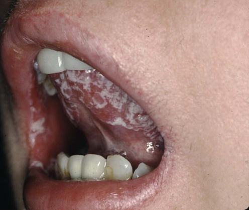
Cutaneous candidiasis


First-line therapies
 Topical antifungal
 Topical antifungal combined with topical corticosteroids
Second-line therapies
 Systemic azoles
 Lavender oil
 Topical mupirocin
Chronic mucocutaneous candidiasis


First-line therapies
 Systemic azole antimycotics
Second-line therapies
 Echinocandins
 Oral amphotericin B
 Intravenous amphotericin B
 Transfer factor
 Cimetidine and zinc sulfate
[/level-membership-for-dermatology-category][not-level-membership-for-dermatology-category]
Cutaneous candidiasis and chronic mucocutaneous candidiasis
Cutaneous candidiasis
First-line therapies
Second-line therapies
Fluconazole versus ketoconazole in the treatment of dermatophytoses and cutaneous candidiasis.
Stengel F, Robles-Soto M, Galimberti R, Suchil P. Int J Dermatol 1994; 33: 726–9.
Buy Membership for Dermatology Category to continue reading. Learn more here
[/not-level-membership-for-dermatology-category]
