Colon and Rectal Cancer

Published on 09/04/2015 by admin

Filed under Hematology, Oncology and Palliative Medicine

Last modified 22/04/2025

Print this page

rate 1 star rate 2 star rate 3 star rate 4 star rate 5 star
Your rating: none, Average: 3 (20 votes)

This article have been viewed 4591 times

PMS2, MSH6

Peutz-Jeghers syndrome Hamartomatous polyps throughout the gastrointestinal tract; mucocutaneous pigmentation; estimated 9-to 13-fold increased risk of gastrointestinal (GI) and non-GI cancers LKB1/STK11 Cowden disease Multiple hamartomas involving breast, thyroid, skin, central nervous system (CNS), and GI tract; increased risk of breast, uterus, and thyroid cancer; risk of GI cancer unclear PTEN Juvenile polyposis syndrome Multiple hamartomatous/juvenile polyps with predominance in colon and stomach; variable increase in colorectal and stomach cancer risk; facial changes DPC4
BMPR1A
PTEN MYH-associated polyposis (MAP) Multiple adenomatous gastrointestinal polyps, autosomal recessive often associated with somatic K-Ras mutations MYH Multiple adenoma and colorectal cancer Multiple colorectal adenomas and colorectal cancer; endometrial cancer and brain tumors in some individuals and families POLD1, POLE Hereditary mixed polyp syndrome Multiple types of colorectal polyps (e.g., Peutz-Jeghers polyps, juvenile polyps, serrated lesions, conventional adenomas) and colorectal cancer GREM1

Hereditary Colorectal Cancer Syndromes and Molecular Pathways of Colorectal Carcinogenesis

It is now generally accepted that the accumulation of somatic mutations and epigenetic defects drives the initiation and progression of tumor development. The constellation of somatic mutations that commonly accumulate in a clonal fashion in CRCs has largely been defined. 1720 However, only a limited subset of this collection of gene defects is usually present in any individual tumor. Thus far, the somatic mutations of greatest initial interest to the field have been those that are recurrent and clonal (i.e., present in all, or nearly all, neoplastic cells of a primary tumor at a given stage, but not present in the normal cells of the patient). It is inferred that such recurrent, clonal mutations are causal in promoting further tumor outgrowth/progression, because somatic mutations can only become clonal by a limited number of mechanisms. The genetic alteration itself could have been selected for because it provided the cell with a growth advantage, allowing it to become the predominant cell type in the tumor (clonal expansion). Alternatively, the specific alteration detected might have arisen coincident with another, perhaps undetected, alteration that was the crucial change underlying clonal outgrowth.
As noted previously, only about 5% of all CRC cases are associated with defined highly penetrant cancer syndromes. The bulk of these cases are attributable to hereditary nonpolyposis colorectal cancer syndromes (HNPCCs), with another significant subset associated with the familial adenomatous polyposis (FAP) syndrome (Table 34-1 ). A few other syndromes comprise the remaining highly penetrant CRC syndrome cases.

Adenomatous polyposis coli Gene: Gatekeeper in Familial Adenomatous Polyposis and Sporadic Cancer

Familial Adenomatous Polyposis

FAP is an autosomal dominant syndrome affecting about 1 in 8000 individuals in the United States and accounting for about 0.5% of CRCs. 4,5 Hundreds to thousands of adenomas arise in the large bowel and rectum beginning in the second decade of life. Although only a fraction of the adenomas may progress to cancer, the lifetime incidence of colorectal cancer in untreated FAP patients approaches 100% with a mean age at diagnosis of 39 years, necessitating the prophylactic removal of the patient’s colon early in adult life. 4 The gene that when mutated underlies FAP is the adenomatous polyposis coli (APC) tumor suppressor gene on chromosome 5q21. Germline mutations have been identified in one APC allele of affected individuals in 90% to 95% of families with FAP studied. More than 95% of mutations lead to premature truncation of APC protein synthesis—about two thirds by small insertions or deletions leading to a frameshift, and the remainder by introducing a stop codon 21 (Figure 34-1 ). Up to 25% of FAP cases seem to be caused by de novo germline mutations and therefore do not show the characteristic autosomal pattern of inheritance. 4 Moreover, about 20% of individuals with a de novo APC mutation display somatic mosaicism for the mutation in their tissues, with the mutant APC allele present in only subsets of cells within a given tissue and/or in germ cells. 4 Some of these cases with somatic mosaicism may manifest milder polyposis phenotypes than individuals carrying inherited germline APC mutations, as the defective APC allele is present in only a fraction of cells in the somatic mosaicism situation.
image
Figure 34-1 Schematic representation of the adenomatous polyposis coli (APC) protein and mutation histograms (A) Selected sequence motifs of the 2943-amino-acid APC protein and its interaction partners are indicated. The amino-terminal region has a domain that regulates its oligomerization. Repeated sequences with similarity to the Drosophila armadillo protein and its vertebrate homologue β-catenin (so-called armadillo repeats) are localized in the amino-terminal third of APC. Multiple independent 20-amino-acid repeats mediating binding to β-catenin and several binding sites for the Axin protein (termed SAMP repeats) are localized in the central third of APC. The carboxy-terminal third contains a basic region that is involved in microtubule (MT) binding and mediates interactions with the proteins EB1 and hDlg. (B) The frequency and distribution of germline mutations in familial adenomatous polyposis (FAP) patients are indicated with respect to the APC coding region. Virtually all mutations result in premature truncation at or very close to the mutation position. Two apparent mutational hotspots are seen at codons 1061 and 1309. (C) Frequency and distribution of somatic APC mutations in sporadic colorectal cancers are indicated. The mutations appear to predominate in the “mutation cluster region” (MCR), and mutations at codons 1309 and 1450 are most common. (A-C with permission from Polakis P. Mutations in the APC gene and their implications for protein structure and function. Curr Opin Genet Develop 1995;5:66).
Several extracolonic tumors and symptoms are associated with FAP (see Table 34-1). The combination of polyposis with brain tumors (in particular, medulloblastoma in pediatric cases) has been termed Turcot syndrome. 22 Gardner syndrome comprises extensive polyposis with epidermoid cysts, desmoid tumors, and osteomas and seems to be correlated with mutations between APC codons 1403 and 1578. 23,24 Upper gastrointestinal polyps are responsible for a large proportion of morbidity and mortality in FAP patients after prophylactic total colectomy. This is particularly true for the duodenal cancers that develop in 4% to 12% of patients. 4,25,26 Benign gastric fundic gland polyps and gastric adenomas, potential premalignant precursor lesions of gastric cancer, are observed with increased frequency, ultimately leading to stomach cancer in 0.5% of cases. 4,25,26 In only a small percentage of FAP cases, thyroid cancer, bile duct cancer, hepatoblastoma (pediatric), and central nervous system tumors such as medulloblastoma are observed. In addition to FAP, rare germline APC variants appear to play a role in other familial CRC cases. 27 Epidemiologic studies have revealed that the APC I1307K (isoleucine-to-lysine substitution at codon 1307) allele is present only in individuals of Ashkenazi Jewish origin, and those who carried the I1307K allele had a roughly twofold increased risk of developing CRC. 27 The mechanism underlying increased risk of adenomas and CRC in carriers of the APC I1307K allele is that the allele appears to be more susceptible than the wild-type APC allele to somatic mutations—particularly insertion and deletion of one or a few nucleotides near the homopolymeric adenine tract created by the lysine 1307 codon substitution, resulting in frameshift mutations. 27
image
Figure 34-2 Model of adenomatous polyposis coli (APC) and β-catenin function (A) The APC protein is part of a “destruction complex” containing glycogen synthase kinase 3β (GSK3β), Axin, and casein kinase 1α (CK1α), which phosphorylates β-catenin in conserved serine and threonine residues in the N terminus. This phosphorylation creates an epitope for recognition by the F-box protein β-TrCP as part of a ubiquitin ligase complex, which leads to polyubiquitination and proteasomal degradation of β-catenin. Hence, in most cells free β-catenin levels are kept low and transcription of target genes is repressed by recruitment of repressors of the transducin-like enhancer of split family (TLE). (B) On binding of Wnt ligands to a cognate receptor complex consisting of one member of the Frizzled (Fz) family and one member of the low-density-lipoprotein receptor-related protein family (i.e., LRP5 or LRP6), the destruction complex is disassembled in part by recruitment of Axin to the LRP receptors and in part by not clearly defined action of disheveled proteins (Dvls). Similarly inactivating mutations of the APC protein or the Axin1 protein or mutations of the conserved phosphorylation sites of β-catenin lead to the accumulation of free β-catenin, which enters the nucleus and activates transcription of target genes by displacement of co-repressors and recruitment of coactivators such as CBP/p300 and a complex of the homologues of Drosophila Legless (Lgs) and Pygopus (Pygo).

Somatic APC Mutations in Sporadic Tumors

Notwithstanding the critical role of the APC gene in FAP and related variant syndromes, the APC gene has an even more prominent role in sporadic colorectal tumors, as about 80% of sporadic colorectal adenomas and carcinomas have somatic mutations inactivating APC, 28 the overwhelming majority of which lead to premature truncation of the APC protein. APC mutations have been found in a number of the earliest sporadic lesions analyzed, including microscopic adenomas composed of only a few dysplastic glands. 29 As predicted by Knudson’s two-hit model for tumor suppressor genes, both APC alleles appear to be inactivated in the majority of colorectal adenomas and carcinoma, as a result of localized mutations in both alleles or localized mutation in one allele and chromosomal loss of the remaining allele. 28

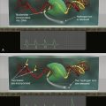
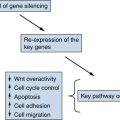
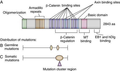
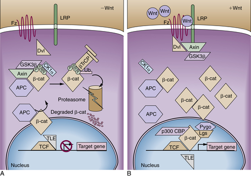
APC Protein Function

The APC tumor suppressor gene encodes a large protein of approximately 300 kDa (see Figure 34-1). The APC protein likely has multiple functions in the cell, 30 but the best understood function of APC is as a major binding partner and regulator of the β-catenin protein. 31 β-Catenin was first identified because of its role in linking the cytoplasmic domain of the E-cadherin cell-cell adhesion molecule to the cortical actin cytoskeleton, via binding to the adaptor molecule α-catenin. Based on studies from many different investigators, a model has been developed to explain the biologic significance of APC’s interaction with β-catenin. 31 The model proposes that in the absence of an activating Wnt ligand signal, APC binds to and collaborates with the scaffold protein Axin to promote sequential phosphorylation by casein kinase I and glycogen synthase kinase-3β (GSK3β) of several conserved serine/threonine residues in the N-terminal region of β-catenin, thereby targeting β-catenin for ubiquitination and proteasomal degradation (Figure 34-2 ). In a physiologic setting, the activating Wnt ligands inhibit degradation of β-catenin via binding to their cognate receptor complex of a Frizzled and an LRP5/6 protein.
In the roughly 80% of CRCs where both APC alleles are defective, the coordinated destruction of β-catenin is severely impaired, essentially mimicking constitutive activation of Wnt ligand-mediated signaling (see Figure 34-2). As a result, β-catenin accumulates in the cytoplasm, complexes with DNA binding proteins of the TCF (T-cell factor family)/Lef (lymphoid enhancer family) family, and translocates to the nucleus. Once there, β-catenin functions as a transcriptional co-activator, activating the expression of TCF-regulated genes. In a subset of the cancers that lack mutations in APC, somatic mutations in β-catenin have been found. 32 A critical net consequence of β-catenin stabilization by APC inactivation or other somatic mutations in CRCs is that the constitutive activation of β-catenin/TCF transcription appears to promote a stem or progenitor cell phenotype in the affected epithelial cells independent of their position in the crypt compartment. 31 Normally, β-catenin/TCF activation is restricted to the crypt base, especially in the so-called crypt base columnar stem cells that are characterized by expression of the Wnt-regulated Lgr5 gene and protein. 31
Further work has implicated β-catenin stabilization not only in the establishment of a crypt progenitor program but also in the spatial organization and migratory pattern of the cells in the continuous renewal of crypt. 31 Strikingly, feedback inhibitors functioning in the Wnt/β-catenin/TCF pathway are prominent among the critical proteins encoded by β-catenin/TCF-regulated genes, such as the AXIN2, DKK1, NKD1, APCDD1, and WIF-1 proteins, all of which function in some fashion to antagonize Wnt signaling. 31 In CRCs with APC mutations, the ability of the induced feedback regulator proteins to interfere with stabilized β-catenin is largely or entirely abrogated, because the induced Wnt-pathway feedback regulators function upstream of or at the level of the APC protein in the pathway. 31 Interestingly, some of the induced Wnt pathway feedback regulators that are highly expressed in CRCs with APC mutations, such as AXIN2, may play important contributing roles in CRC progression, such as through the ability of AXIN2 to interact with other proteins and pathways in the promotion of epithelial-mesenchymal transition and invasive phenotypes. 33

Mouse Models of FAP and Genetic and Epigenetic Modifiers

An important mouse genetic model of intestinal tumorigenesis known as the Min (for multiple intestinal neoplasia) mouse was described more than two decades ago. 34 The Min mouse carries a germline mutation in the murine homologue of the APC gene, resulting in truncation of the murine Apc protein at codon 850. The intestinal tumor phenotype of Min or Apc(Min) mice is similar, but not identical, to that seen in FAP patients, with Apc(Min) mice developing largely small intestinal tumors, in contrast to the colorectal lesions seen in FAP patients.
Other cellular genes can significantly influence the number of polyps that arise in mice that are heterozygous for the Apc(Min) mutant allele or other mutant Apc alleles. For instance, when the Apc(Min) mutation was introduced into mice of varying genetic backgrounds, significant variability in the number of intestinal tumors was seen. This finding was attributable to the effects of strain-specific modifier genes unlinked to the Apc locus, particularly the modifier gene Pla2g2a, which encodes a secreted phospholipase A2 protein. 35 Other genes that substantially modify intestinal tumorigenesis in Apc-mutant mice include genes encoding DNA methyltransferases. Specifically, mice that carry the Apc(Min) mutation and a germline defect in one allele of the maintenance DNA methyltransferase Dnmt1 or a gut-specific deletion of both Dnmt3 alleles have a two- to threefold reduction in macroscopic polyp number. 36,37 Treatment of Apc(Min) mice with 5-azacytidine, a pharmacologic inhibitor of DNA methyltransferases, resulted in a more than fivefold reduction in polyp number. 36 Combining the genetic and pharmacologic manipulations of DNA methyltransferase activity synergistically reduced the polyp number roughly 50-fold. 36 Treatment of mice with NSAIDs that inhibit COX-1 and COX-2 or agents that specifically inhibit COX-2 activity also markedly inhibited adenoma formation. 38,39 The findings highlight the utility of the mouse genetic models for identifying novel genes and pharmacologic agents that modify intestinal tumor development.

Other Forms of Intestinal Polyposis

Some patients with germline mutations in the APC gene have a milder or attenuated FAP (AFAP) phenotype, due to the nature of the germline APC mutation that they carry and/or other genetic variants that they may carry that modify the severity of their polyposis phenotype. 4,5 However, many of the individuals who have an AFAP phenotype and typically present to clinical attention between 40 and 60 years of age with fewer than a dozen to a hundred polyps do not carry a germline APC mutation. 4,5 Rather, these affected individuals carry homozygous mutations in the MYH gene encoding the base-excision repair pathway protein MutYH. The syndrome is often termed MAP for MYH-associated polyposis syndrome. 4,5 The mutations in the MYH gene lead to constitutional defects in the repair of oxidative DNA damage in cells, with increased GC-to-AT base-pair transversions arising. As a result of the increased mutation frequency, in the colon, somatic mutations in various genes likely arise at increased frequency, with the somatic mutations contributing to adenoma formation.
Some individuals and families who develop 10 to 100 or more adenomas during adult life, and who also develop CRC at an increased rate unless managed clinically, have been found to lack germline mutations in the APC or MYH genes. Recent studies have shown that some of these affected individuals and families carry heterozygous germline mutations in the POLD1 and POLE genes encoding DNA polymerases δ and ε, respectively. 40 The mutations appear to affect the proofreading domains of the polymerases. In the case of POLD1 mutations, affected individuals appear to be predisposed not only to adenomas and CRC but also to endometrial cancer and perhaps brain tumors. 40 The colon tumors arising in POLD1 and POLE mutation carriers were found to have inactivated the remaining wild-type copy of the gene by loss of heterozygosity (LOH). As a result, the tumors lack intact polymerases δ or ε function, depending on the nature of the patient’s germline mutation, and the tumors manifest a markedly greater frequency of base-substitution mutations relative to similar stage colon tumors with intact polymerase δ and ε function, presumably as a direct result of the deficits in polymerase δ or ε function. 40
Other intestinal polyposis syndromes in which patients manifest numerous nonadenomatous lesions have also been described. 4,5 Several of these syndromes have an increased risk of gastrointestinal and/or non-gastrointestinal cancers (see Table 34-1). Starting from childhood, patients with the autosomal dominant juvenile polyposis syndrome (JPS) develop multiple hamartomatous polyps throughout the gastrointestinal tract with some preference for the colon and the stomach. JPS is a genetically heterogeneous disease, with inactivating mutations in the SMAD4 and BMPR1A genes among the underlying genetic bases. Cowden syndrome is an autosomal dominant syndrome in which affected individuals develop macrocephaly and hamartomas in many organ sites, including the breast, thyroid, skin, central nervous system, and gastrointestinal tract. 4,5 The gene responsible for Cowden syndrome is the PTEN tumor suppressor gene on chromosome 10q23. Acting as a phospholipid-phosphatase, the PTEN protein is a major antagonist of the phosphatidylinositol-3-kinase (PI3K) survival pathway. In a small number of nonfamilial cases of juvenile polyposis lacking other features of Cowden syndrome, PTEN germline mutations have been reported. 4,5 Peutz-Jeghers syndrome, a rare autosomal dominant condition affecting fewer than 1 in 25,000, is characterized by gastrointestinal hamartomatous polyps and mucocutaneous melanin deposition. 4,5 The hamartomatous polyps contain essentially normal epithelial cells, but the mucosal components are arranged abnormally. Germline mutations in the LKB1/STK11 tumor suppressor gene on chromosome 19p can be seen in a significant fraction of cases. 4,5 Inactivation of this tumor suppressor gene leads to the hyperactivation of the mammalian target of rapamycin (mTOR) pathway, which is responsible for integrating the nutritional supply with cell proliferation and growth. Finally, recent studies of a rare inherited syndrome known as hereditary mixed polyposis syndrome (HMPS), in which individuals are predisposed to develop a range of lesions, including Peutz-Jeghers polyps, juvenile polyps, serrated lesions, conventional adenomas, and CRC, is due in some families to germline mutations that lead to overexpression of the bone morphogenetic protein (BMP) antagonist GREM1. 41 Thus, although HMPS is connected to JPS, based on the fact that BMPs signal through the transforming growth factor β (TGF-β) pathway, further work is needed to understand how derangements in TGF-β and BMP signaling contribute to predisposition to colorectal tumors.

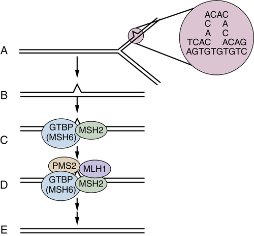
DNA Mismatch Repair Deficiency and Lynch Syndrome

Hereditary nonpolyposis colorectal cancer (HNPCC) was arguably the first inherited cancer syndrome to be well described in the literature. In 1913, Warthin presented a particularly striking example of a three-generation family with CRC and other cancers, including gastric cancer and tumors of the female reproductive tract. 42 Following Warthin’s lead, Lynch and others described kindreds with autosomal dominant patterns of CRC, not accompanied by extensive polyposis. 43 In such families, CRCs of early onset were seen, often along with cancers in some other organs including gastric, uterine endometrial, ovarian, small bowel, renal, and hepatobiliary cancers. 44 In recognition of Dr. Henry Lynch’s major contributions to the field, HNPCC is often referred to as Lynch syndrome.
Criteria for the diagnosis of Lynch syndrome in families have often included the following: affected families must show Lynch syndrome–typical tumors in three relatives (one being a first-degree relative to the other two) in at least two successive generations, with one of the tumors occurring before age 50 (so-called 3-2-1 rule). The sensitivity of these diagnostic criteria is considerably less than 100% for identifying those affected by Lynch syndrome, and the specificity of the criteria is also an issue. 4 However, the diagnostic criteria proved critical for the initial discovery of the germline mutations underlying Lynch syndrome, and the criteria have also been useful in clinical assessment over the past 15 to 20 years since the genetic bases of Lynch syndrome were uncovered. By way of a brief background, the early genetic work indicated genetic heterogeneity for Lynch syndrome, with mapping of the predisposing gene to chromosome 2p in some families 45 and to chromosome 3p in other families. 46 In yet other families with Lynch syndrome, no evidence for linkage to chromosome 2p or 3p was found.
To explore the relevance of Knudson’s two-hit hypothesis for Lynch syndrome genes, investigators initially sought to demonstrate loss of parental heterozygosity for chromosome 2p in cancers from individuals carrying a defect in that particular predisposition gene. 45 However, not only was there no LOH of chromosome 2p sequences in the cancers, but microsatellite DNA sequences examined in the analyses demonstrated marked length variations in tumor tissues compared with the patient’s normal tissue. Microsatellite changes were seen at many different loci scattered throughout the genome and in all tumors from patients with Lynch syndrome. 45 The phenotype was termed the microsatellite instability” (MSI) or replication error (RER) phenotype. Cancers with evidence of microsatellite instability at more than 40% of a panel of mononucleotide and dinucleotide sequences are termed high-frequency MSI (MSI-H) cancers. Although most CRCs display no instability when a panel of microsatellite tracts are studied—so-called microsatellite stable (MSS) cases—a subset of cancers show low-frequency instability of the microsatellite markers, termed MSI-L.
image
Figure 34-3 Mismatch repair pathway in human cells (A, B) During DNA replication, DNA mismatches may arise, such as from strand slippage (shown) or misincorporation of bases (not shown). (C) The mismatch is recognized by MutS homologues, perhaps most often MSH2 and GTBP/MSH6, although MSH5 may substitute for GTBP/MSH6 in some cases. (D, E) MutL homologues, such as MLH1 and PMS2, are recruited to the complex, and the mismatch is repaired through the action of a number of proteins, including an exonuclease, helicase, DNA polymerase, and ligase. A-E with permission from Kinzler KW, Vogelstein B. Lessons from hereditary colorectal cancer. Cell. 1996;87:159-170.
The finding of MSI-H in the cancers was noteworthy because similar DNA instability phenotypes had previously been seen in mutant yeast strains with defective DNA mismatch repair genes. The prediction that defects in one or more DNA mismatch repair genes might underlie Lynch syndrome was quickly borne out (Figure 34-3 ). A human homologue of the bacterial mutS mismatch repair gene, designated MSH2, was mapped to chromosome 2p, and one allele was found to be mutated in the germline of a subset of Lynch syndrome patients, with the remaining allele inactivated in cancers arising in the mutation carriers. Other genes involved in DNA mismatch repair were studied and found mutated in other groups of Lynch syndrome patients, including the MLH1 gene on chromosome 3p, the PMS2 gene on 7q, and the GTBP/MSH6 gene on chromosome 2p. 47 Mutations in the MSH2 and MLH1 genes are a far more common cause of Lynch syndrome than mutations in the other mismatch repair genes, with MSH2 and MLH1 mutations together accounting for about 70% to 75% of the known mutations in Lynch syndrome patients. 47
image
Figure 34-4 Relative effects of germline mutations on tumor initiation and progression (A) In sporadic cancers, both initiation of a neoplastic lesion (e.g., the adenoma) and progression to an advanced lesion (i.e., the carcinoma) are rate-limiting events because two somatic mutations are required for inactivation of tumor suppressors such as adenomatous polyposis coli (APC) (initiation of adenoma) and p53 (tumor progression). (B) In the case of familial adenomatous polyposis (FAP), germline inactivation of one APC allele markedly increases the formation of adenomas, because inactivation of both APC alleles is a critical (rate-limiting) event in adenoma formation, and those with inactivation of one allele in all colonic cells need only inactivate the remaining APC allele to initiate adenoma formation. (C) In the case of Lynch syndrome (hereditary nonpolyposis colorectal cancer [HNPCC]), germline inactivation of one of the mismatch-repair genes (e.g., MSH2 or MLH1) coupled with somatic inactivation of the remaining allele in an initiated lesion (e.g., an early adenoma) greatly increases the mutation rate, and subsequently the rate and speed of progression to more advanced lesions. With permission from Bettingon et al. The serrated pathway to colorectal cancer: current concepts and challenges. Histopathology. 2013; 62:367-386.
In the normal cells of a patient with Lynch syndrome, DNA repair is rarely impaired, because the cells have a wild-type copy of the gene (i.e., the copy inherited from the nonaffected parent). However, during cancer development, the remaining wild-type allele is inactivated by a somatic mutation. Following inactivation of the wild-type allele, full mismatch repair activity is lost (see Figure 34-3). Then, affected cells manifest a mutator phenotype and accumulate mutations in a much more rapid fashion. Lynch syndrome is, therefore, a disease with more rapid tumor progression from a benign, initiated clone to frank malignancy (Figure 34-4 ).
Although germline mutations in the known mismatch repair genes have been detected in only 2% to 4% of CRC patients, roughly 15% of all CRCs display the MSI-H phenotype. 47 The somatic inactivation of MLH1 via epigenetic mechanisms, including hypermethylation of its promoter, seems to be responsible for most MSI-H CRC cases outside of Lynch syndrome families. 47
Many of the mutations that arise in cells with the MSI-H phenotype may be detrimental to cell growth or may exert no positive selection pressure. However, a subset of mutations could potentially activate oncogenes or inactivate tumor suppressor genes. An example of a gene containing a mononucleotide tract in its coding sequence that is frequently mutated in MSI-H CRCs is the TGF-β type II receptor. 47 Some other genes commonly altered in MSI-H CRCs are described later.

Inflammation and Colon Cancer

Chronic inflammatory bowel disease (IBD), especially ulcerative colitis, confers an increased risk of CRC. In the case of UC, the risk of CRC is associated with both the duration and extent of the inflammatory disease. From a pathogenetic standpoint it is notable that UC-associated CRCs often develop without the formation of a polyp as a precursor lesion, although dysplasia is a common factor. 15,48 Reducing the degree of inflammation in IBC seems to reduce the risk for CRC. 49 Some insights into the factors and pathways by which inflammation contributes to colitis-associated CRC (CAC) have begun to emerge 50 and likely include important roles for NF-κB signaling in both immune cells and epithelial cells and local activation of various cytokines and lymphokines, including tumor necrosis factor, interleukin 1 (IL-1), IL-6, IL-17, and IL-23. 50
The responses of the innate and cellular immune systems to variations in intestinal microbial communities as well as inflammatory mediators all seem to have important and potentially much broader contributing roles in intestinal neoplasia beyond the setting of CAC. For instance, a human colonic commensal—enterotoxigenic Bacteroides fragilis—can affect tumor progression in the Apc(Min) mouse model of intestinal tumorigenesis in part via inflammation and the generation of IL-17–expressing T helper 17 [T(H)17] cells. 51 Recent studies have further highlighted the role of microbes and microbial products, along with IL-23 and T(H)17 cells in Apc-mutation–dependent colon tumor progression in the mouse. 52 Future studies are likely to further clarify the likely complex relationships among the intestinal microbiota, host immune cells, and epithelial cells in the initiation and progression of colon neoplasia in mouse models and in humans.

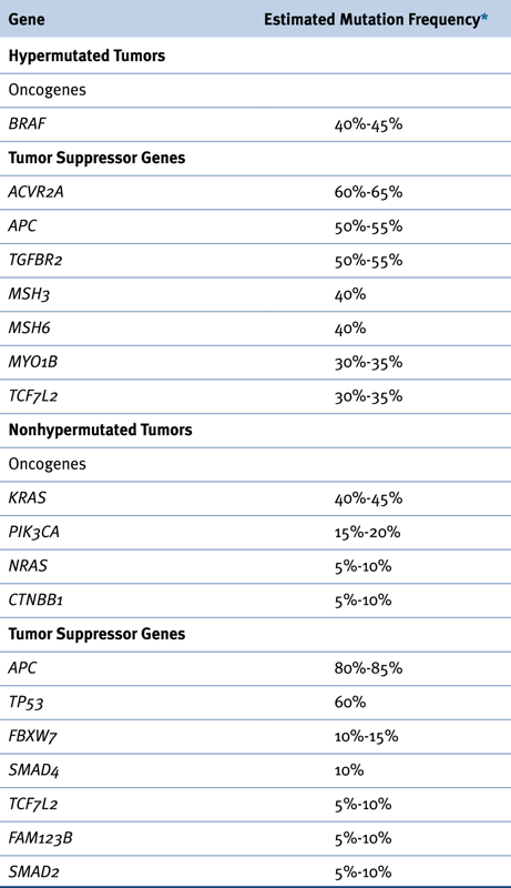
Recurrent Somatic Alterations in Colorectal Cancer

Somatic alterations have key roles in the initiation and progression of colorectal tumors, in patients with inherited predispositions to CRC as well as in the majority of CRC patients whose tumors are apparently sporadic. A sizeable number of mutational alterations in CRCs were identified over the past 30 years, largely by various combinations of genetic and genomic approaches. 18 Among the major targets for somatic mutations in CRCs identified before whole genome sequence-based approaches included oncogenic (activating) point mutations affecting KRAS (about 40% of CRCs), NRAS (about 5%), PIK3CA (20%), and BRAF (5% to 10%), as well as amplifications of the EGFR (5% to 10%), CDK8 (10% to 15%), CMYC (5% to 10%), and ERBB2 (5%) genes. 18 Similarly, among the tumor suppressor genes known to be frequently affected by somatic mutations in CRCs as a result of the “old-school,” focused sequencing approaches were the following: APC (80%), TP53 (60%), FBXW7 (20%), SMAD4 (10% to 15%), PTEN (10%), SMAD2 (5% to 10%), ACVR2A (10%), and TGFBR2 (10%). 18
Over the past few years, comprehensive sequencing approaches have allowed for expansive analyses of the nature and spectrum of mutational alterations in CRCs. 17,19,20 One of the major advances of the comprehensive sequencing-based approaches has been knowledge of the variation in total somatic mutations among CRCs. For example, in an exome-sequence analysis of 224 tumor and normal pairs from CRC patients, it was found that 84% of the tumors had a median number of nonsilent (predicted to change protein-coding sequence) somatic mutations of 58 per tumor; the remaining 16% of the tumors were reported to manifest a “hypermutated” phenotype, with a median of 728 somatic nonsilent mutations per tumor. 19 These findings that about 16% of CRCs had a hypermutated phenotype was not entirely unexpected. As described earlier in the discussion of mismatch repair gene defects in Lynch syndrome and also apparently sporadic CRCs, about 15% of all CRCs manifest the MSI-H phenotype, in the majority of MSI-H CRCs because of epigenetic silencing of the MLH1 gene. 18,47 As a result of their defective mismatch repair pathway function, the MSI-H CRCs were known to have a hypermutable phenotype. However, what was a bit unexpected about the hypermutated CRCs identified via the comprehensive sequencing approaches was that about 25% of the hypermutated tumors lacked the MSI-H phenotype and had intact function of MLH1. Instead, the hypermutated non-MSI-H cases had somatic mutations in other mismatch repair genes (e.g., MSH2, MSH3, MLH3, MSH6, PMS2) or in the POLE gene encoding DNA polymerase ε. 19 One somewhat puzzling issue is why somatic mutations in certain mismatch repair genes do not invariably lead to demonstrable instability in the microsatellite sequences that define the MSI-H phenotype, whereas epigenetic silencing of MLH1 does apparently lead to the MSI-H phenotype in almost all cases. Perhaps part of the explanation might be the relative timing of the defects in mismatch repair function in the natural history of a given CRC.

Table 34-2

Localized Somatic Mutations in Oncogenes and Tumor Suppressor Genes in Hypermutated and Non hypermutated CRCs

image

Mutation estimates from References 18 and 19.

Besides clarifying the mutation rate in CRCs and illuminating the molecular basis for the markedly increased mutation rate in the roughly 16% of CRCs that are hypermutated, comprehensive molecular characterization efforts have yielded additional insights, such as refinements in the estimated frequencies for known oncogene and tumor suppressor gene defects in CRC, the identification of some new oncogene and tumor suppressor gene defects contributing to CRC, and some initial insights into how epigenetic changes and gene expression patterns may be associated with certain mutation patterns as well as biologic and clinicopathologic subsets of CRC. Table 34-2 summarizes some of the genes most frequently affected by somatic mutations in nonhypermutated and hypermutated CRCs. 19,20
In addition to the extensive data on localized mutation frequencies, comprehensive molecular characterization efforts have begun to further inform understanding of recurrent gene amplification, deletion, and translocation in CRCs. Besides largely confirming the gene amplification events previously uncovered in CRCs, such as CDK8, EGFR, CMYC, and ERBB2 gene amplifications, 18 the work uncovered gene amplification and overexpression of the IGF2 gene in about 10% to 15% of CRCs. 19,20 Gene deletions affecting well-defined tumor suppressor genes were confirmed in a subset of CRCs, such as those affecting APC, PTEN, or SMAD3. 19,20 In addition, frequent deletions affecting the FHIT presumptive tumor suppressor gene were seen in about 20% to 30% of CRCs. 19,20 Recurrent translocations activating two different genes encoding R-spondin protein family members that cooperate with Wnt ligands to activate canonical β-catenin-dependent Wnt signaling were seen in 5% to 10% of CRCs. 20

Genetic Instability: Chromosomal Instability versus DNA Mismatch Repair Deficiency

In general, the roughly 15% of CRCs that manifest the MSI-H phenotype tend to have largely diploid or near-diploid karyotypes, whereas the remaining 85% of CRCs are aneuploid. 53,54 Key factors and mechanisms underlying the numerical chromosome instability (CIN) phenotype in CRC and other cancers are poorly defined, but some clues have emerged. Rare mutations and more commonly changes in the expression of genes that encode proteins that regulate chromosome segregation in mitosis or other genes that encode DNA repair response proteins have been suggested to play roles in the CIN phenotype in CRC. 54 However, because the mutations appear to be quite rare in primary CRCs and the changes in gene expression observed have been difficult to conclusively implicate in the CIN phenotype, unambiguous data on the gene defects with significant roles in the CIN phenotype in CRC are lacking. 54
Recently, three genes from a region of chromosome 18q affected by frequent copy number loss in CRC—PIGN, MEX3C, and ZNF516—have been implicated as potential contributing factors in CIN. 55 Specifically, experimental silencing of any one of the three genes led to the CIN phenotype in CRC cells in culture. 55 The genes appear to regulate the cellular response to DNA replication stress, and reduced DNA replication stress appears linked to reduced chromosome segregation defects. 55 Nonetheless, although the recent work on these three chromosome 18q genes appears to be an encouraging lead in terms of defining common molecular defects that may play important contributing roles in the CIN phenotype in CRC, much further work is needed to substantiate the contribution of the genes as well as the means by which loss of their function might lead to CIN.

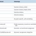
Epigenetic Changes in Colorectal Carcinogenesis: the CpG Island Hypermethylation Phenotype

In mammalian genomes, most 5′-CpG-3′ dinucleotides have been lost during evolution. 56 DNA methylation covalently modifies more than 80% of the remaining CpG sites, except for localized regions of high CpG-dinucleotide content, which have been termed CpG islands. 56 The promoter and proximal regulatory regions of about 50% of all genes contain CpG islands. Dense methylation or hypermethylation of these CpG islands seems to be associated with gene silencing, implicating CpG methylation as a potentially quite significant epigenetic mechanism to reinforce or “lock in” long-term gene silencing following more transient posttranslational chromatin modifications that are linked to inhibition of transcription (e.g., certain histone tail methylation and deacetylation changes). 56 Although the global trend in the majority of CRCs is in fact a decrease in total DNA methylation—that is, global DNA hypomethylation—the CpG island hypermethylation of selected promoters has been implicated in transcriptional silencing in CRC and other cancers. 56,57 Indeed, it is likely that a large fraction of CRCs reduce or silence expression of one or more critical growth-controlling genes (e.g., tumor suppressor genes) in part through epigenetic mechanisms, such as promoter DNA hypermethylation. 57
The functional significance of global DNA hypomethylation in CRC is more enigmatic than DNA hypermethylation, but some findings have suggested a potential contributing role for global DNA hypomethylation in chromosome missegregation events and the CIN phenotype. 57 Other work has suggested that global DNA hypomethylation might also lead to altered gene expression, such as the reactivation of certain imprinted genes that are normally silenced during early development. 18
A subset of CRCs manifest DNA hypermethylation at a sizeable number of distinct CpG islands scattered around the genome, compared to the DNA methylation patterns seen in adjacent normal colon epithelium or the bulk of other CRCs that lack the significant increase in DNA methylation. This phenotype has been termed the CpG island hypermethylation phenotype (CIMP). 57,58 A subset of CIMP CRCs shows hypermethylation of the MLH1 mismatch repair gene, and this group of CRCs constitutes the major fraction of sporadic MSI-H CRCs discussed at several earlier points. The specific factors and mechanisms that underlie CIMP status in CRC are not understood. However, it is noteworthy that the MSI-H CIMP subset of CRCs often harbors B-RAF oncogenic mutations. 9,10,57

Altered Gene Expression in Colorectal Cancer

Besides the genes whose expression is affected by DNA methylation, many other genes show major differences in gene expression in CRCs relative to normal tissues as well as among CRCs. This is likely a result of complex and heterogeneous mechanisms, such as the following: (1) somatic mutations deregulating key cellular signaling pathways that affect downstream gene expression in CRCs, along with significant intertumoral heterogeneity among CRCs with respect to the specific collection of signaling pathways and factors affected by oncogene and tumor suppressor gene mutations in any given CRC; (2) a subset of CRCs with mutations or epigenetic defects affecting specific chromatin regulatory factors, such as ARID1A 19,58 ; and (3) potential intertumoral heterogeneity in the specific cell of origin, with some CRCs arising from presumptive crypt base columnar stem cells and other CRCs potentially arising from more differentiated cell types. As a result of the potential differences in cell of origin and/or the specific signaling pathways and chromatin regulatory factors that are dysregulated in one CRC versus another, there are likely to be major variations among CRCs in terms of the expression of key transcriptional activators and repressors that directly regulate gene expression. There are also likely to be major variations in the structure and posttranslational modifications of chromatin and chromatin-associated proteins in CRCs. It will not be possible to review in any detail the very large collection of published studies on differentially expressed genes in CRC relative to normal tissues or the variations in gene expression among different subsets of CRC. Rather, one goal here is to offer some thoughts about the potential significance of gene expression changes in CRC that are not directly linked to specific mutations. A second goal is to introduce the notion that gene expression changes in CRCs affect not only mRNAs encoding proteins, but also various noncoding RNAs, such as miRNAs and long noncoding RNAs (lncRNAs).
The key somatic mutations in CRC highlighted earlier are clonal and recurrent, and the mutations lead to novel or increased function of oncogenes or inactivation of tumor suppressor genes. As described for the MLH1 gene, epigenetic changes, such as promoter hypermethylation and perhaps posttranslational modifications of histone or other chromatin-associated proteins, silence MLH1 expression in nearly all of the apparently sporadic MSI-H CRCs. Based on this powerful example, it could be that many other genes with dysregulated expression in CRC—either increased or decreased expression—might have a functionally significant role as an oncogene or TSG, respectively. However, the altered expression of many genes in CRCs may largely reflect rather than cause the altered growth and differentiation properties of cancer cells compared to normal cells. Hence, all available data on the function of any differentially expressed gene must be evaluated to establish whether the gene likely has a major contributing role in CRC and whether it might appropriately be designated as an oncogene or tumor suppressor gene.
The stability of mRNA transcripts as well as their ability to be translated into proteins is regulated by microRNAs (miRNAs), which are processed from longer precursor transcripts by the Drosha and Dicer proteins to roughly 18 to 24 nucleotides in length. Recognition of target transcripts predominantly occurs by binding of the miRNA to imperfect complementary sequences in the 3′ untranslated region (3′UTR) of various transcripts. Not unexpectedly, because the primary RNA transcripts that are ultimately processed to mature miRNAs are transcribed by RNA polymerase II (as are mRNAs), miRNA levels in CRCs vary considerably from levels in normal colonic mucosa. 57 miRNA expression differences have been reported in comparisons of MSS CRCs to MSI-H CRCs. 57,59 Many of the reported differences in miRNA levels in CRCs versus normal colon tissues and among different subsets of CRCs may be due to differential expression of the primary transcripts that are processed to miRNAs. However, p53 missense mutations have been hypothesized to exert specific effects on the processing of certain miRNAs. 60 Other somatic mutations in CRC could also conceivably exert major effects on the processing and/or nuclear-cytoplasmic transport of miRNAs. Similar to the situation discussed earlier for protein-coding mRNAs, in most cases it is largely uncertain which miRNA changes have causal roles in CRC development and which are secondary events associated with the process. Unlike the situation in some other cancer types where the genomic location of certain miRNAs with altered expression maps close to recurrent chromosomal breakpoints or deletions, few if any of the miRNAs with altered expression in CRC have been linked to recurrent chromosome lesions in CRC. 61 Besides the likely contribution of oncogene and TSG defects to changes in the levels of various miRNAs in CRC, it is likely that the expression of key components in critical oncogene and TSG pathways in CRC is modulated by many different miRNAs. Finally, although there are no conclusive data to implicate alterations in the function or expression of lncRNAs in CRC development and progression, it seems likely that at least a few of the estimated 5000 or more lncRNAs will have major contributing roles in CRC, such as through the ability of some lncRNAs to function as scaffolds for various chromatin-regulatory proteins. 62 Similar to the situation for both protein-encoding mRNAs, critical assessment of the collective expression and functional data will be required to implicate any given miRNA or lncRNA as a causal factor in CRC development and progression.

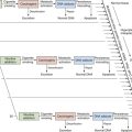
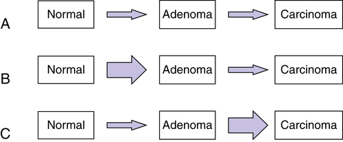
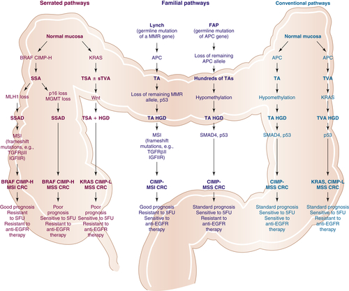
Multistep Genetic Models of Colorectal Tumor Development

Based on the accumulation of selected somatic oncogene and tumor suppressor gene defects together with methylation changes in CRCs, an initial genetic model of colorectal tumorigenesis was proposed more than 20 years ago. 63 The model relied on the assumption that many carcinomas arise from preexisting adenomas and that certain genetic alterations tended to accumulate at particular stages of tumor development, such as APC mutations in adenoma formation and p53 mutations in the transition to carcinomas. Hence, this was the basis for assigning a relative order to the alterations in a multistep pathway. However, the order of the mutations in the development of any given CRC was not viewed as invariant, as a few small adenomas with p53 mutations had been identified and KRAS mutations were sometimes associated with progression to carcinoma in some late-stage adenomas. 18,63
Since the initial genetic model for CRC development was proposed, other distinct molecular pathways to CRC have been suggested, some of which involve other precursor lesions than adenomatous polyps. 9,10 Some of the alternative molecular scenarios are presented in Figure 34-5 . The initial genetic model suggested in Reference 63 is outlined at the far right as one of the so-called conventional pathways. As indicated in Figure 34-5, the roughly 2% to 4% of CRC cases arising in individuals carrying germline mutations in a DNA mismatch repair (MMR) gene—Lynch syndrome—likely share some similarities with conventional pathway lesions in terms of the somatically acquired gene lesions that give rise to adenomatous lesions, such as APC mutations. As for the approximately 10% to 12% of apparently sporadic cancers that manifest the MSI-H phenotype, many of these are presumed to arise from serrated adenomatous lesions, such as sessile serrated adenomas (SSAs). 9,10 Some of the molecular lesions associated with the genesis of SSAs and the subsequent progression of SSAs to CRCs are distinct from those somatic defects that contribute to the conventional CRCs and the Lynch-type MSI-H CRCs, including frequent B-RAF mutations and the silencing of certain genes via promoter hypermethylation (i.e., CIMP CRCs). Finally, though not depicted in Figure 34-5, it is worth noting that the nature and order of mutational events seem to be different in UC-associated CRCs than in sporadic CRCs. For instance, p53 mutations are typically observed at an earlier time point, perhaps even occurring in the nonneoplastic inflamed mucosa of some patients. 48
image
Figure 34-5 Putative pathways to colorectal cancer (CRC) Shown in the figure are potential precursor lesions and selected molecular defects suggested to play important roles in the serrated pathways, familial pathways, or conventional pathways for CRC. The conventional pathways highlight the fact that the majority of CRCs are believed to arise from adenomatous precursor lesions over a period of years or even decades, with progression from a TA or TVA lesion to one that has high grade-dysplasia (TA HGD or TVA HGD). Some of the potential somatic alterations in tumor suppressor genes (APC, SMAD4, p53) and oncogenes (KRAS) that might contribute to tumor initiation and/or progression are illustrated. Possible global phenotypic changes in the CRC cells that are discussed in the text are noted, such as CpG island methylator phenotype-negative (CIMP-) or CIMP-low (CIMP-L) and microsatellite stable (MSS), along with the potential prognostic and therapeutic generalizations. Two familial pathways to CRC that together account for about 2% to 4% of CRC are highlighted in the scheme. The role of gene lesions and presumptive mutational mechanisms are extensively discussed in the text. The figure indicates the relationship of molecular lesions to prognosis and therapy response. A significant fraction of CRCs, especially lesions in the proximal colon, are thought to arise from a serrated precursor lesion, such as a sessile serrated adenoma (SSA) or a traditional serrated adenoma (TSA), with progression through lesions that manifest dysplasia or high-grade dysplasia (SSAD and TSA + HGD). A sizeable subset of SSAs and the resultant CRCs that arose from SSAs manifest the CIMP-high (CIMP-H) phenotype. The possible relationships of the various serrated pathway lesions to prognosis and therapeutic response are noted. anti-EGFR, Epidermal growth factor receptor inhibitors; 5-FU, 5-fluorouracil therapy; IGFIIR, insulin-like growth factor receptor II; p16, p16 inhibitor of cyclin-dependent kinase 4; TA, tubular adenoma; TGFRβII, TGF-β receptor II; TVA, tubulovillous adenoma. With permission from Bettingon et al. The serrated pathway to colorectal cancer: current concepts and challenges. Histopathology. 2013; 62:367-386.

Clinical Applications of Molecular Genetic Insights

The advances in our understanding of inherited and somatic defects in CRCs have made possible certain clinical applications and have highlighted the potential for additional clinical strategies that collectively should greatly improve the diagnosis and care of patients and families affected by CRC. Although many possible future clinical applications can be envisioned, just a few that are being actively pursued are described in the following sections.

Risk Assessment

The accurate presymptomatic diagnosis of FAP or Lynch syndrome is of significant value to individuals and families affected by these syndromes. The identification of germline mutations in the APC gene in more than 80% of families with FAP and Gardner syndrome provides the basis for genetic counseling and clinical management of families and individuals at risk for polyposis. 4,5 Similar major progress has been made in defining the germline mutations in Lynch syndrome patients and families. 4,5 Given the progress reviewed here and summarized in Table 34-1 in defining other genetic variants that confer a significantly increased risk of CRC development, the early identification of individuals and families at greatly elevated risk of CRC should lead to improved clinical management of such individuals and families, in large part via the incorporation of optimal screening and prophylactic surgery approaches and potentially even new chemopreventive agents and regimens.

Early Detection

The results of clinical trials indicate that the colonoscopic removal of larger (greater than 1 cm) adenomas and early CRCs has a major impact on CRC incidence. 8 The development of highly specific and sensitive molecular tests for early detection of CRC is an important goal, given the reduced specificity and sensitivity of current noninvasive tests, such as fecal occult blood testing. 8 If inexpensive and reliable molecular diagnostic tests could be developed to detect the presence of advanced adenomas or early-stage CRCs, such as through the analysis of analytes in the blood or in stool specimens, then such tests might serve an adjunctive role along with more invasive and expensive methods for early detection, such as colonoscopy. Findings from studies of DNA isolated from stool samples of patients known to have CRCs or large, advanced adenomas indicate that stool-based tests for mutant oncogenes and tumor suppressor genes may have utility. 64 More recent stool-based DNA tests incorporate detection of mutant KRAS alleles and DNA methylation of specific sequences, along with hemoglobin quantification, and the results seem potentially quite promising compared to earlier generation stool DNA tests. 65 Some early work analyzing DNA methylation of multiple genes in cell-free DNA in plasma from patients with colorectal cancer and adenomatous polyps suggests the future potential for using a panel of DNA methylation and/or somatic mutation markers for screening. 66 Extensive further studies are needed to clarify whether any plasma-based tests for circulating nucleic acid markers (e.g., DNA hypermethylation and somatic mutations) are strong competitors for the stool-based DNA tests and what the optimal strategies will be for deploying any stool-based and/or plasma-based DNA tests in populations at high risk and/or average risk for adenomas and CRC.

Prognostic and Predictive Markers

In addition to presymptomatic diagnosis (risk assessment) and early detection of tumors, several studies indicate that characterization of the specific genetic alterations in a cancer may provide improved/increased prognostic information about the likelihood of local and distant tumor recurrence. Perhaps among the most robust prognostic markers defined thus far for colorectal cancer are those for the MSI-H phenotype. In particular, this phenotype has been convincingly associated with improved survival in stage II and stage III colorectal cancer patients. 67 Interestingly, the use of 5-fluorouracil (5-FU)-based adjuvant chemotherapy did not appear to show any benefit in survival for patients with MSI-H tumors. 68 In fact, although not a statistically significant result in the initial study, the trend was for poorer survival in 5-FU–treated patients whose tumors displayed the MSI-H phenotype. Some of the data on molecular pathways to CRC and the relationship of the pathways and somatic alterations to prognosis and response to therapies are highlighted in Figure 34-5. Moreover, a number of studies have suggested that oncogenic mutations in KRAS are associated with resistance to EGFR-based therapies, 69 and some recent studies indicate that there is strong biologic selection for the outgrowth of KRAS-mutant CRCs when EGFR blockade is used therapeutically in CRC. 70

Summary and Future Directions

Molecular genetic studies of colorectal tumors have yielded profound insights into inherited predispositions to colorectal cancer as well its pathogenesis. A relatively limited number of oncogenes and tumor suppressor genes—the KRAS, APC, and TP53 genes, and a few others—have been found to be frequently mutated in CRCs, and intensive studies of the function of these critical genes in normal and neoplastic cell growth continue. The relative significance to the cancer cell phenotype of each of the various inherited and somatic mutations has not been well defined. Comprehensive sequence analyses indicate that a considerable number of additional oncogenes and tumor suppressor genes with roles in the development and progression of subsets of CRC likely exist. Identification of these genes and characterization of their contribution to cancer will be an important, albeit a challenging, task. At present, there is little understanding of the relationship between dietary and environmental agents associated with increased risk of colorectal cancer and the mutation rate and nature of the mutations that arise in normal and neoplastic cells in the colon and rectum. Only limited insights have been obtained into the true significance and generality of the findings from the clinical correlative studies undertaken to date. Nevertheless, it is clear that further efforts will yield insights into the molecular basis of CRC and can be expected to result in advances in the diagnosis and clinical care of patients with colorectal tumors.
References

1. Siegel R. , Naishadham D. , Jemal A. Cancer statistics, 2013 . CA: Cancer J Clin . 2013 ; 63 : 11 30 .

2. Polite B.N. , Dignam J.J. , Olopade O.I. Colorectal cancer model of health disparities: understanding mortality differences in minority populations . J Clin Oncol . 2006 ; 24 : 2179 2187 .

3. Dimou A. , Syrigos K.N. , Saif M.W. Disparities in colorectal cancer in African-Americans vs whites: before and after diagnosis . World J Gastroenterol . 2009 ; 15 : 3734 3743 .

4. Jasperson K.W. , Tuohy T.M. , Neklason D.W. et al. Hereditary and familial colon cancer . Gastroenterology . 2010 ; 138 : 2044 2058 .

5. Patel S.G. , Ahnen D.J. Familial colon cancer syndromes: an update of a rapidly evolving field . Curr Gastroenterol Rep . 2012 ; 14 : 428 438 .

6. Chan A.T. , Giovannucci E.L. Primary prevention of colorectal cancer . Gastroenterology . 2010 ; 138 : 2029 2043 .

7. Watson A.J.M. , Collins P.D. Colon cancer: a civilization disorder . Dig Dis . 2011 ; 29 : 222 228 .

8. Ransohoff D.F. Colon cancer screening in 2005: status and challenges . Gastroenterology . 2005 ; 128 : 1685 1695 .

9. Jass J.R. Classification of colorectal cancer based on correlation of clinical, morphological and molecular features . Histopathology . 2007 ; 50 : 113 130 .

10. Bettington M. , Walker N. , Clouston A. et al. The serrated pathway to colorectal carcinoma: current concepts and challenges . Histopathology . 2013 ; 62 : 367 386 .

11. Murakami R. , Tsukuma H. , Kanamori S. et al. Natural history of colorectal polyps and the effect of polypectomy on occurrence of subsequent cancer . Int J Cancer . 1990 ; 46 : 159 164 .

12. Winawer S.J. , Zauber A.G. , Fletcher R.H. et al. Guidelines for colonoscopy surveillance after polypectomy: a consensus update by the US Multi-Society Task Force on Colorectal Cancer and the American Cancer Society . Gastroenterology . 2006 ; 130 : 1885 .

13. Lin O.S. , Gerson L.B. , Soon M.S. et al. Risk of proximal colon neoplasia with distal hyperplastic polyps: a meta-analysis . Arch Int Med . 2005 ; 165 : 382 390 .

14. Lin O.S. , Schembre D.B. , McCormick S.E. et al. Risk of proximal colorectal neoplasia among asymptomatic patients with distal hyperplastic polyps . Am J Med . 2005 ; 118 : 1113 1119 .

15. Ullman T. , Croog V. , Harpaz N. et al. Progression of flat low-grade dysplasia to advanced neoplasia in patients with ulcerative colitis . Gastroenterology . 2003 ; 125 : 1311 1319 .

16. Soetikno R. , Friedlnad S. , Kaltenbach T. et al. Nonpolypoid (flat and depressed) colorectal neoplasms . Gastroenterology . 2006 ; 130 : 566 576 .

17. Sjoblom T. , Jones S. , Wood L.D. et al. The consensus coding sequences of human breast and colorectal cancers . Science . 2006 ; 314 : 268 274 .

18. Fearon E.R. Molecular genetics of colorectal cancer . Annu Rev Pathol Mech Dis . 2011 ; 6 : 479 507 .

19. The Cancer Genome Atlas Network . Comprehensive molecular characterization of human colon and rectal cancer . Nature . 2012 ; 487 : 330 337 .

20. Seshagiri S. , Stawiski E.W. , Durinck S. Recurrent R-spondin fusions in colon cancer . Nature . 2012 ; 488 : 660 664 .

21. Polakis P. Mutations in the APC gene and their implications for protein structure and function . Curr Opin Genet Dev . 1995 ; 5 : 66 71 .

22. Hamilton S.R. , Liu B. , Parsons R.E. et al. The molecular basis of Turcot’s syndrome . N Engl J Med . 1995 ; 332 : 839 847 .

23. Davies D.R. , Armstrong J.G. , Thakker N. et al. Severe Gardner syndrome in families with mutations restricted to a specific region of the APC gene . Am J Hum Genet . 1995 ; 57 : 1151 1158 .

24. Niewuwenhuis M.H. , Vasen H.F. Correlation between mutation site in APC and phenotype of familial adenomatous polyposis (FAP): a review of the literature . Crit Rev Oncol Hematol . 2007 ; 61 : 153 161 .

25. Brosens L.A. , Keller J.J. et al. Prevention and management of duodenal polyps in familial adenomatous polyposis . Gut . 2005 ; 54 : 1034 1043 .

26. Gallagher M.C. , Phillips R.K. , Bulow S. Surveillance and management of upper gastrointestinal disease in familial adenomatous polyposis . Fam Cancer . 2006 ; 5 : 263 273 .

27. Laken S.J. , Petersen G.M. , Gruber S.B. et al. Familial colorectal cancer in Ashkenazim due to a hypermutable tract in APC . Nat Genet . 1997 ; 17 : 79 83 .

28. Kinzler K.W. , Vogelstein B. Lessons from hereditary colorectal cancer . Cell . 1996 ; 87 : 159 170 .

29. Jen J. , Powell S.M. , Papadopoulos N. et al. Molecular determinants of dysplasia in colorectal lesions . Cancer Res . 1994 ; 54 : 5523 5526 .

30. Nathke I.S. The adenomatous polyposis coli protein: the Achilles heel of the gut epithelium . Annu Rev Cell Dev Biol . 2004 ; 20 : 337 366 .

31. Clevers H. , Nusse R. Wnt/β-catenin signaling and disease . Cell . 2012 ; 149 : 1192 1205 .

32. Morin P.J. , Sparks A.B. , Korinek V. et al. Activation of beta-catenin-Tcf signaling in colon cancer by mutations in beta-catenin or APC . Science . 1997 ; 275 : 1787 1790 .

33. Wu Z.Q. , Brabletz T. , Fearon E. et al. Canonical Wnt suppressor, Axin2, promotes colon carcinoma oncogenic activity . Proc Natl Acad Sci U S A . 2012 ; 109 : 11312 11317 .

34. Moser A.R. , Pitot H.C. , Dove W.F. A dominant mutation that predisposes to multiple intestinal neoplasia in the mouse . Science . 1990 ; 247 : 322 324 .

35. Cormier R.T. , Hong K.H. , Halbert R.B. et al. Secretory phospholipase Pla2g2a confers resistance to intestinal tumorigenesis . Nat Genet . 1997 ; 17 : 88 91 .

36. Laird P.W. , Jackson-Grusby L. , Fazeli A. et al. Suppression of intestinal neoplasia by DNA hypomethylation . Cell . 1995 ; 81 : 197 205 .

37. Lin H. , Yamada Y. , Nguyen S. et al. Suppression of intestinal neoplasia by deletion of Dnmt3b . Mol Cell Biol . 2006 ; 26 : 2976 2983 .

38. Oshima M. , Dinchuk J.E. , Kargman S.L. et al. Suppression of intestinal polyposis in Apcdelta716 knockout mice by inhibition of cyclo-oxygenase 2 (COX-2) . Cell . 1996 ; 87 : 803 809 .

39. Peek Jr. R.M. Prevention of colorectal cancer through the use of COX-2 selective inhibitors . Cancer Chemother Pharmacol . 2004 ; 54 : S50 S56 .

40. Palles C. , Cazier J.B. , Howarth K.M. et al. Germline mutations affecting the proofreading domains of POLE and POLD1 predispose to colorectal adenomas and carcinomas . Nat Genet . 2013 ; 45 : 136 144 .

41. Jaeger E. , Leedham S. , Lewis A. et al. Hereditary mixed polyposis syndrome is caused by a 40-kb upstream duplication that leads to increased and ectopic expression of the BMP antagonist GREM1 . Nat Genet . 2012 ; 44 : 699 703 .

42. Warthin A.S. Heredity with respect to carcinoma . Arch Int Med . 1913 ; 12 : 546 .

43. Lynch H.T. , Shaw M.W. , Magnuson C.W. et al. Hereditary factors in cancer: study of two large Midwestern kindreds . Arch Int Med . 1966 ; 117 : 206 212 .

44. Watson P. , Riley B. The tumor spectrum in the Lynch syndrome . Fam Cancer . 2005 ; 4 : 245 248 .

45. Peltomaki P. , Aaltonen L.A. , Sistonen P. et al. Genetic mapping of a locus predisposing to human colorectal cancer . Science . 1993 ; 260 : 810 812 .

46. Lindblom A. , Tannergard P. , Werelius B. et al. Genetic mapping of a second locus predisposing to hereditary non-polyposis colon cancer . Nat Genet . 1993 ; 5 : 279 282 .

47. Pino M.S. , Chung D.C. Microsatellite instability in the management of colorectal cancer . Expert Rev Gastroenterol Hepatol . 2011 ; 5 : 385 399 .

48. Ullman T.A. , Itzkowitz S.H. Intestinal inflammation and cancer . Gastroenterology . 2011 ; 140 : 1807 1816 .

49. Beaugerie L. Inflammatory bowel disease therapies and cancer risk: where are we and where are we going . Gut . 2012 ; 61 : 476 483 .

50. Grivennikov S.I. Inflammation and colorectal cancer: colitis-associated neoplasia . Semin Immunopathol . 2013 ; 35 : 229 244 .

51. Wu S. , Rhee K.J. , Albesiano E. et al. A human colonic commensal promotes colon tumorigenesis via activation of T helper type 17 T cell responses . Nat Med . 2009 ; 15 : 1016 1022 .

52. Grivennikov S.I. , Wang K. , Mucida D. et al. Adenoma-linked barrier defects and microbial products drive IL-23/IL-17-mediated tumour growth . Nature . 2012 ; 491 : 254 258 .

53. Lengauer C. , Kinzler K.W. , Vogelstein G. Genetic instability in human cancers . Nature . 1998 ; 396 : 643 649 .

54. Pino M.S. , Chung D.C. The chromosomal instability pathway in colon cancer . Gastroenterology . 2010 ; 138 : 2059 2072 .

55. Burrell R.A. , McClelland S.E. , Endesfelder D. et al. Replication stress links structural and numerical cancer chromosome instability . Nature . 2013 ; 494 : 492 496 .

56. Jones P.A. Functions of DNA methylation: islands, start sites, gene bodies and beyond . Nat Rev Genet . 2012 ; 13 : 484 492 .

57. Goel A. , Boland C.R. Epigenetics of colorectal cancer . Gastroenterology . 2012 ; 143 : 1442 1460 .

58. Jones S. , Li M. , Parsons D.W. et al. Somatic mutations in the chromatin remodeling gene ARID1A occur in several tumor types . Hum Mutat . 2012 ; 33 : 100 103 .

59. Lanza G. , Ferracin M. , Gafa R. et al. mRNA/microRNA gene expression profile in microsatellite unstable colorectal cancer . Mol Cancer . 2007 ; 6 : 54 .

60. Suzuki H.I. , Yamagata K. , Sugimoto K. et al. Modulation of microRNA processing by p53 . Nature . 2009 ; 460 : 529 533 .

61. Lu J. , Getz G. , Miska E.A. et al. MicroRNA expression profiles classify human cancers . Nature . 2005 ; 435 : 834 838 .

62. Rinn J.L. , Chang H.Y. Genome regulation by long noncoding RNAs . Annu Rev Biochem . 2012 ; 81 : 145 166 .

63. Fearon E.R. Vogelstein. A genetic model for colorectal tumorigenesis . Cell . 1990 ; 61 : 759 767 .

64. Imperiale T.F. , Ransohoff D.F. , Itzkowitz S.H. et al. Fecal DNA versus fecal occult blood for colorectal-cancer screening in an average-risk population . N Engl J Med . 2004 ; 351 : 2704 2714 .

65. Ahlquist D.A. , Zhou H. , Domanico M. et al. Next-generation stool DNA test accurately detects colorectal cancer and large adenomas . Gastroenterology . 2012 ; 142 : 248 256 .

66. Cassinotti E. , Melson J. , Liggett T. et al. DNA methylation patterns in blood of patients with colorectal cancer and adenomatous polyps . Int J Cancer . 2012 ; 131 : 1153 1157 .

67. Gryfe R. , Kim H. , Hsieh E.T. et al. Tumor microsatellite instability and clinical outcome in young patients with colorectal cancer . N Engl J Med . 2000 ; 342 : 69 77 .

68. Carethers J.M. , Smith E.J. , Behling C.A. et al. Use of 5-fluorouracil and survival in patients with microsatellite-unstable colorectal cancer . Gastroenterology . 2004 ; 126 : 394 401 .

69. Markman B. , Javier Ramos F. , Capdevila J. et al. EGFR and KRAS in colorectal cancer . Adv Clin Chem . 2010 ; 51 : 71 119 .

70. Diaz Jr. L.A. , Williams R.T. , Wu J. et al. The molecular evolution of acquired resistance to targeted EGFR blockade in colorectal cancers . Nature . 2012 ; 486 : 537 540 .

71. Bettingon et al. , et al. The serrated pathway to colorectal cancer: current concepts and challenges . Histopathology . 2013 ; 62 : 367 386 .