Published on 18/03/2015 by admin
Filed under Dermatology
Last modified 22/04/2025
This article have been viewed 2552 times
Clifford M. Lawrence
Evidence Levels: A Double-blind study B Clinical trial ≥ 20 subjects C Clinical trial < 20 subjects D Series ≥ 5 subjects E Anecdotal case reports
Chondrodermatitis is a benign condition; the only indication for treatment is pain causing sleep disturbance. Painless areas of chondrodermatitis can be ignored or managed conservatively. Lesions on the helix are easier to treat surgically than antihelix lesions and give better cure results. A pressure relieving cushion is a good first choice alternative to surgery for antihelix lesions.
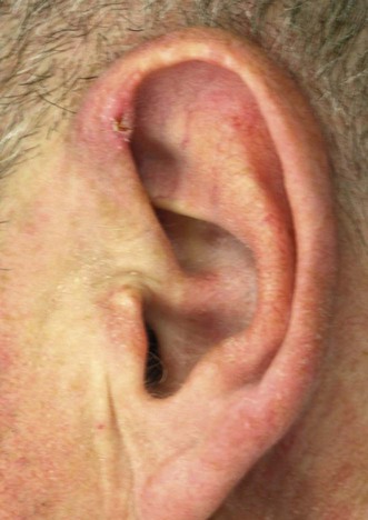
Chondrodermatitis usually occurs on the lateral portion of the ear on the preferred sleeping side. It is generally caused by the weight of the head crushing the ear against the pillow during sleep. Ear injury or surgery may leave an irregular ear margin that becomes a focus for sleep related pressure. The most protuberant part of the ear is affected; this is generally the helix in men and the antihelix in women. Patients who can only adopt one sleeping position due to arthritis, etc., are particularly vulnerable. The incidence increases with age because ear cartilage becomes less flexible with time. Patients should be reassured that it is not skin cancer, advised to use a soft pillow that is still compressible when the head is resting on it, and to change their sleeping position. Conservative or medical treatment, such as lidocaine (lignocaine) gel, a potent topical or intralesional corticosteroid or pressure-relieving cushion can be tried in all patients. If sleep is not disturbed there is really no need for any further intervention unless cosmesis is a problem.
Numerous surgical strategies have been described to treat chondrodermatitis and most work to some degree; it is tempting to suggest that some work by making the ear so painful that the subject is forced to adapt their sleeping position until the lesion resolves spontaneously. Most authors believe that the principle of surgical treatment is excision of the affected area of cartilage without the need for skin or ulcer excision. Other destructive therapies have been advocated but overall are less effective. Antihelix lesions respond so well to cartilage excision that many recommend surgery as a first line treatment.
None required
Biopsy of a lesion only necessary if surgery is indicated. Lesions managed conservatively do not require biopsy
Beck MH. Br J Dermatol 1985; 113: 504–5.
Topical corticosteroids (betamethasone valerate cream 0.025%) used twice a day for 6 weeks and intralesional triamcinolone (0.2–0.5 mL, 10 mg/mL) are effective in almost 25% of patients.
Cox NH, Denham PF. Br J Dermatol 2002; 146: 712–13.
A retrospective analysis of 60 patients with CNH treated with 0.1 ml intralesional triamcinolone acetonide 10 mg/mL or triamcinolone hexacetonide 5 mg/mL showed good response on 43% of helix and 31% of antihelix lesions.
Flynn V, Chisholm C, Grimwood R. J Am Acad Dermatol 2011; 65: 531–6.
Topical 2% nitroglycerin ointment twice daily resulted in complete clearance and resolution of symptoms in eight of 13 CNH lesions. Four gained only symptomatic improvement, and one showed no benefit.
Reassurance that this is not a tumor puts the patient’s mind at rest. In many instances the symptoms are mild and can be tolerated without any major intervention. Initially, conservative therapy should be tried. Recommend a change in sleeping position and a soft pillow that can still be further compressed when the head is resting on it.
Lawrence CM. Arch Dermatol 1991; 127: 530–5.
Cartilage excision without skin removal resulted in cure rates of 84% for helix and 75% for antihelix lesions. Local anesthesia with lidocaine and epinephrine (adrenaline) reduced bleeding and improved the visibility without ear skin necrosis. On the helix the skin was reflected back from a helix rim incision to expose the cartilage. On the antihelix, a medially based skin flap was raised to expose cartilage. After exposed cartilage excision, all remaining cartilage edges were left smooth and gently shelving up to the uninvolved cartilage to prevent recurrences which may develop on rough or protuberant cartilage edges. Cartilage excision alone is not disfiguring and further cartilage edges can be removed if recurrences occur.
de Ru JA, Lohuis PJ, Saleh HA, Vuyk HD. J Laryngol Otol 2002; 116: 677–81.
Most otolaryngologists treat patients with chondrodermatitis nodularis by wedge excision. This can leave the patient with an asymmetric deformed ear. The authors described their results after changing to removal of cartilage alone. In a mean follow-up of 30 months, all 34 patients remained symptom free and only one required revision surgery.
Hussain W, Chalmers RJ. Simplified surgical treatment of chondrodermatitis nodularis by cartilage trimming and sutureless skin closure. Br J Dermatol 2009; 160: 116–18.
After skin incision mosquito forceps were used to ‘nibble’ away fragments of cartilage to leave a smooth cartilage contour without the need for full exposure of the cartilage edge. Skin nodules or ulcers were not excised but left to resolve spontaneously. Tape strips were used for skin closure. Thirty-two of 34 (94%) patients with chondrodermatitis, seven on the antihelix and 27 on the helix, showed no discomfort or clinical recurrence 4 months after this procedure.
Munnoch DA, Herbert KJ, Morris AM. Br J Plast Surg 1996; 49: 473–6.
Fifty-four chondrodermatitis lesions (36 helix, 18 antihelix) including 23 recurrent after previous surgery, were treated by minimal skin excision combined with extensive cartilage resection. There were no recurrences and minimal postoperative deformity.
Rex J, Ribera M, Bielsa I, Mangas C, Xifra A, Ferrándiz C. Dermatol Surg 2006; 32: 400–4.
A narrow elliptical excision of the papule followed by a slice of the underlying cartilage was taken and trimmed carefully to remove any cartilage spikes. Good cosmetic results were obtained in all 74 patients retrospectively analyzed. The recurrence rate ranged from 10% for helical and 37% for antihelical lesions after a median follow-up of 54 and 50 months, respectively.
Ormond P, Collins P. Dermatol Surg 2004; 30: 208–10.
Excision of the cartilage alone is therapeutically and cosmetically effective. To simplify the surgical procedure, a narrow ellipse of skin over the nodule was excised and cold steel dissection of the adjacent skin replaced by hydrodissection to create a plane of cleavage between the skin and cartilage. These two refinements maintained the clinical and cosmetic efficacy but simplified the surgical technique.
All the above techniques are based around removal of damaged cartilage and avoidance of leaving irregular cartilage margins as these may act as a focus for recurrence. Skin excision can be included if this makes the procedure easier to perform but this is not required for surgical success.
Allen DL, Swinson PA, Arnstein PM. J Maxillofac Prosthet Technol 1998; 2: 5–10.
Thirty-five of 46 patients treated in this way had complete resolution of their symptoms.
Moncrieff M, Sassoon EM. Br J Dermatol 2004; 150: 892–4.
A simple to construct ear cushion design was used made up of a bath sponge with the center removed, held in place with a head band. Thirteen out of 15 patients followed for 1 month responded.
The bath sponge pressure relieving device is commercially available (CNH ear protector, Delasco).
Sanu A, Koppana R, Snow DG. J Laryngol Otol 2007; 121: 1096–8.
Patients used a doughnut-shaped pillow made of a modern orthopedic ‘memory foam.’ This distributes the weight of the recumbent head more evenly and is designed to relieve the pressure on the affected ear. Thirteen of 23 (56%) patients treated remained pain free after a 1-year follow-up.
Belgi A, Logan RA. Br J Dermatol 2007; 157: 68.
Seventy-eight patients with CNH used a custom made ear pressure–relieving device designed and built by the local maxillofacial technicians. Forty-eight patients responded to a questionnaire examining the outcome. Only seven (15%) found it helpful, 20 (42%) reported no benefit, and 20 (42%) some benefit.
Timoney N, Davison PM. Br J Plast Surg 2002; 55: 387–9.
Forteen patients with CNH were prospectively enrolled and used protective padding of the ear at night. Most patients were rapidly relieved of their symptoms, although healing was frequently prolonged.
Kuen-Spiegl M, Ratzinger G, Sepp N, Fritsch P. J Dtsch Dermatol Ges 2011; 9: 292–6.
Using a self-made bandage of foam plastic applied during the night, 11 out of 12 patients reported substantial reduction of pain within the first month and were pain free after an average of 1.75 months.
Various types of ear cushion are described. They all share the same aim, i.e., to transfer the weight of the sleeping head from the ear to the surrounding scalp. Authors report a wide range of benefits, with between 15% and 80% of patients not requiring surgery. In the hands of this writer the results are generally disappointing. This technique can be helpful for patients in whom surgery has been unsuccessful and in frail patients with thin skin and antihelix lesions who are unable to adapt their sleeping position, because recurrences at this site are more common after surgery.
Kromann N, Hoyer H, Reymann F. Acta Derm Venereol 1983; 63: 85–7.
A total of 142 cases of chondrodermatitis were principally treated by curettage followed by electrocauterization. Seventy-eight patients were re-examined after an average interval of 7.1 years. This simple surgical technique produced a relapse rate of 31%.
Coldiron BM. J Dermatol Surg Oncol 1991; 17: 902–4.
An ellipse of skin is removed around the ulcerated area. The central necrotic or damaged cartilage is removed using a curette and the skin edges sutured. A series is not reported.
Taylor MB. J Dermatol Surg Oncol 1991; 17: 862–4.
The CO2 laser was used to vaporize the cutaneous nodules and involved cartilage. The wounds were allowed to heal with only minimal care using hydrogen peroxide cleansing and applications of topical antibiotic ointment. Twelve lesions have been treated with no recurrences after 2 to 15 months.
Rajan N, Langtry JA. Br J Dermatol 2007; 157: 744–7.
Twenty-three lesions (15 helix, three antihelix, five not recorded) were treated by punch removal of the nodule and underlying cartilage. The small defect was closed with a full thickness skin graft taken from behind the ear using the same punch.
This novel ‘punch and graft’ technique eschews all the dogma about the need to just remove cartilage and leave smooth cartilage margins but still produces a cure rate of 83%.
Cryotherapy has been advocated, but there are no published outcomes.
Treatment of Skin Disease Comprehensive Therapeutic Strategies 4e
WhatsApp us


 Reassurance not cancer
 Conservative management
 Topical corticosteroids
 Intralesional corticosteroids
 2% topical nitroglycerin
 Helix lesions: excision of cartilage without skin excision
 Antihelix lesions: pressure relieving cushion or device
 Curettage
 Incision and cartilage curettage
 CO2 laser ablation
 Punch and graft