Published on 18/03/2015 by admin
Filed under Dermatology
Last modified 22/04/2025
This article have been viewed 2589 times
29
Irshad Zaki
Evidence Levels: A Double-blind study B Clinical trial ≥ 20 subjects C Clinical trial < 20 subjects D Series ≥ 5 subjects E Anecdotal case reports
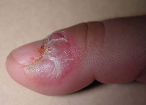
Blistering distal dactylitis (BDD) is a superficial, tender, blistering infection seen in childhood and the early teens. It is usually caused by group A β-hemolytic streptococci, although group B organisms, staphylococci and more recently MRSA have also been implicated. The distal volar fat pads of the fingers are the most common site of infection, but involvement of the nailfolds and toes can occasionally occur.
Blistering distal dactylitis can cause considerable alarm to parents as large tense blisters rapidly develop. Despite the absence of constitutional symptoms, patients usually seek help soon after the onset of the infection. The condition does not resolve spontaneously, but prompt treatment results in rapid improvement. Blisters should be incised to release fluid, which can vary from clear and watery to frank pus. Subsequent application of topical antibiotics can be helpful, but systemic treatment is usually also required. Penicillin V is the treatment of choice for streptococcal infection, but erythromycin is an effective alternative for patients allergic to penicillin.
The differential diagnosis of the condition includes traumatic blisters, herpetic whitlow, staphylococcal bullous impetigo, and the Weber–Cockayne variant of epidermolysis bullosa.
Gram stain of blister fluid
Culture of blister fluid
Swab of nasopharynx for bacteriology
Hays GC, Mullard JE. Paediatrics 1975; 56: 129–31.
First large series report describing 13 patients with BDD. Streptococci were found on culture of blister fluid in all cases, and Gram-positive cocci were usually found on Gram staining. This report suggests a link with infection of the nasopharynx, but this has not been confirmed in other case reports.
Lyon M, Doehring MC. J Emerg Med 2004; 26: 421–3.
Although uncommon under the age of 2 years, this paper reports three cases under 9 months of age.
These reports highlight the importance of initiating bacteriology prior to commencing treatment. Staphylococcal infection is a relatively rare but recognized cause of BDD.
Scheinfeld NS. Clin Exp Dermatol 2007; 32: 314–16.
Good review of literature and treatment. Confirms the importance of incision and drainage, adequate dressings, and the need to consider Staphylococcus aureus as a cause if penicillin is ineffective.
Schneider JA, Parlette HL. Arch Dermatol 1982; 118: 879–80.
Short report of the authors’ personal experience, suggesting that this is a relatively common problem. Systemic penicillin and erythromycin were both found to be effective. It is likely that many cases of BDD are wrongly diagnosed as bullous impetigo by clinicians not familiar with this disorder.
Fretzayas A, Moustaki M, Tsagris V, Brozou T, Nicolaidou P. Pediatr Dermatol 2011; 28: 433–5.
First report of BDD due to MRSA infection. This case highlights the importance of culture of blister fluid as infection with this organism may increase in the future.
Scheinfeld N. Dermatol Online J 2007; 13: 8.
Staphylococcus aureus was found to be the etiologic agent in two HIV-positive patients. The condition improved following treatment with a proprietary mixture of amoxicillin trihydrate and clavulanate potassium. The report highlights that blisters may not always be present and that BDD may present with erosions.
Ney AC, English JC 3rd, Greer KE. Cutis 2002; 69: 46–8.
Consider comorbidity if BDD does not respond to antibiotics.
Treatment of Skin Disease Comprehensive Therapeutic Strategies 4e
WhatsApp us


 Incision and drainage of blister
 Topical antibiotics
 Systemic penicillin
 Systemic erythromycin
 Intravenous vancomycin
 Amoxicillin/clavulanic acid
 Conservative measures for herpetic whitlow