Back pain quietly changes everyday routines before people fully realize how much it affects them. Sitting through meetings becomes distracting, driving feels exhausting, and even simple things like putting on shoes or getting out of bed start taking more effort than they used to. Most people try stretching, rest, or physical therapy first because many back problems improve without major treatment.
The difficult part is that some pain behaves differently. Symptoms lasting for months, spreading into the legs, or causing numbness and weakness can point toward deeper spinal issues instead of simple muscle strain. That is usually when doctors begin considering more detailed imaging rather than continuing to rely only on symptoms and guesswork alone.
Pain Patterns Sometimes Reveal More Than the Initial Exam
Back pain often sounds similar from patient to patient at first. Stiffness after sitting too long. Sharp pain while bending. Tightness that improves slightly, then returns again a few days later. Those symptoms can come from muscles, joints, irritated nerves, or spinal discs, which makes diagnosis harder than people expect sometimes.
Physical exams help narrow things down, but they do not always show what is happening internally. A patient may describe numbness down one leg, burning pain near the hip, or weakness in the foot, while basic movement tests still appear fairly normal in the office. That disconnect is one reason doctors sometimes recommend more detailed imaging even when outward symptoms seem manageable initially.
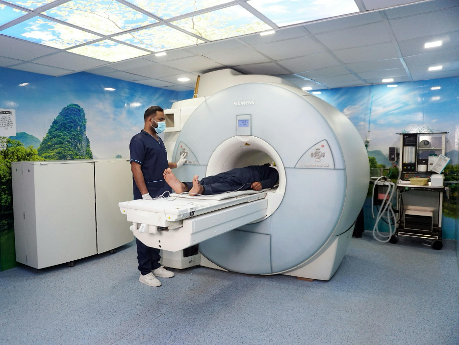
Disc problems create especially confusing symptoms because pain does not always stay near the spine itself. Pressure against spinal nerves can travel outward into the legs, feet, or hips while the actual source remains deeper inside the lower back. Some patients feel tingling. Others describe electric shock sensations or heaviness in one leg after standing too long. In these cases, doctors recommend an MRI for herniated disc diagnosis.
Imaging becomes more useful because it helps doctors see whether nerve compression, disc damage, or narrowing around the spine may actually be contributing to symptoms that physical exams alone cannot fully confirm. An MRI provides clearer views of soft tissues, spinal discs, and surrounding nerves compared to standard X-rays, which mainly show bones rather than the structures more commonly involved in nerve-related back pain.
Some Symptoms Suggest Nerves May Be Involved
Muscle pain usually improves slowly with rest and movement changes. Nerve-related pain behaves differently. Tingling in the foot, numbness, burning pain down the leg, or weakness while walking often suggests irritation somewhere along the spine instead of simple strain. Some people describe sharp electrical sensations, while others just notice their leg feels strangely heavy or unreliable during normal movement.
The body adapts quietly, too. People avoid bending certain ways, sit differently, or stop lifting heavier objects without fully noticing how much their movement has changed over time. That adjustment can hide worsening problems underneath, which is why detailed imaging becomes more important once nerve symptoms start appearing consistently.
Not Every Herniated Disc Causes Severe Pain
One frustrating thing about back pain is that scan results do not always match how severe symptoms feel. Some people have noticeable disc damage with only mild discomfort, while others deal with intense nerve pain even when imaging looks less dramatic. The spine adapts strangely sometimes. Muscles tighten around weak areas, inflammation changes pressure on nerves, and pain tolerance varies from person to person.
That is why doctors usually combine imaging with physical exams and symptom history instead of relying on scans alone. A herniated disc does not automatically mean surgery, either. Many cases improve through physical therapy, medication, injections, movement changes, or minimally invasive treatments, depending on the actual cause and severity.
Modern Imaging Helps Avoid Guesswork
Years ago, diagnosing spine problems relied more heavily on symptoms and limited imaging tools. Today, MRI technology gives much clearer views of discs, nerves, inflammation, and soft tissue structures surrounding the spine.
That matters because back pain is not always caused by the obvious thing patients initially suspect. Someone may assume they pulled a muscle lifting boxes when the real issue involves nerve compression from an existing disc problem that only became noticeable afterward.
Detailed imaging also helps rule out more serious conditions occasionally. Infections, fractures, tumors, or severe spinal narrowing sometimes mimic ordinary back pain early on, before more obvious symptoms develop later. Most back pain still turns out to be mechanical or degenerative rather than dangerous. The challenge is identifying which cases need more attention before symptoms worsen unnecessarily.
Technology improved treatment planning, too. Doctors can now match imaging results more precisely with targeted therapies, including laser-assisted and plasma-based spine procedures designed to minimize disruption to surrounding tissue whenever possible. Still, imaging works best when combined with a full understanding of the patient’s symptoms rather than being treated like the only answer by itself.
Timing Matters More Than People Realize
Not every episode of back pain needs immediate MRI imaging. Most short-term back pain improves within several weeks through conservative treatment alone. That is why doctors usually recommend rest modification, anti-inflammatory treatment, stretching, or physical therapy before jumping directly into advanced scans for routine cases.
The timing changes once symptoms persist longer than expected or start affecting nerve function, though. Ongoing numbness, worsening weakness, difficulty walking, or pain continuing despite treatment often pushes doctors toward a more detailed evaluation sooner.
Patients sometimes feel frustrated by that waiting period initially because pain feels urgent while imaging gets delayed. The reasoning usually comes down to avoiding unnecessary procedures and focusing on symptoms most likely to indicate deeper structural problems underneath. Still, there is a balance. Waiting too long when nerve compression is actively worsening can create longer recovery periods later.
Better Imaging Usually Leads to More Precise Treatment
The goal of detailed imaging is not simply finding abnormalities. Many adults have spinal changes visible on scans without experiencing serious symptoms at all. The real value comes from matching imaging findings with the patient’s actual experience. When doctors understand exactly where nerve irritation, disc damage, or spinal narrowing exists, treatment becomes more targeted instead of relying on broad guesswork.
That precision matters because spine problems affect movement, sleep, work performance, and daily routines in ways that gradually wear people down over time. Someone living with persistent back pain often adapts quietly for months before realizing how much the condition has already changed normal life. More detailed imaging does not always lead to surgery or major intervention. Often, it simply provides clarity. Sometimes that clarity alone helps patients finally understand why symptoms behaved the way they did for so long.
