Published on 19/03/2015 by admin
Filed under Dermatology
Last modified 22/04/2025
This article have been viewed 2152 times
Yasaman Mansouri and John Berth-Jones
Evidence Levels: A Double-blind study B Clinical trial ≥ 20 subjects C Clinical trial < 20 subjects D Series ≥ 5 subjects E Anecdotal case reports
Cutaneous manifestations of infection with Mycobacterium tuberculosis are varied and are influenced considerably by the immune status of the patient. Cutaneous tuberculosis (TB) is often misdiagnosed due to its diverse clinical features and the difficulty demonstrating acid-fast bacilli on histology specimens. Cutaneous inoculation with M. tuberculosis can occur from an endogenous source, via lymphatic, hematogenous or contiguous spread, and causes periorificial TB, lupus vulgaris (LV), scrofuloderma, acute miliary TB, and tuberculous gumma. LV can also result from primary inoculation of the skin from an exogenous source, although this usually leads to tuberculous chancre and tuberculosis verrucosa cutis (warty TB). The tuberculids are ‘hypersensitivity’ reactions to extracutaneous sources of M. tuberculosis (tuberculous antigen) in individuals with high levels of immunity. Skin biopsy specimens from tuberculids seldom reveal mycobacteria histologically, although more sensitive PCR techniques do sometimes detect mycobacterial antigens. The tuberculids include erythema induratum of Bazin, erythema nodosum, lichen scrofulosorum, papulonecrotic tuberculid, and nodular tuberculid. Tuberculids respond to treatment of the underlying tuberculosis.
Clinical examination, typical histological findings and the presence of M. tuberculosis in tissue culture remains the gold standard method for diagnosis of TB. Early diagnosis is essential, to prevent further spread of disease.
Newer tests have overtaken the tuberculin skin tests (TST), which can show false positive results in individuals with previous bacille Calmette–Guérin (BCG) vaccination, as well as those infected with M. avium, and be falsely negative in the presence of immunosuppression. Interferon-γ release assays (IGRA) such as T-SPOT assay TB test (T-SPOT), and QuantiFERON-TB Gold test (QTF-G) have high specificity for identification of M. tuberculosis infection and require only one patient visit.
Identification of acid-fast bacilli in tissue cultures often fails as cutaneous TB cases are often paucibacillary. Polymerase chain reaction (PCR) is being used more frequently because of its high sensitivity and specificity.
Once cutaneous TB has been diagnosed, it is imperative to search for an extracutaneous focus of infection. Contact tracing is an important component of efficient TB management. HIV testing is recommended in all patients with diagnosed or suspected TB.
The aims of treatment are to rapidly cure the patient, to reduce transmission of TB to others, and to prevent the development and transmission of drug resistance. The standard treatment regimens recommended are based on controlled trials carried out for pulmonary tuberculosis. The standard 6-month regimen for adults comprises rifampicin (10 mg/kg), isoniazid (INH) (5 mg/kg), pyrazinamide (35 mg/kg), and ethambutol (15 mg/kg) for the initial 2 months of ‘intensive phase treatment’, followed by rifampicin and INH for a further 4 months in the ‘continuation phase’, ideally in fixed-dose combinations. Daily dosing throughout treatment is preferred. Occasionally longer treatment regimens may be necessary to achieve a complete cure, such as in HIV positive cases. Multidrug-resistant tuberculosis should be managed at specialist centers.
Skin biopsy for histopathology and tissue or pus for culture for M. tuberculosis
Interferon-γ-based assays
Polymerase chain reaction for M. tuberculosis DNA in skin
Screening for tuberculosis at other sites: chest X-ray; cultures of sputum, early morning urine, etc.
Tuberculin skin test
HIV testing
Vera-Kellet C, Peters L, Elwood K, Dutz JP. Arch Dermatol 2011; 147: 949–52.
IGRAs confirmed the diagnosis in five patients and supported initiation of anti-TB treatment in four of five patients, who had positive TST with a history of prior BCG vaccination.
Yilmaz N, Zehra Aydin S, Inanc N, Karakurt S, Direskeneli H, Yavuz S. Lupus 2012; 21: 491–5.
QTF-G test and TST were performed in 78 patients with SLE and 49 healthy subjects. Twenty-four percent had positive QFT-G test results compared with 50% positive TST results; 29% were TST(+)/QTF-G(−) while only 4% were TST(−)/QTF-G(+). The QuantiFERON-TB Gold assay seemed to be a more accurate test for the detection of latent TB infection in SLE patients.
Frevel T, Schäfer KL, Tötsch M, Böcker W, Dockhorn-Dworniczak B. Mol Pathol 1999; 52: 283–8.
PCR-based techniques are sensitive, specific, and rapid methods for the detection of mycobacteria in routinely processed, formalin fixed, paraffin wax embedded, histological samples.
Victor T, Jordaans HF, van Niekerk DJ, Louw M, Jordaan A, Van Helden PD. Am J Dermatopathol 1992; 14: 491–5.
Mycobacterial antigens may also be detected in tuberculids using PCR.
World Health Organization, Geneva. 2009.
New patients with pulmonary TB should receive a regimen containing 6 months of rifampicin. The essential anti-TB drugs recommended are isoniazid (H) (5 mg/kg), rifampicin (R) (10 mg/kg), pyrazinamide (Z) (25 mg/kg), streptomycin (S) (15 mg/kg), and ethambutol (E) (15 mg/kg). The recommended regimen is 2 months of HRZE followed by 4 months of HR. The WHO recommends that daily dosing throughout the duration of therapy (two HRZE/four HR) is optimal for all newly diagnosed patients with TB. The WHO no longer recommends omission of ethambutol during the intensive phase of treatment for patients with a low risk of resistance to INH. In tuberculous meningitis, ethambutol should be replaced by streptomycin.
National Institute for Health and Clinical Excellence. March 2011. http://guidance.nice.org.uk/CG117/NICEGuidance/pdf/English.
The National Institute for Health and Clinical Excellence (UK) recommends the same regimen as the WHO: i.e., 6 months of isoniazid and rifampicin supplemented in the first 2 months with pyrazinamide and ethambutol.
Ramesh V, Misra RS, Saxena U, Mukherjee A. Clin Exp Dermatol 1991; 16: 106–9.
Three antituberculous drug regimens were employed to study the response in 90 patients with cutaneous TB. The first two regimens contained rifampin (adults 450 mg, children 15 mg/kg), INH (adults 300 mg, children 5 mg/kg), and either pyrazinamide (adults 1500®mg, children 30 mg/kg) or thiacetazone (adults 150 mg, children 4 mg/kg); the third regimen had rifampin and INH only. The patients with lupus vulgaris and warty TB cleared with all three regimens in 4 and 5 months for localized and generalized disease, respectively. Patients with scrofuloderma responded well to both triple-drug regimens, with the skin lesions subsiding completely within 5 months in the localized and 6 months in the widespread forms of the disease. However, 9 to 10 months, treatment was necessary in the group receiving isoniazid and rifampin.
Asano Y, Kano Y, Shiohara T. Acta Derm Venereol 2008; 88: 183–4.
A 73-year-old woman developed cellulitis-like cutaneous TB following infliximab initiation for rheumatoid arthritis. Quadruple therapy, comprising isoniazid, rifampicin, pyrazinamide, and ethambutol, was started and the lesion resolved after 5 months of treatment.
Wong S, Rizvi H, Cerio R, O’Toole EA. Clin Exp Dermatol 2011; 36: 277–80.
A 26-year-old Indian woman with lymphatic and pulmonary tuberculosis in association with localized papulonecrotic tuberculid of the vulva is described. Standard quadruple antituberculous treatment (rifampicin, isoniazid, pyrazinamide and ethambutol) resulted in complete resolution of the ulcers and papules within 3 weeks of treatment.
Arunachalam M, Scarfi F, Galeone M, Maio V, Bellandi S, Difonzo E. Arch Dermatol 2012; 148: 531–6.
A 63-year-old butcher presented with tuberculosis verrucosa cutis of two fingers of the left hand. This was successfully treated with rifampicin (450 mg/day) and isoniazid (300 mg/day) for 4 months.
Connolly B, Pitcher Jr, JD, Roth B, Youngberg RA, Devine J. Am J Orthop 1999; 28: 417–20.
A report on an immunocompromised patient who presented with scrofuloderma of the lower extremity. This failed to resolve with the standard antituberculous regimen consisting of four drugs – INH, rifampin, pyrazinamide, and ethambutol for 2 months, followed by INH and rifampin for 3 months, but was then successfully treated with wide resection under spinal anesthesia.
Okazaki M, Sakurai A. Ann Plast Surg 1997; 39: 643–6.
A 59-year-old woman with an initial diagnosis of hemangioma had surgical treatment followed by antituberculous therapy (INH, rifampin, and pyridoxine for 9 months) for lupus vulgaris of the earlobe.
Moriarty B, Kennedy C, Bourke JF, Fitzgibbon J. J Am Acad Dermatol 2009; 60: AB106.
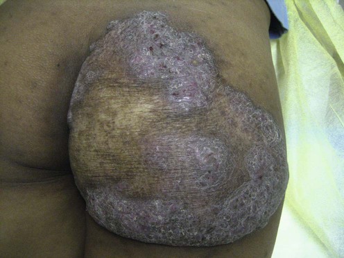
A 73-year-old male with plaque form lupus vulgaris of the buttock developed drug-induced hepatitis and fulminant hepatic failure due to antituberculous medication. A 6-month trial of topical calcipotriol 50 µg/g ointment then led to resolution of the lesion without recurrence.
Dowling GB, Gauvain S, Macrae DE. Br Med J 1948; 6; 1(4548): 430–5, 438–2.
The use of vitamin D to treat tuberculosis dates back to the earliest availability of commercial formulations and, even prior to this, to the use of cod liver oil!
This remedy was dropped after the advent of antituberculous antibiotics, so presumably its efficacy is limited.
Treatment of Skin Disease Comprehensive Therapeutic Strategies 4e
WhatsApp us


 Antituberculous drugs
 Excision
 Calcipotriol