[level-membership-for-dermatology-category]
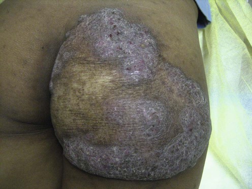
Tuberculosis and tuberculids
Specific investigations


First-line therapies
 Antituberculous drugs
Second-line therapies
 Excision
 Calcipotriol
[/level-membership-for-dermatology-category][not-level-membership-for-dermatology-category]
Tuberculosis and tuberculids
