[level-membership-for-dermatology-category]
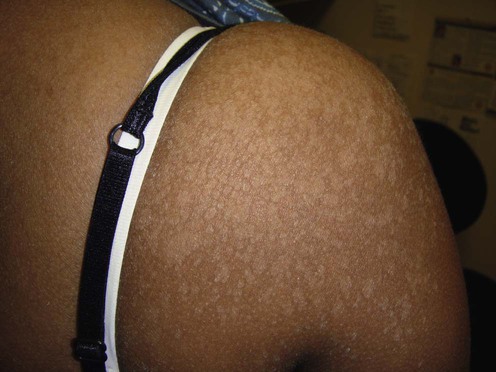
Tinea versicolor (pityriasis versicolor)


First-line therapy
 Ketoconazole
 Bifonazole
 Terbinafine
 Clotrimazole
 Econazole
 Oxiconazole
 Butenafine
 Ciclopirox
 Fluconazole shampoo
 Selenium sulfide 2.5%
 Tioconazole
 Zinc pyrithione shampoo
Second-line therapy
 Itraconazole
 Ketoconazole
 Fluconazole
 Itraconazole
Third-line therapy
 Pramiconazole (oral triazole)
 Adapalene
 Naftifine (topical allylamine)
 Isotretinoin
[/level-membership-for-dermatology-category][not-level-membership-for-dermatology-category]
Tinea versicolor (pityriasis versicolor)
