Published on 18/03/2015 by admin
Filed under Dermatology
Last modified 22/04/2025
This article have been viewed 2676 times
Eavan G. Muldoon and Derek Freedman
Evidence Levels: A Double-blind study B Clinical trial ≥ 20 subjects C Clinical trial < 20 subjects D Series ≥ 5 subjects E Anecdotal case reports
Syphilis is an ancient disease that many mistakenly believe to be on the decline. A century ago, Osler said ‘Know syphilis in all its manifestations and relations, and all things clinical will be added unto you.’ Syphilis’ manifestations are protean, hence the reason it is often known as ‘the great impostor’ or ‘great pretender.’ Syphilis facilitates the transmission of human immunodeficiency virus (HIV) by a number of mechanisms, including increasing HIV viral load, disruption of the mucous membrane barrier and increased HIV shedding. Prompt recognition and treatment have significant public and personal health benefits. Syphilis is an indicator disease for HIV, and a syphilis diagnosis provides opportunities for the screening and early diagnosis of HIV, which will have impact on patients’ prognosis and further transmission.
Men who have sex with men (MSM), commercial sex workers, or patients from areas of the world where there is a high prevalence of syphilis have a higher risk of syphilis acquisition. A thorough history is essential and may uncover past potential risks that may not be immediately evident, such as woman with past partners who are MSM. However, many are unaware of their risk status, and routine testing is required.
Transmission of syphilis is by direct contact with an infectious individual. Sexual contact is the most common mode of transmission. However, in the current era of so-called ‘safer sex practices’ transmission by kissing, or other close contact with an active lesion, such as oral sex, have become more common. Transplacental transmission, blood transfusion or accidental inoculation can all also rarely occur.
The appropriate management is determined by the stage of syphilis infection. Infection is divided into:
Incubation period
Primary syphilis characterized by a primary chancre(s)
Secondary syphilis
A period of sub-clinical infection or latent syphilis
Tertiary syphilis, which does not occur in every patient
The patient is most infectious in the early stages of disease, particularly when the primary chancre, mucous patch or condyloma lata are present. An immunologically intact person cannot spread syphilis after 4 years of infection.
Treponema pallidum penetrates the intact mucous membrane or gains access through abraded skin. Within hours to days it enters the lymphatic system and/or bloodstream and disseminates throughout the body. This occurs soon after contact; patients have been infected by blood transfusions from donors with negative serology who are in the incubation period. The incubation period is directly proportional to the size of the inoculum: in rabbit models as few as four to eight spirochetes are needed to establish infection. The median incubation period is 3 weeks, but can vary from 3 to 90 days.
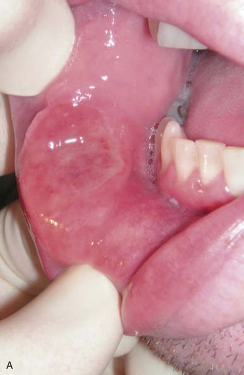
Classically, a primary chancre is described as a single painless papule that occurs at the site of inoculation. The site of a chancre is not limited to the genital region. Any area where contact with an infectious lesion has occurred can be involved. The mouth and anal area are common extragenital sites (see Figure A).
The chancre appears after the incubation period, erodes and becomes indurated. The base is usually smooth, and the borders raised and firm with a cartilaginous consistency. The ulcer has a clean appearance with no exudate, unless it is super-infected. It is usually round; however, it may be oblong following tissue lines. There is little pain or bleeding when the ulcer is scraped, as may occur while obtaining samples for dark-field microscopy. Multiple chancres can occur, particularly in the setting of HIV infection. Atypical lesions are said to occur in 60% of patients, and many patients may have no primary lesion. The appearance of the lesion depends on the size of the inoculum, the immune status of the patient, and concurrent antibiotic usage. Regional lymphadenopathy of moderately enlarged, firm non-suppurative painless lymph nodes frequently accompanies the primary lesion.
The differential diagnosis of a primary chancre is herpes simplex, chancroid, and traumatic super-infected lesions. Other diagnoses that should be considered are early warts caused by human papilloma virus, granuloma inguinale, tuberculosis or atypical mycobacterial infections. Perianal lesions may be dismissed as hemorrhoids.
Secondary syphilis occurs when the spirochete multiplies and disseminates throughout the body. It lasts until a sufficient host response develops to control the organism. It begins 2 to 8 weeks after the appearance of the chancre; however, this is highly variable and the signs of primary and secondary infection may overlap. Many patients presenting with secondary syphilis, do not recall a primary chancre. The manifestations of secondary syphilis are protean.
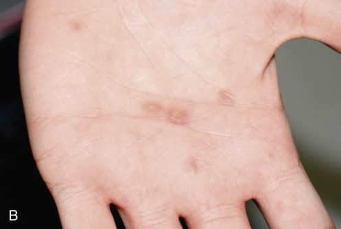
The most commonly recognized signs of secondary syphilis are dermatological. The rash of secondary syphilis may be macular, maculopapular, papular, pustular or a combination of the above. Vesicular lesions occur only in congenital syphilis. Skin lesions usually begin on the trunk and proximal extremities. Classically, they are pink-red macular lesions 3–10 mm in diameter. Any surface area can become involved. The rash can last from a few days to up to 8 weeks. The lesions may evolve into papules and in some patients into pustules, when they are known as pustular syphilids. All types of rash may be present at the same time and the rash can involve any body surface. Palmar and plantar rashes (see Figure B) are highly suggestive of syphilis. Fever can accompany the rash, and the clinical syndrome can be mistaken for acute HIV infection, infectious mononucleosis or a non-specific viral syndrome.
Alopecia occurs when the hair follicles become involved. In warm moist areas, such as the genital and perianal areas, inner aspects of the thighs, the skin under breasts, nasolabial folds, axilla, antecubital fossae, and webs of fingers and toes, the papules can coalesce and erode resulting in painless, broad, gray-white, highly infectious plaques called condylomata lata. They can be mistaken for warts or hemorrhoids. Lesions may also develop on mucous membranes where they are called mucous patches. Associated enlargement of the epitrochlear lymph nodes is a unique finding that should always suggest the diagnosis.
Neurosyphilis, i.e., syphilitic infection of the central nervous system, can present at any time after infection. It can be classified into early and late disease. Early in the course of syphilis most forms of neurosyphilis will involve the cerebrospinal fluid (CSF), meninges and vasculature, while later disease that occurs in tertiary syphilis tends to affect the brain parenchyma and spinal cord. Currently neurosyphilis is most common in patients with HIV infection, and these patients almost exclusively have the early forms of neurosyphilis, often presenting as concomitant eye disease. Ocular syphilis is part of the clinical spectrum of neurosyphilis, and the initial presentation may be to an unsuspecting ophthalmologist. The presentation of ocular involvement is varied, the most common presentation being uveitis.
Latent syphilis is the asymptomatic period following infection. It is usually divided into early latent disease, within 2 years of infection, and late latent disease after 2 years of infection. Careful history is required to attempt to stage asymptomatic disease. Sexual history, obstetric history, and travel history will all offer valuable clues.
Tertiary syphilis is only seen in approximately one-third of untreated patients. It takes the form of neurological disease, cardiovascular disease, and gummatous disease. It is rarely seen today due to antibiotic therapy, often inadvertent; however, it may be more prevalent in certain immigrant populations.
The diagnosis of syphilis depends on the stage of the disease. T. pallidum is too slender to be visualized by direct microscopy and the bacterium cannot be cultured. Dark-field or dark-ground microscopy, a specialized technique that utilizes an oblique light to visualize the organism, can be used in cases when patients have demonstrable clinical lesions; it has a sensitivity of 50% in experienced hands. Moist lesions containing large numbers of treponemes such as primary chancres, condyloma lata or mucous patches are particularly amenable to dark-field microscopy. It is not recommended for oral lesions, as commensal oral spirochetes are indistinguishable from T. pallidum. The vast majority of cases of syphilis are diagnosed by serology, an indirect diagnostic method. Serological tests fall into two categories, non-treponemal tests and treponemal tests. Use of non-treponemal tests such as rapid plasma reagin (RPR) or venereal disease research laboratory (VDRL) tests are used for screening. Positive tests are then confirmed with a treponemal test such as Treponema pallidum particle agglutination (TPPA) or fluorescent treponemal antibody absorbed (FTA-ABS). False positive reactions may be seen with the non-treponemal tests in autoimmune conditions and various other infections, including HIV. Quantitative non-treponemal tests are useful for providing a baseline from which change can be measured. Re-infection can be demonstrated by a fourfold rise in titre, for example. The reactivity of the test can differ depending on variation in antigen preparation and for this reason it is recommended that sequential tests should be performed using the same laboratory method and preferably performed by the same laboratory. If the clinical suspicion for syphilis is high, and the non-treponemal tests are negative, the ‘prozone’ phenomenon caused by very high antibody titers should be considered. Dilution of the serum converts the test to positive.
Guidelines on treatment differ; however, the basic principles are the same. Long-acting penicillin is the treatment of choice in all patients. Patients with neurological or ocular involvement will need daily treatment for a minimum of 10 to 14 days with parenteral penicillin. Doxycycline is an alternative to penicillin in those allergic to penicillin; however, in the case of neurological disease and pregnant patients, penicillin desensitization is preferred. Patients need to be followed with regular serology post treatment to ensure adequate response to therapy.
The Jarisch–Herxheimer reaction may be noted within hours of treatment manifested by fever, chills, headache, myalgias, exacerbation of skin lesions, and other systemic symptoms. Treatment with non-steroidal anti-inflammatory agents or systemic steroids may be necessary.
Syphilis is an easily treatable disease with diverse manifestations. It is an indicator disease for HIV: consequently all patients diagnosed with syphilis must be screened for HIV and other sexually transmitted infections. If syphilis is not considered during the evaluation of patients presenting with symptoms of unclear etiology, the diagnosis may easily be missed. Careful history taking regarding risks factors for the acquisition of the infection, possible chancrous lesions, systemic illness, and rash may offer vital clues in making the diagnosis of this curable disease, but routine serological testing of all patients is a safeguard to establish the diagnosis.
Workowski KA, Berman S. Centers for Disease Control and Prevention (CDC). MMWR Recomm Rep 2010; 59(RR-12): 1–110.
The definitive diagnostic test is dark-field microscopy; however, this is obviously limited to those patients who have a lesion amenable to dark-field microscopy, such as a genital lesion. Most diagnoses are made using serological methods. The combination of a non-treponemal test (e.g., VDRL or RPR) and a treponemal test (e.g., FTA-ABS or TPPA) should be used to confirm the diagnosis. Non-treponemal antibody titers correlate with disease activity: a fourfold rise in titer is necessary to demonstrate a clinically significant difference. Tests should be performed using the same method and preferably the same laboratory. VDRL and RPR are equally valid assays; however, quantitative results from the two tests cannot be compared directly. Non-treponemal titers should decline post treatment, and may become non-reactive with time, particularly in patients treated for primary syphilis. However, most will have a reactive non-treponemal test for life. A positive non-treponemal test should prompt a treponemal test. HIV positive patients may have atypical serological tests; however, serological tests are reliable and accurate for the diagnosis of syphilis in HIV infected individuals. Follow-up serology should be performed at 3, 6, 9, 12, and 24 months.
If the patient has clinical signs of neurosyphilis (e.g., cranial nerve dysfunction, meningitis, stroke, altered mental status, loss of vibration sensation, or hearing or visual changes) further investigation and treatment is warranted. CSF VDRL is highly specific, but not sensitive: most other tests lack sensitivity and specificity, and diagnosis depends of a combination of abnormal CSF white cell count, elevated protein, and VDRL. HIV infected patients with CD4 count of <350 cells/mL and or an RPR >1 : 32 are more likely to have neurosyphilis regardless of symptoms.
Søgaard OS, Lohse N, Østergaard L, Kronborg G, Røge B, Gerstoft J, et al. PLoS One 2012; 7: e32538.
A population-based study to identify indicators for subsequent HIV infection. A total of 2036 HIV positive patients were matched to 35 718 controls. In those patients who had a hospital-based diagnosis of syphilis in the preceding 5 years, their adjusted odds ratio of being diagnosed with HIV was 94.7.
Binnicker MJ, Jespersen DJ, Rollins LO. J Clin Microbiol 2012; 50: 148–50.
Traditionally, serum for syphilis serology was screened by use of a non-treponemal test such as the RPR and positives confirmed with a treponemal test. However, more recently laboratories have been using automated treponemal tests such as an enzyme immunoassay (EIA) to deal with the large volumes of samples that need to be screened. Direct comparison of this method indicates that it will pick up more false positives by EIA (0.6%); however, additional cases are also likely to be detected.
Guy R, Wand H, Holt M, Mao L, Wilson DP, Bourne C, et al. Sex Transm Dis 2012; 39: 268–75.
Testing for syphilis is now recommended every 3 months in HIV positive MSM and every 6 months for MSM not infected with HIV. In this survey of 6329 MSM, 14% had never tested for syphilis. Risks for not having tested were older age, lower number of sexual partners, and not being aware that syphilis may be asymptomatic. In men that were deemed high risk, not living in a metropolitan area, and not being aware that syphilis could be spread through oral sex were additional risks.
T. pallidum penicillin resistance has never been reported, despite>60 years of use.
Fitzgerald MR, Ahmed-Jushuf I, Radcliffe KW, Rooney G, Welch J, Wilson JD. Sex Transm Infect 2002; 78: 81–2.
Penicillin is first-line therapy recommended in international guidelines, for all stages of syphilis infection. The duration of therapy may differ however. Penicillin desensitization is recommended for pregnant patients or those diagnosed with neurosyphilis who have a penicillin allergy.
Idsøe O, Guthe T, Willcox RR. Bull WHO 1972; 47: 1–68.
In studies totalling 1381 patients with seronegative primary syphilis, 97% of treated patients were clinically well and serologically negative. Treatment failures were attributed to re-infection.
Myint M, Bashiri H, Harrington RD, Marra CM. Sex Transm Dis 2004; 31: 196–9.
Relapses of infectious syphilis after treatment with the recommended doses of penicillin is rare but does occur. For this reason, it is important to follow treated patients both clinically and serologically. These patients respond to higher and more prolonged doses of penicillin.
All patients not receiving penicillin require close follow-up to ensure response to therapy. Pregnant patients and patients diagnosed with neurosyphilis should be treated with penicillin and desensitized if they report a penicillin allergy.
Clinical Effectiveness Group (Association of Genitourinary Medicine and the Medical Society for the Study of Venereal Diseases). Sex Transm Infect 1999; 75: S29–33.
The recommended treatment for primary, secondary, or early latent syphilis in immunocompetent persons who are allergic to penicillin is orally administered doxycycline, 100 mg twice daily, or tetracycline 500 mg four times daily for 14 days. Amoxicillin 500 mg four times daily plus probenecid 500 mg four times daily for 14 days is not as effective as parenteral penicillin owing to poorer compliance with this regimen. Doxycycline 100 mg by mouth twice daily for 28 days is recommended for immunocompetent patients with uncomplicated late latent syphilis or syphilis of unknown duration. Some experts prefer a dose of 200 mg twice daily.
Ghanem KG, Workowski KA. Clin Infect Dis 2011; 53(Suppl 3): S110–28.
The data for using alternative agents for the treatment of neurosyphilis is limited. There are two retrospective trials looking at doxycycline in early disease; however, no patients with late syphilis infection were included and there were few HIV positive patients. Careful follow-up of HIV positive patients to ensure serological response is essential if these agents are used.
Augenbraun M, Workowski K. Clin Infect Dis 1999; 29: 1337–8.
A single injection of 1 g ceftriaxone is not effective for treating infectious syphilis. Daily or alternate-day injections for 8–10 days appear to be efficacious, but more data are needed to evaluate late failures.
Spornraft-Ragaller P, Abraham S, Lueck C, Meurer M. Eur J Med Res 2011; 16: 47–51.
In a retrospective study, 24 consecutive HIV positive patients with early syphilis infection treated with either benzathine penicillin or ceftriaxone were compared. After 20 months of follow-up 23 of 24 patients had a serological response to therapy, suggesting that ceftraixone may have a comparable efficacy in HIV positive patients with higher CD4 counts.
Azithromycin should not be used for the treatment of pregnant patients or MSM due to high rates of resistance.
Lukehart SA, Godornes C, Molini BJ, Sonnett P, Hopkins S, Mulcahy F, et al. N Engl J Med 2004; 351: 154–8.
A mutation that makes T. pallidum resistant to azithromycin was identified with the use of a restriction-digestion assay in 15 of 17 samples (88%) from Dublin, 12 of 55 samples (22%) from San Francisco, three of 23 samples (13%) from Seattle, and two of 19 samples (11%) from Baltimore. The study suggests that a mutated strain was either introduced into a sexual network or has been selected for among persons who engage in high-risk behavior.
Katz KA, Klausner JD. Curr Opin Infect Dis 2008; 21: 83–91.
Azithromycin resistance in T. pallidum is increasing in the US, Canada, and Ireland. Closer observation for treatment failures is needed in patients treated with azithromycin.
Lewis DA, Lukehart SA. Sex Transm Infect 2011; 87(Suppl 2): ii39–43.
As macrolide mutations can be identified in strains by restriction digestion of PCR products, these techniques are only available in a limited number of settings, meaning that samples from most geographical regions cannot be tested. While there has been no reported resistance in samples from Africa, the trials were conducted before or early in the era of the recognition of resistant strains. Azithromycin should only be used in regions where the prevalence of resistant strains is known to be low.
Treatment of Skin Disease Comprehensive Therapeutic Strategies 4e
WhatsApp us


 Benzathine penicillin
 Procaine plus probenecid
 Doxcycline
 Tetracycline
 Amoxicillin plus probenecid
 Ceftriaxone
 Azithromycin
 Erythromycin