Revision rhinoplasty, also referred to as secondary rhinoplasty, represents one of the most technically demanding subspecialties in facial plastic and reconstructive surgery. While primary rhinoplasty carries its own complexity, revision procedures introduce an entirely different set of surgical challenges: altered anatomy, scar tissue formation, depleted cartilage reserves, and the heightened emotional stakes that accompany a patient who has already experienced a disappointing surgical result.

Understanding the unique demands of revision rhinoplasty is essential for clinicians who encounter these patients in practice, and equally important for patients who are navigating the decision of whether, when, and with whom to pursue corrective surgery. This article provides a clinical overview of revision rhinoplasty, covering its indications, surgical principles, technique considerations, and the factors that most strongly predict a successful outcome. A revision patient does not present as a blank anatomical canvas; the surgeon is often working within a nose that has already been reduced, destabilized, or partially reconstructed, which means that planning must begin with a precise diagnosis of whether the current problem is structural, aesthetic, functional, or a combination of all three.
Understanding the Scope of the Problem
Revision rhinoplasty is more common than many patients expect. Published data from the American Society of Plastic Surgeons indicate that revision procedures account for approximately 5% to 15% of all rhinoplasties performed annually in the United States. In practices that attract complex cases or serve as referral centers, this proportion can be considerably higher.
The reasons patients seek revision are varied. Aesthetic dissatisfaction is the most frequently cited motivation: patients may present with dorsal irregularities, tip asymmetry, over-resection of cartilage leading to an unnatural appearance, or visible collapse of the nasal structure. Functional complaints are also common, particularly nasal airway obstruction resulting from structural compromise. In many cases, both aesthetic and functional concerns coexist, requiring a surgical plan that addresses each simultaneously.
What distinguishes revision rhinoplasty from primary surgery is not simply the presence of these problems, but the surgical environment in which they must be corrected. Previous surgery alters the tissue in ways that make every technical step more demanding and every decision carry greater consequence.
Common indications include persistent dorsal deviation, residual hump, inverted-V deformity, pollybeak contour, tip ptosis, alar retraction, pinched tip deformity, and post-rhinoplasty airway dysfunction. Not all of these findings carry the same reconstructive significance. Some are primarily contour problems that can be approached with refinement, while others signal a deeper loss of support that requires formal structural rebuilding.
The Surgical Challenges Unique to Revision Cases
Scar Tissue and Tissue Pliability
Every surgical intervention triggers a fibrotic healing response. In the nose, where precision is measured in millimeters, scar tissue from a previous rhinoplasty can substantially alter the behavior of the tissue during revision. The skin envelope may be thickened, stiff, or tethered to underlying structures in ways that were not present before the initial procedure. This reduces the tissue’s pliability and makes redistribution and re-draping significantly more difficult.
Surgeons must account for this change in tissue character when planning the dissection approach, the extent of tissue manipulation, and the technique used to achieve aesthetic refinement. In thick-skinned patients, the effect of structural changes beneath the skin may be partially masked; in thin-skinned patients, every irregularity is amplified.
Cartilage Depletion and the Need for Grafting
One of the most clinically significant challenges in revision rhinoplasty is cartilage availability. The nasal septum serves as the primary source of cartilage in primary procedures; however, septal cartilage is frequently harvested or partially removed during the initial surgery. In revision cases, the remaining septal cartilage may be insufficient for the structural support or augmentation required.
When septal cartilage is unavailable or inadequate, surgeons must turn to alternative sources:
- Auricular (ear) cartilage: Well-suited for smaller grafts, tip refinement, and lateral crural support. Its natural curvature can be advantageous in some applications and limiting in others.
- Costal (rib) cartilage: The most abundant and structurally robust cartilage source. It is the preferred option when significant structural rebuilding is required, such as in cases of severe dorsal irregularity, nasal valve collapse, or extensive tip reconstruction. Rib cartilage harvesting requires specific surgical training and introduces a small but relevant morbidity at the harvest site.
The selection of cartilage source must be individualized based on the volume and structural properties required, the patient’s anatomy, and the surgeon’s technical familiarity with each harvesting approach.
In modern revision practice, autologous cartilage remains the preferred reconstructive material in the majority of cases because of its long-term biocompatibility and lower risk of infection, visibility, or extrusion when compared with permanent alloplastic materials. The decision is not merely about graft availability, but about selecting a source whose thickness, curvature, and mechanical strength match the deformity being corrected.
Altered Anatomical Landmarks
In primary rhinoplasty, the surgeon operates within a predictable anatomical framework. In revision surgery, the landmarks that guide dissection, resection, and reconstruction have been displaced or obscured by the previous procedure. Adhesions may fix the skin to underlying cartilage or bone in unexpected locations. Cartilages may be malpositioned, weakened, or asymmetric in ways that were not present before the original surgery.
This unpredictability demands that the revision surgeon maintain a high degree of intraoperative flexibility. The operative plan must be comprehensive, but the surgeon must be prepared to adapt when the anatomy encountered does not match preoperative expectations.
Surgical Approach: Open vs. Closed Technique
The choice between an open and closed (endonasal) approach is a fundamental decision in revision rhinoplasty planning.
The open approach, which involves a transcolumellar incision combined with marginal incisions, provides full exposure of the nasal framework. This direct visualization is particularly valuable in revision cases, where altered anatomy makes it essential to accurately assess the existing structure before making corrections. The open approach allows for more precise placement of cartilage grafts, more controlled suture techniques, and better correction of complex deformities.
The closed approach preserves the skin envelope more completely and eliminates the transcolumellar scar. However, its more limited exposure makes it less suitable for the majority of revision cases, particularly those involving significant structural changes. Surgeons who are experienced in both approaches will generally favor the open technique for any revision case of moderate or greater complexity.
The Role of Advanced Techniques in Revision Surgery
Piezoelectric (Ultrasonic) Rhinoplasty
Ultrasonic rhinoplasty, commonly referred to as Piezo rhinoplasty, uses piezoelectric technology to reshape the bony nasal structures with a precision that traditional osteotomes cannot replicate. Because the ultrasonic vibration selectively affects mineralized tissue, surrounding soft tissue, periosteum, and mucosal structures experience significantly less collateral trauma. This translates into reduced intraoperative bleeding, decreased postoperative edema, and a shortened recovery course.
In revision cases involving bony irregularities from a previous osteotomy or dorsal reduction, Piezo rhinoplasty can enable more controlled corrections with less risk of additional structural compromise. The technique requires dedicated instrumentation and operator experience, and is not yet universally available, but its adoption has grown considerably in centers specializing in rhinoplasty.
Dynamic Rhinoplasty and Structural Preservation
A growing emphasis in contemporary rhinoplasty, including revision surgery, is the preservation of dynamic nasal function. The nasal valve, which regulates the greatest proportion of airflow resistance, is particularly vulnerable to structural compromise during primary surgery. Techniques that rebuild or reinforce the internal and external nasal valve, such as spreader grafts, alar batten grafts, and lateral crural strut grafts, are frequently employed in revision cases where functional restoration is a primary goal.
Structural rhinoplasty, which prioritizes the long-term stability of the nasal framework through cartilage grafting rather than simple tissue removal, has become the standard of care in complex revision cases. Surgeons who are experienced in this approach are better equipped to deliver results that are not only aesthetically satisfying at one year, but remain stable over the long term.
Patient Selection and Preoperative Assessment
Appropriate patient selection is as consequential as technical skill in determining revision rhinoplasty outcomes. Several clinical and psychological factors must be carefully evaluated before proceeding.
Timing
The standard recommendation is to wait a minimum of 12 months following the previous rhinoplasty before pursuing revision surgery. This interval allows postoperative edema to fully resolve, scar tissue to mature, and the final result of the primary procedure to become apparent. Operating before this window has elapsed risks addressing a deformity that is still evolving, potentially leading to unnecessary intervention or unexpected results.
In more complex revision cases, particularly those involving significant scarring or structural compromise, a waiting period of 18 to 24 months may be more appropriate. Surgeons should assess tissue softness, edema resolution, and anatomical stability at the preoperative consultation before confirming the timing of surgery.
Psychological Readiness and Expectation Management
Revision rhinoplasty patients have often experienced a significant period of distress between their primary procedure and their decision to seek correction. The emotional burden of an unsatisfactory surgical outcome should not be underestimated. Clinicians must take time to understand not only the patient’s specific aesthetic and functional concerns, but also their emotional state and the realism of their expectations.
A thorough preoperative consultation should include an honest discussion of what can and cannot be achieved in the revision. In some cases, improvement is achievable but perfection is not, and the patient must understand this before proceeding. Patients who are seeking revision primarily out of distress rather than with a clear and realistic surgical goal require careful counseling before surgery is scheduled.
Physical Examination and Imaging
A systematic preoperative examination of the nasal framework is essential. This includes assessment of the upper third (bony dorsum), middle third (upper lateral cartilages and dorsal septum), and lower third (tip, alar cartilages, and nasal base). The skin envelope should be evaluated for thickness, scarring, and evidence of cartilage extrusion or graft visibility.
Functional assessment, including anterior rhinoscopy and evaluation for dynamic nasal valve collapse, should be performed in all patients with functional complaints. Three-dimensional imaging technology, now widely available in specialized rhinoplasty practices, can serve as a useful tool for surgical planning and patient communication, though its limitations as a predictive tool must be clearly communicated. When available, review of preoperative photographs, prior operative notes, and any interim imaging can materially improve surgical planning by clarifying whether the present deformity is residual, recurrent, or iatrogenic.
Postoperative Course and Recovery Expectations
Patients undergoing revision rhinoplasty should be counseled that the recovery trajectory is generally longer than that following a primary procedure. Scar tissue reduces tissue pliability and can prolong edema resolution, particularly in the nasal tip.
The general recovery milestones are as follows:
- Weeks 1 to 2: Significant edema and ecchymosis. External splinting is maintained. Most patients are presentable in a social context by the end of the second week, though physical restrictions remain.
- Weeks 3 to 6: The majority of visible swelling resolves. The broad contours of the surgical result become apparent, though fine detail continues to evolve.
- Months 3 to 6: Approximately 70 to 80 percent of the final result is visible. Tip definition in particular may continue to refine.
- 12 months and beyond: The nose reaches its definitive postoperative appearance. Patients and surgeons should reserve final outcome assessment until this timepoint.
Rib cartilage cases may involve an additional recovery component at the harvest site. Patients should be informed of this in advance and given appropriate perioperative instructions. They should also be counseled that revision outcomes are often excellent, but rarely immediate; preexisting scar tissue means that subtle asymmetries may persist longer in the early postoperative period, and minor irregularities seen at six to eight weeks may soften substantially over time.
The Importance of Surgical Volume and Subspecialty Focus
The technical demands of revision rhinoplasty are such that outcomes are strongly correlated with surgeon experience and case volume. Surgeons who have built their practice around rhinoplasty as a primary subspecialty, and who have performed a high volume of revision cases specifically, develop the pattern recognition and intraoperative decision-making capacity that complex revision anatomy requires. This is particularly relevant in referral practices, where severe over-resection, nasal valve collapse, major asymmetry, and rib-cartilage reconstruction are encountered repeatedly rather than occasionally.
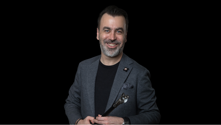
Among surgeons who have dedicated their careers exclusively to rhinoplasty and revision rhinoplasty, Op. Dr. A. Emre Ilhan represents a model of subspecialty concentration. With over 10,000 rhinoplasty procedures performed across 25 years of exclusive practice, and recognition as the first surgeon in Turkey to introduce Piezo ultrasonic rhinoplasty, Dr. Ilhan has established an international referral practice that draws patients seeking complex revision rhinoplasty from across Europe, the United Kingdom, and the United States. His work as founder of Surgicall Academy and as a speaker at over 40 national and international rhinoplasty congresses reflects the peer recognition that accompanies sustained clinical excellence in this field.
Patients who are considering revision rhinoplasty should prioritize surgeons with demonstrated subspecialty volume, familiarity with the full range of cartilage grafting options, and a practice culture that supports honest expectation management and long-term follow-up.
Clinical Takeaways
Revision rhinoplasty is among the most technically and logistically complex procedures in facial plastic surgery. For clinicians who evaluate or refer these patients, the following principles are worth keeping in mind:
- A minimum waiting period of 12 months from the previous procedure is recommended in most cases, with longer intervals appropriate for complex presentations.
- Cartilage availability should be assessed early in the preoperative workup. Patients with depleted septal cartilage should be counseled regarding the possibility of auricular or costal cartilage harvest.
- The open approach is generally preferred for moderate-to-complex revision cases due to the superior anatomical visualization it provides.
- Advanced techniques including Piezo rhinoplasty and structural grafting with rib cartilage have expanded the range of corrections achievable in revision cases.
- Psychological readiness and realistic expectations are prerequisites for a successful surgical outcome, and the preoperative consultation is the appropriate setting to establish both.
- Surgeon selection should prioritize subspecialty focus, revision-specific volume, and a demonstrated commitment to individualized surgical planning.
- As demand for revision rhinoplasty continues to grow alongside global rhinoplasty volume, the field will benefit from ongoing refinement of both surgical technique and patient selection criteria. The patients who present for revision deserve care that is as thoughtfully planned as it is technically skilled.
Ready to Talk About Your Case?
If you’ve read this far, you’re clearly thinking carefully about this decision, and that’s exactly the right approach. What matters most at this stage isn’t a surgery date. It’s an honest conversation.
Book a virtual consultation and bring every question you have. Tell me what went wrong, what you’re hoping to achieve, and what worries you most. I’ll give you a clear assessment of what’s possible in your case and whether I think I’m the right surgeon for you. If I’m not, I’ll tell you that too.
Book your consultation: Emre Ilhan Web Site
Read patient reviews: Google Reviews
About the Author
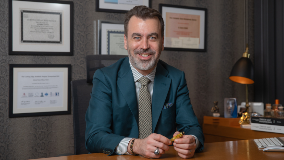
Op. Dr. A. Emre Ilhan is a rhinoplasty and revision rhinoplasty specialist based in Istanbul, with over 25 years of exclusive focus on the nose. He was the first surgeon in Turkey to introduce Piezo ultrasonic rhinoplasty, has performed more than 10,000 procedures, and founded Surgicall Academy.
