Published on 18/03/2015 by admin
Filed under Dermatology
Last modified 18/03/2015
This article have been viewed 1140 times
Karolyn A. Wanat, Joel M. Gelfand and William D. James
Evidence Levels: A Double-blind study B Clinical trial ≥ 20 subjects C Clinical trial < 20 subjects D Series ≥ 5 subjects E Anecdotal case reports
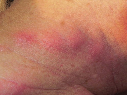
(Courtsey of Misha Rosenbach MD.)
Sweet syndrome is a neutrophilic dermatosis, characterized clinically by multiple painful, well-demarcated, non-scarring erythematous plaques or pustules on the face, neck, upper trunk, and extremities. There can be a pseudovesicular appearance. Fever, leukocytosis, arthralgias, myalgias, headaches, and general malaise may occur. Oral, ocular, and internal organ involvement is rare. On histopathology, there is a diffuse neutrophilic infiltrate in the upper dermis without evidence of primary leukocytoclastic vasculitis. Neutrophilic dermatosis of the dorsal hands (NDDH) is classified as an anatomically limited subset of Sweet syndrome.
Sweet syndrome is often idiopathic, but may be associated with malignancy (most commonly acute myelogenous leukemia, but also lymphomas, dysproteinemias, and carcinomas), inflammatory bowel disease, infection (commonly Streptococcus or Yersinia), medication, radiation, and pregnancy. Work-up includes skin biopsy, complete physical examination, and laboratory studies. Treatment of the underlying associated condition or discontinuation of the causative medication may lead to resolution of skin lesions. The standard treatment is corticosteroids. For patients with contraindications to corticosteroids, or for therapeutic failures, second-line therapies may be used.
History and physical examination
Complete blood count with differential
Erythrocyte sedimentation rate or C-reactive protein
Cultures as indicated by history and physical examination
Pregnancy test (in women of childbearing potential)
Medication history
Cohen PR, Kurzrock R. Int J Dermatol 2003; 42: 761–78.
An excellent review of the literature.
Walling HW, Snipes CJ, Gerami P, Piette WW. Arch Dermatol 2006; 142: 57–63.
A report of nine cases of NDDH and review of all 43 cases reported.
Heymann WR. J Am Acad Dermatol 2009; 61: 693–4.
The term ‘histiocytoid’ refers to the histiologic presence of immature neutrophils; the clinical implications are identical to those of the classical Sweet syndrome.
Vignon-Pennamen MD, Julliard C, Rybojad M, Wallach D, Danie MT, et al. Arch Dermatol 2006; 142: 1170–6.
Nine cases of chronic Sweet syndrome in which the initial biopsies demonstrated a lymphocytic infiltrate with atypical mononuclear cells positive for CD68 and myeloperoxidase, suggesting a myeloid origin; this presentation appeared to be a predictive marker of underlying myelodysplasia.
Kroshinsky D, Alloo A, Rothschild B, Cummins J, Tan J, Montecino R, et al. J Am Acad Dermatol 2012; 67: 945–54.
Three cases of acute necrotizing neutrophilic dermatosis characterized by deep tissue neutrophilic infiltration and soft tissue necrosis. The authors state that ‘the diagnosis of NSS should be entertained in all cases of suspected necrotizing fasciitis failing to respond to therapy and lacking a causative organism.’
Gray PE, Bock V, Ziegler DS, Wargon O. Pediatrics 2012; 129: e1353–9.
Sweet syndrome in the neonatal period requires thorough investigation as it can be associated with a serious underlying disorder.
Treatment of Skin Disease Comprehensive Therapeutic Strategies 4e
WhatsApp us