Published on 18/03/2015 by admin
Filed under Dermatology
Last modified 22/04/2025
This article have been viewed 1647 times
Roy A. Palmer, Ian Coulson and Martin Keefe
Evidence Levels: A Double-blind study B Clinical trial ≥ 20 subjects C Clinical trial < 20 subjects D Series ≥ 5 subjects E Anecdotal case reports
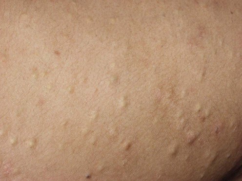
Although probably genetically heterogeneous, steatocystoma multiplex often demonstrates an autosomal dominant pattern of inheritance. Some pedigrees have demonstrable keratin 17 abnormalities. It is characterized by the development in adolescence or early adulthood of cysts on the trunk and proximal limbs, or in some patients on the face and scalp. These are true ‘sebaceous cysts’: they contain sebum, and sebaceous gland lobules are present in the walls. Overlap with eruptive vellus cysts and association with pachyonychia congenita type II have been reported.
The cysts persist indefinitely. Although usually a minor cosmetic problem, they can be highly disfiguring. Paradoxically, those patients who would benefit the most from treatment are sometimes regarded as being unsuitable for surgery because they have too many cysts to excise. The surgical technique described below is quick, and so can be used on large numbers of lesions in one session. It produces good cosmetic results.
Lesions can become inflamed due to rupture of the cyst wall with leakage of the contents into the dermis, or because of bacterial infection. Suppuration and scarring may follow. The clinical picture then resembles cystic acne and is called steatocystoma multiplex suppurativum. Oral isotretinoin is an effective treatment for inflammatory lesions but not for non-inflamed cysts. This suggests it operates by a direct anti-inflammatory effect rather than by reducing the sebum excretion rate. Alternatively, inflamed cysts can be treated with incision and drainage, intralesional triamcinolone, tetracycline 1 g/day, or minocycline 100–200 mg/day.
Topical treatment is largely ineffective because it does not penetrate to reach the cyst wall.
Skin biopsy if diagnosis is in doubt
Keratin gene testing
Zang D, Zhou C, He M, Ma X, Zhang J. Eur J Dermatol 2011; 21: 142–4.
Gass JK, Wilson NJ, Smith FJ, Lane EB, McLean WH, Rytina E, et al. Br J Dermatol 2009; 161: 1396–8.
Schwartz JL, Goldsmith LA. Cutis 1984; 34: 149–53.
Statham BN, Cunliffe WJ. Br J Dermatol 1984; 111: 246.
Rosen BL, Broadkin RH. Cutis 1986; 115: 115–20.
Mortiz DL, Silverman RA. Cutis 1988; 42: 437–9.
Friedman SJ. Cutis 1987; 39: 506–7.
These five papers report a total of seven patients treated with oral isotretinoin, at a dose of approximately 1 mg/kg/day for about 20 weeks. Inflammation of cysts was greatly reduced. Non-inflammatory lesions were unaffected, and in one patient appeared to increase in size and number. One successfully treated patient relapsed 10 weeks after ceasing therapy, but other patients did not relapse during a follow-up period of up to 8 months.
Keefe M, Leppard BJ, Royle G. Br J Dermatol 1992; 127: 41–4.
Pamoukian VN, Westreich M. Plast Reconstruct Surg 1997; 99: 1142–6.
Schmook T, Burg G, Hafner J. J Am Acad Dermatol 2001; 44: 1041–2.
Kaya TI, Ikizoglu G, Kokturk A, Tursen U. Int J Dermatol 2001; 40: 785–8.
Duzova AN, Senturk GB. Int J Dermatol 2004; 43: 60–2.
Lee SJ, Choe YS, Park BC, Lee WJ, Kim do W. Dermatol Surg 2007; 33: 82–4.
Madan V, August PJ. Int J Dermatol 2009; 48: 329–30.
These seven reports describe variants of a simple surgical technique for non-inflamed cysts. Local, regional, general or no anesthesia is used, depending on the exact technique and the number of cysts being treated. In most cases a 1–10 mm incision is made with a surgical blade, the contents of the cyst are expressed, then fine artery forceps are passed through the opening to grasp the base of the cyst, which is pulled out. The incisions heal by secondary intention. Good cosmetic results and a very low recurrence rate are reported.
Excision of cysts and aspiration have also been described.
Krahenbuhl A, Eichmann A, Pfaltz M. Dermatologica 1991; 183: 294–6.
‘Fairly good’ results were reported.
Moody MN, Landau JM, Goldberg LH, Friedman PM. Dermatol Surg 2012: 38(7 Pt 1): 1104–6.
A single case report showing substantial clearance of chest and abdominal steatocystomata after two laser treatment sessions using two complementary lasers: a 1450 nm diode laser to target the abnormal sebaceous glands and a 1550 nm fractionated erbium-doped fiber laser to target the dermal cysts.
Notowicz A. J Dermatol Surg Oncol 1980; 6: 98–9.
Three or 4 days after cryotherapy, the necrotic skin overlying the cyst was removed and the intact cyst expressed through the opening.
Treatment of Skin Disease Comprehensive Therapeutic Strategies 4e
WhatsApp us


 Isotretinon
 Antibiotics
 Incision and drainage
 Surgical incision and extraction of cyst wall
 CO2 laser therapy
 Combination laser therapy
 Cryotherapy