Published on 18/03/2015 by admin
Filed under Dermatology
Last modified 22/04/2025
This article have been viewed 1645 times
Jason J. Emer and Heidi A. Waldorf
Evidence Levels: A Double-blind study B Clinical trial ≥ 20 subjects C Clinical trial < 20 subjects D Series ≥ 5 subjects E Anecdotal case reports
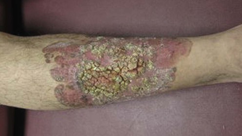
Squamous cell carcinoma (SCC) is a malignant neoplasm arising from epithelial keratinocytes of the skin and mucous membranes that generally appears as an erythematous, keratotic papule or nodule that may become ulcerated. There may be a history of tenderness as the lesion enlarges and becomes nodular. Although it is most commonly associated with chronic sun exposure in light-skinned individuals, SCC can arise secondary to scarring processes (burns, chronic ulcers, hidradenitis suppurativa), chemical carcinogens (arsenic, tobacco tar, hydrocarbons), human papillomavirus (types 16, 18, 30, 33, 35), ionizing radiation exposure (X-rays, γ-rays, radium), and in those with a genetic predisposition (xeroderma pigmentosum). Immunosuppression due to disease or drug therapy is an instigating factor.
The management of SCC depends on the histopathologic classification and clinical setting. The risk of local recurrence and of metastasis is correlated with poor histologic differentiation, perineural invasion, tumor size (≥2 cm), tumor depth (≥4 mm), tumor location (lip, ear, temple, genitalia), treatment modality, history of recurrence, host immunosuppression, rate of growth, presence of neurological symptoms such as pain, paresthesias, or paralysis, and precipitating factors other than UV light (radiation, site of inflammatory process). Regional lymphadenopathy is a poor prognostic sign.
Accurate histopathologic diagnosis is the most critical investigation. The differential diagnosis may include verruca vulgaris, actinic keratosis, SCC in situ (Bowen disease), keratoacanthoma (KA), irritated seborrheic keratosis, and superficial basal cell carcinoma (BCC) (these conditions are described in separate chapters). Specimens for histopathologic diagnosis should include epidermis and deep reticular dermis. A deep curette or scoop shave biopsy is generally sufficient, but punch biopsy to subcutaneous fat, or an incisional or excisional biopsy, is useful if there is concern regarding obtaining adequate tissue.
Regional lymph node palpation is performed when an aggressive tumor is suspected, based on the criteria specified above. In the absence of palpable lymphadenopathy, additional radiologic and surgical studies are performed if deep invasion (bone, cartilage, parotid) or perineural spread is suspected.
Small superficial SCC may be effectively treated using a destructive modality. Cryosurgery destroys tumor if it is frozen to −40°C to −70°C. Cryosurgery has the advantage of being fast, simple, and inexpensive, although a serious consequence is recurrence that may become extensive due to concealment in a previous scar. Curettage and electrodesiccation involves a sequence of three curette scrapings and electrodesiccations and can be used for well-differentiated primary SCC and SCC in situ <1 cm in diameter. A margin of 3–4 mm around the tumor should be treated with cryosurgery and with curettage and desiccation. Adequately treated lesions often require several weeks to heal and may leave hypopigmented, atrophic, or hypertrophic scars. Cosmetically sensitive areas, concave surfaces, and skin prone to keloid formation or poor wound healing should be avoided. Cure rates are highly technique dependent.
Standard surgical excision, utilizing 3–4 mm margins beyond the clinically apparent tumor, is a reasonable option for a well-demarcated SCC, particularly of the trunk and extremities. Higher-risk SCC (size ≥2 cm, poor histologic differentiation, invasion to fat, high risk areas) requires at least 6 mm margins. Recent studies have suggested that light curettage of the lesion prior to excision, traditionally used to better define tumor margins, may not be efficacious. Extensive tumors of the limbs involving bone may require amputation.
The gold standard for treatment of SCC is Mohs micrographic surgery, a technique in which the surgeon also acts as pathologist. The tumor is extirpated in a series of thin layers that are precisely oriented, horizontally sectioned, and immediately processed for evaluation. The process is continued until all margins are clear to maximize tumor eradication and tissue conservation. Indications for Mohs micrographic surgery include tumor size, location (cosmetically or functionally sensitive, high recurrence rate), indistinct margins, a history of recurrence, aggressive histopathology, and young patient age. Adjuvant radiation therapy following Mohs micrographic surgery should be considered for facial SCC ≥2 cm in diameter and those with perineural spread.
Although the use of radiation therapy as a primary modality has decreased, it remains a good treatment option for selected patients, particularly those of advanced age or poor surgical risk, with uncomplicated tumors of the head and neck. A total of 4000–7000 cGy is given sequentially over several weeks. One standard schedule is administration of 500 cGy three to five times per week over 2 to 6 weeks. Complications include hypopigmentation, telangiectasia, loss of adnexae, radiation dermatitis, and late (10–20 years) tumor recurrence.
For patients with SCC not amenable to surgical extirpation or radiation due to advanced disease stage or large number of lesions, topical therapy with imiquimod or 5-fluorouracil (5-FU), intralesional therapy with bleomycin, 5-FU or interferon (IFN), or systemic therapy with retinoids or IFN may be effective. Photodynamic therapy (PDT), most commonly using topical porphyrins followed by selective irradiation with visible light, is another alternative. Systemic regimens, including monoclonal antibodies and protein kinase inhibitors, are also being studied for inoperable tumors. Recent studies focusing on combining more than one therapeutic modality have shown potential for increased cure rates while minimizing adverse reactions and maximizing cosmetic results.
Histopathology
Physical examination for lymphadenopathy
Radiologic examination (MRI, CT scan, ultrasound)
Sentinel lymph node biopsy
Madan V, Dawn G. J Am Acad Dermatol 2007; 56: S103–4.
Standard curette biopsy may produce fragmented tissue and hence be suboptimal for diagnosis of SCC.
Jagdeo J, Weinstock MA, Piepkorn M, Bingham S. J Am Acad Dermatol 2007; 57: 279–84.
A prospective blinded study of 3926 specimens assigned a diagnosis by local pathologists and one or both (355 slides) central dermatopathologists. Intraobserver agreement was highest for BCC and lowest in the categories of invasive SCC, SCC in situ, and actinic keratosis, suggesting that the diagnostic boundaries of these diagnoses are imprecisely defined.
Jackson JE, Kelly B, Petitt M, Uchida T, Wagner RF Jr. J Am Acad Dermatol 2012; 67: 122–7.
Retrospective study of 235 diagnostic non-melanomatous skin cancer biopsies and their corresponding excisions for margin status and the presence of residual tumor, respectively. Twelve of 148 SCCs (8.1%) had negative biopsy margins and were associated with excisional specimens free of residual tumor. The SCCs with negative biopsy margins were predominantly non-facial, superficial tumors of the well-differentiated and KA subtype. The results suggest that negative-margin diagnostic biopsies may be therapeutic for well-differentiated or KA subtypes of SCC.
Ch’ng S, Low I, Ng D, Brasch H, Sullivan M, Davis P, et al. Hum Pathol 2008; 39: 344–9.
Epidermal growth factor (EGF) receptor protein over-expression was detected in 36% of primary SCCs without metastases, 79% of primary SCCs with subsequent metastases, and 47% of SCCs with metastatic nodal disease. EGF receptor over-expression was concluded to be an independent prognostic factor for subsequent metastases and could have important prognostic and therapeutic implications for high-risk tumors.
Su F, Viros A, Milagre C, Trunzer K, Bollag G, Spleiss O, et al. N Engl J Med 2012; 366: 207–15.
Molecular analysis to identify oncogenic mutations (HRAS, KRAS, NRAS, CDKN2A, and TP53) in patients treated with the BRAF inhibitor vemurafenib was performed. Among 21 tumor samples, 13 had RAS mutations (12 in HRAS). In a validation set of 14 samples, eight had RAS mutations (four in HRAS). Thus, 60% (21 of 35) of the specimens harbored RAS mutations. Mutations in RAS, particularly HRAS, are frequent in SCC that develops in patients treated with vemurafenib.
Marinez JC, Cook JL. Dermatol Surg 2007; 33: 410–20.
A literature review suggested that patients with cutaneous SCC who underwent elective node dissection had no proven survival benefit over those who are initially staged as node negative and undergo therapeutic neck dissection after the development of apparent regional disease.
Renzi C, Caggiati A, Manooranperampil TJ, Passarelli F, Tartaglione G, Pennasilico G, et al. Eur J Surg Oncol 2007; 33: 364–9.
In a consecutive series of 22 clinically node-negative high-risk cutaneous SCC patients who underwent sentinel lymph node biopsy (SLNB), 4.5% (one patient) had histologically positive nodes and developed a recurrence during follow-up. SLNB-negative patients had no metastases at a median follow-up of 17 months. Literature review revealed an incidence of positive sentinel nodes ranging from 12.5% to 44.4%. Analysis combining the results of this series and 61 patients from previous reports found in the literature supported the concept of SLNB for high-risk cutaneous SCC.
Park JJ, Emmerling O, Westhofen M. Acta Otolaryngol 2012; 132: 218–24.
A retrospective analysis of 140 patients with primarily surgically treated head and neck SCC. Each patient received neck ultrasound during follow-up visits. Recurrence rate, survival rate, and outcome of salvage therapy were determined. Recurrences occurred in 35.0% of all studied cases. Local, regional, and distant recurrences were found in 11.4%, 7.9%, and 15.7%.
CT is the preferred imaging technique of choice during oncologic follow-up, but is limited in terms of frequency of applications because of radiation exposure. Compared with CT, neck ultrasound is an easily feasible and cost-effective examination that can be performed during any routine visit in an outpatient clinic setting.
Almeida Goncalves JC. G Ital Dermatol Venereol 2011; 146: 249–55.
Forty-eight patients (31 women and 17 men) with 50 advanced SCCs of the extremities were treated with liquid nitrogen spray (two freeze–thaw cycles) reaching a temperature around −50°C inside the tumor. The overall cure rate was 88%, with follow-up between 1 and 8 years (mean of 2.64 years). The authors concluded that cryosurgical method is an effective treatment for advanced cancers of the extremities, yielding a high cure rate and, in many cases, preventing amputation.
Reschly MJ, Shenefelt PD. J Drugs Dermatol 2010; 9: 773–6.
Two retrospective studies investigating the efficacy of curettage and electrodesiccation in the treatment of low-risk SCC. One was a small controlled study in a Veterans Administration teaching hospital dermatology clinic that compared cure rates of low-risk SCCs at 1 year by curettage and electrodesiccation to those of surgical excision. A second study examined the cure rate of low-risk SCCs treated by curettage and electrodesiccation alone in a private practice. The first study found no significant difference in cure rates between curettage and electrodesiccation (14 of 14 cases successfully treated) and excision (15 of 16 successfully treated and one recurrence). The second study found the curettage and electrodesiccation cure rate (106 of 106 successfully treated) to be significantly greater than an arbitrary cure rate of 95%.
De Graaf YG, Basdew VR, van Zwan-Kralt N, Willemze R, Bavinck JN. Br J Dermatol 2006; 154: 493–7.
Retrospective review of 211 histologically confirmed SCCs in 48 organ transplant recipients treated by curettage and electrodesiccation. The authors concluded organ transplant recipients with many low-risk SCCs, curettage and electrodesiccation is a safe therapy with acceptable cure rates.
Peikert JM. Int J Dermatol 2011; 50: 1135–8.
Sixty-nine patients with 100 non-facial tumors, ≤2 cm in diameter, consisting of superficial BCC, superficial nodular BCC with papillary dermal invasion, SCC in situ, and SCC with papillary dermal invasion were prospectively treated with curettage and cryotherapy, and subsequently evaluated at 1- and 5-year intervals. No tumor recurred after 1 year of follow-up, and one recurrence occurred within the 5-year interval, for a 99% recurrence-free end-point.
Brodland DG, Zitelli JA. J Am Acad Dermatol 1992; 27: 241–8.
Based on a prospective study of subclinical microscopic tumor extension, it was recommended that minimal margins of 4 mm be taken around clinical borders of SCC. Margins of at least 6 mm were proposed for tumors ≥2 cm, histologic grade ≥2, with invasion of subcutaneous tissue, and/or location in high-risk areas.
Huang CC, Boyce SM. Semin Cutan Med Surg 2004; 23: 167–73.
Review article discussing the optimal use of standard surgical excision for the treatment of non-melanomatous skin cancers, in particular an excellent discussion on the relationship of electrodesiccation and curettage to excision, patient characteristics that are optimal for excision, excisional technique, recommended margins, implications of positive margins on excision, and the potential for metastasis.
Leibovitch I, Huilgol S, Selva D, Hill D, Richards S, Paver R. J Am Acad Dermatol 2005; 53: 253–60.
In a prospective multicenter case series of 1263 patients with cutaneous SCC treated with Mohs micrographic surgery, recurrent tumors were larger than the primary tumors, had larger post-excision defects, required more Mohs stages, and had more cases of subclinical extension. Recurrence rate was 2.6% for primary SCC and 5.9% for previously recurrent SCC.
Leibovitch I, Huilgol SC, Selva D, Hill D, Richards S, Paver R. J Am Acad Dermatol 2005; 53: 2612–16.
Of 1177 SCC patients treated with Mohs micrographic surgery in a prospective multicenter case series, 70 were found to have perineural invasion. Perineural invasion was more common in men, in previously recurrent tumors, and in moderately and poorly differentiated histologic subtypes. Perineural invasion was associated with larger tumor size, postoperative defect size, subclinical extension, and mean number of Mohs stages.
Chren MM, Torres JS, Stuart SE, Bertenthal D, Labrador RJ, Boscardin WJ. Arch Dermatol 2011; 147: 540–6.
This was a prospective, long-term study (median 6.6 years after treatment) to determine tumor recurrence rates after treatment. Consecutive samples were taken from all 495 patients with 616 primary non-melanoma skin cancers diagnosed in 1999 and 2000 and treated with curettage and electrodesiccation, excision, or Mohs surgery. A total of 127 tumors were treated with curettage and electrodesiccation (20.9%), 309 with excision (50.8%), and 172 with Mohs surgery (28.3%). Over the course of the study, 21 tumors recurred (3.5%): two (two BCC) after curettage and electrodesiccation (1.6%), 13 (nine BCC, four SCC) after excision (4.2%), and six (four BCC, two SCC) after Mohs surgery (3.5%). Recurrent tumors were detected earliest after curette and electrodesiccation (1.5 years) and latest after Mohs surgery (6.0 years). Median time to detection of recurrence after excision was 3.8 years. The recurrence rate after curettage and electrodesiccation was lower than expected, and the recurrence rate after Mohs surgery was higher than expected.
Hernandez-Machin B, Borrego L, Gil-Garcia M, Hernandez BH. Int J Dermatol 2007; 46: 453–9.
A retrospective study of 604 BCCs and 106 SCCs irradiated between 1971 and 1996 revealed cure rates for SCC of 92.7% at 5 years and 78.6% at 15 years, suggesting that radiation therapy is an effective first option in many cases.
Alam M, Nanda S, Mittal BB, Kim NA, Yoo S. J Am Acad Dermatol 2011; 65: 377–88.
This article reviews the indications, efficacy, and adverse effects of brachytherapy in the treatment of non-melanomatous skin cancers.
Peris K, Micantonio T, Fargnoli MC, Lozzi GP, Chimenti S. J Am Acad Dermatol 2006; 55: 324–7.
This open-label clinical trial used imiquimod 5% cream five times a week for up to 16 weeks to treat seven invasive SCC lesions on 10 patients. Five sites (71.4%) showed complete clinicopathologic regression and two (28.6%) partial regression at 16 weeks. There were no recurrences at a mean of 31 months follow-up.
Love WE, Bernhard JD, Bordeaux JS. Arch Dermatol 2009; 145: 1431–8.
A systematic review to determine clearance rates and adverse effects of topical imiquimod or FU therapy in the treatment of non-melanoma skin cancers. Clearance rates varied by drug regimen, and most of the studies lacked long-term follow-up. Imiquimod produced clearance rates of 73% to 88% for SCC in situ and 71% for invasive SCC. FU use produced 27–85% clearance for SCC in situ. Up to 100% and 97% of patients applying imiquimod and FU, respectively, experienced at least one adverse event such as erythema, pruritus, and pain.
Rodríguez-Cuevas S, Barroso-Bravo S, Almanza-Estrada J, Cristóbal-Martínez L, González-Rodríguez E. Arch Med Res 2001; 32: 273–6.
Fifteen patients with 38 skin lesions participated in a phase II prospective clinical trial, using intralesional bleomycin plus electric pulses delivered 10 minutes after bleomycin injection, which lasted 100 ms each at field strength of 1300 V/cm and a frequency of 1 Hz. There were BCC (n=9), in-transit metastasis of melanoma (n=2; total 13 nodules), SCC of the upper aerodigestive tract metastatic to the skin (n=2; total two nodules), and skin metastases from breast cancer (n=2; total 14 nodules). Overall objective responses were 98%, with complete responses achieved in 49%, partial responses in 49% (100% of SCCs), and no responses in 2%. The authors concluded that electrochemotherapy treatment represents an alternative to standard surgery or radiotherapy, with an outpatient-based treatment applied in one to three sessions.
Kim KH, Yavel RM, Gross VL, Brody N. Dermatol Surg 2004; 30: 116–20.
Case series of eight cases of seven patients (six men and one woman, 64 to 86 years of age) with biopsy-proven SCC or BCC. Results of the treatment of seven patients with a total of eight skin cancers (five BCC and three SCC). The patients were treated with a series of 1.0–2.5 million units IFN-α2b intralesional injections administered over a 3- to 5-week period, for a total dosage of 9.0–30 million units. The mean follow-up period after treatment was 33 months. There were no recurrences of any of the tumors based on clinical assessment; post-treatment skin biopsies were not performed.
Annest NM, VanBeek MJ, Arpey CJ, Whitaker DC. J Am Acad Dermatol 2007; 56: 989–93.
Thirty-eight cases of KA treated with intralesional methotrexate (MTX) were identified from the author’s institution (n=18) and literature search (n=20). Intralesional MTX achieved resolution in 92%, requiring an average of 2.1 injections an average of 18 days apart. Adverse events were rare, with two reports of pancytopenia in patients with chronic renal failure. The authors concluded that intralesional MTX is a useful non-surgical therapy for the treatment of KA, that histologic diagnosis before initiation of treatment is preferred, and a complete blood cell count at baseline and during treatment should be considered.
Kirby JS, Miller CJ. J Am Acad Dermatol 2010; 63: 689–702.
Excellent review article suggesting reasonable treatment guidelines for the treatment of SCC, KA, and BCC with the most widely available intralesional agents (methotrexate, 5-FU, bleomycin, and IFN).
Good LM, Miller MD, High WA. J Am Acad Dermatol 2011; 64: 413–22.
A good review on intralesional agents and their role in cutaneous malignancies.
Kirsner RS, Spencer J, Falanga V, Garland LE, Kerdel FA. Dermatol Surg 1996; 22: 1015–18.
Patients with extensive local or metastatic SCC of the leg involving bone may require amputation if Mohs micrographic surgery would render the remaining limb unstable.
Fai D, Arpaia N, Romano I, Vestita M, Cassano N, Vena GA. G Ital Dermatol Venereol 2009; 144: 281–5.
Topical PDT with MAL is widely used for the management of actinic keratosis and non-melanoma skin cancers. The authors report the results of a retrospective chart review showing the cumulative 4-year experience with MAL-PDT in a hospital outpatient setting. Case series included a total of 462 patients: 210 patients with actinic keratosis, 228 subjects with 348 BCCs, 17 patients with Bowen disease, and seven with SCC. Bowen’s disease is very responsive to MAL-PDT, unlike microinvasive or invasive SCC.
Marmur ES, Schmults CD, Goldberg DJ. Dermatol Surg 2004; 30: 264–71.
Lebwohl M, Tannis C, Carrasco D. J Dermatol Treat 2003; 14: 3–6.
A 40-year-old man with a history of severe psoriasis began developing numerous cutaneous SCCs after treatment with psoralen and ultraviolet A (PUVA) for 4.5 years. Because of a flare in his psoriasis he was started on 25 mg daily of acitretin, which resulted in a dramatic reduction in the number of SCCs. This report reviews the mechanism by which retinoids are thought to act and describes how oral retinoids may be an important therapy in patients at risk for developing multiple cutaneous malignancies.
Nijsten TEC, Stern RS. J Am Acad Dermatol 2003; 49: 644–50.
To assess whether oral retinoids reduce the risk of developing cutaneous SCC a nested cohort of 135 patients participating in the PUVA follow-up study with at least 1 year of substantial retinoid use was identified. Each patient’s tumor incidence during retinoid therapy was compared to the incidence of SCC development in periods of no use. Overall, a 30% reduction in SCC incidence was noted when patients were on oral retinoid therapy.
Retinoids (isotretinoin, acitretin) may play a role in the treatment of patients with SCC not amenable to surgical extirpation owing to advanced stage of disease or large number of lesions. Several studies have shown an overall response rate of about 70%.
Marquez C, Bair SM, Smithberger E, Cherpelis BS, Glass LF. J Drugs Dermatol 2010; 9: 753–8.
A good review of the literature. Systemic retinoids may play a role in chemoprevention of new and recurrent malignancies in susceptible individuals, such as organ transplant recipients and those with xeroderma pigmentosum.
Maubec E, Petrow P, Scheer-Senyarich I, Duvillard P, Lacroix L Gelly L, et al. J Clin Oncol 2011; 29(25): 3419–26.
A clinical trial to evaluate the safety and efficacy of cetuximab, a monoclonal antibody that binds to human epidermal growth factor receptor, in patients with unresectable SCC of the skin. Thirty-six patients received cetuximab (initial dose of 400 mg/m2 followed by subsequent weekly doses of 250 mg/m2) for at least 6 weeks with a 48-week follow-up. Disease control response at 6 weeks was obtained in 25 of 36 patients (69%) of the intention-to-treat population. The best responses were eight partial responses and two complete responses.
Kondapalli L, Soltani K, Lacouture ME. J Am Acad Dermatol 2005; 53: 291–302.
Protein kinases mediate most signal transduction pathways in malignant cells to increase proliferation, invasion, and metastasis. This review outlines the rationale for the use of protein kinase inhibitors on cutaneous malignancies, including SCC.
Fujisawa T, Umchayashi Y, Ichikawa E, Kawachi Y, Otsuka F. J Am Acad Dermatol 2006; 55: S81–5.
Two case studies of locally invasive SCCs metastatic to regional lymph nodes were treated with intravenous cisplatin and 5-FU concurrent with either conventional fractionated radiation therapy or external beam radiation. Although the primary tumors of both patients initially underwent complete regression, one patient required resection of a local recurrence at 5 years. The other developed lung metastases and died of disseminated disease at 18 months.
Treatment of Skin Disease Comprehensive Therapeutic Strategies 4e
WhatsApp us