Schistosomiasis (Schistosoma)

Published on 27/03/2015 by admin

Filed under Pediatrics

Last modified 22/04/2025

Print this page

rate 1 star rate 2 star rate 3 star rate 4 star rate 5 star
Your rating: none, Average: 0 (0 votes)

This article have been viewed 2782 times

Chapter 292 Schistosomiasis (Schistosoma)

The term schistosomiasis encompasses the acute and chronic inflammatory disorders caused by human infection with Schistosoma spp. parasites. Disease is related to both the systemic and focal effects of schistosome infection and its consequent host immune responses, which often result in disabling morbidity for the affected patient.

Etiology

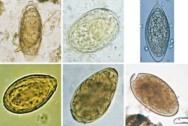
Schistosoma organisms are the trematodes, or flukes, that parasitize the bloodstream. Five schistosome species infect humans: Schistosoma haematobium, S. mansoni, S. japonicum, S. intercalatum, and S. mekongi. Humans are infected through contact with water contaminated with cercariae, the free-living infective stage of the parasite. These motile, forked-tail organisms emerge from infected snails and are capable of penetrating intact human skin. As they reach maturity, adult worms migrate to specific anatomic sites characteristic of each schistosome species: S. haematobium adults are found in the perivesical and periureteral venous plexus, S. mansoni in the inferior mesenteric veins, and S. japonicum in the superior mesenteric veins. S. intercalatum and S. mekongi are usually found in the mesenteric vessels. Adult schistosome worms (1-2 cm long) are clearly adapted for an intravascular existence. The female accompanies the male in a groove formed by the lateral edges of its body. On fertilization, female worms begin oviposition in the small venous tributaries. The eggs of the 3 main schistosome species have characteristic morphologic features: S. haematobium has a terminal spine, S. mansoni has a lateral spine, and S. japonicum has a smaller size with a short, curved spine (Fig. 292-1). Parasite eggs provoke significant granulomatous inflammatory response, which allows them to ulcerate through host tissues to reach the lumen of the urinary tract or intestines. They are carried to the outside environment in urine or feces, where they will hatch if deposited in freshwater. Motile miracidia emerge, infect specific freshwater snail intermediate hosts, and divide asexually. After 4-12 wk, the infective cercariae are released by the snails into the contaminated water.

Epidemiology

Schistosomiasis infects >207 million people worldwide, primarily children and young adults. Prevalence is increasing in many areas as population density increases and new irrigation projects provide broader habitats for vector snails. Humans are the definitive host for the 5 clinically important species of schistosomes, although S. japonicum may infect some animals such as dogs, rats, pigs, and cattle. S. haematobium is prevalent in Africa and the Middle East; S. mansoni is prevalent in Africa, the Middle East, the Caribbean, and South America; and S. japonicum is prevalent in China, the Philippines, and Indonesia, with some sporadic foci in parts of Southeast Asia. The other 2 species are less prevalent. S. intercalatum is found in West and Central Africa, and S. mekongi is found only along the upper Mekong River in the Far East.

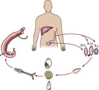
Transmission depends on disposal of excreta, the presence of specific intermediate snail hosts, and the patterns of water contact and social habits of the population (Fig. 292-2). The distribution of infection in endemic areas shows that prevalence increases with age to a peak at 10-20 yr of age. Measuring intensity of infection (by quantitative egg count in urine or feces) demonstrates that the heaviest worm loads are found in the younger age groups. Therefore, schistosomiasis is most prevalent and most severe in children and young adults, who are at maximal risk for suffering from its acute and chronic sequelae.

Pathogenesis

Both the early and late manifestations of schistosomiasis are immunologically mediated. Acute schistosomiasis, known as snail fever or Katayama syndrome, is a febrile illness that represents an immune complex disease associated with early infection and oviposition. The major pathology of infection occurs later, with chronic schistosomiasis, in which retention of eggs in the host tissues is associated with chronic granulomatous injury. Eggs may be trapped at sites of deposition (urinary bladder, ureters, intestine) or be carried by the bloodstream to other organs, most commonly the liver and less often the lungs and central nervous system. The host response to these eggs involves local as well as systemic manifestations. The cell-mediated immune response leads to granulomas composed of lymphocytes, macrophages, and eosinophils that surround the trapped eggs and add significantly to the degree of tissue destruction. Granuloma formation in the bladder wall and at the ureterovesical junction results in the major disease manifestations of schistosomiasis haematobia: hematuria, dysuria, and obstructive uropathy. Intestinal as well as hepatic granulomas underlie the pathologic sequelae of the other schistosome infections: ulcerations and fibrosis of intestinal wall, hepatosplenomegaly, and portal hypertension due to presinusoidal obstruction of blood flow. In terms of systemic disease, antischistosome inflammation increases circulating levels of proinflammatory cytokines such as tumor necrosis factor (TNF)-α and interleukin (IL)-6, associated with elevated levels of C-reactive protein. These responses are associated with hepcidin-mediated inhibition of iron uptake and utilization, leading to anemia of chronic inflammation. Schistosomiasis-related undernutrition may be the result of similar pathways of chronic inflammation. Acquired partial protective immunity against schistosomiasis has been demonstrated in some animal species and may occur in humans.

Clinical Manifestations

Most chronically infected individuals experience mild symptoms and may not seek medical attention; the more severe symptoms of schistosomiasis occur mainly in those who are heavily infected or who have been infected over longer periods of time. In addition to organ-specific morbidities, infected patients may demonstrate anemia, chronic pain, diarrhea, exercise intolerance, and undernutrition. Cercarial penetration of human skin may result in a papular pruritic rash known as schistosomal dermatitis or swimmer’s itch. It is more pronounced in previously exposed individuals and is characterized by edema and massive cellular infiltrates in the dermis and epidermis. Acute schistosomiasis, Katayama syndrome, may occur, particularly in heavily infected individuals 4-8 wk after exposure; this is a serum sickness–like syndrome manifested by the acute onset of fever, chills, sweating, lymphadenopathy, hepatosplenomegaly, and eosinophilia. Acute schistosomiasis most commonly presents in 1st time visitors to endemic areas who experience primary infection at an older age.

Symptomatic children with chronic schistosomiasis haematobia usually complain of frequency, dysuria, and hematuria. Urine examination shows erythrocytes, parasite eggs, and occasional eosinophiluria. In endemic areas, moderate to severe pathologic lesions have been demonstrated in the urinary tract of >20% of infected children. The extent of disease correlates with the intensity of infection, but significant morbidity can occur even in lightly infected children. The advanced stages of schistosomiasis haematobia are associated with chronic renal failure, secondary infections, and cancer of the bladder.

Children with chronic schistosomiasis mansoni, japonica, intercalatum, or mekongi may have intestinal symptoms; colicky abdominal pain and bloody diarrhea are the most common. However, the intestinal phase may remain subclinical, and the late syndrome of hepatosplenomegaly, portal hypertension, ascites, and hematemesis may then be the 1st clinical presentation. Liver disease is due to granuloma formation and subsequent fibrosis; no appreciable liver cell injury occurs, and hepatic function may be preserved for a long time. Schistosome eggs may escape into the lungs, causing pulmonary hypertension and cor pulmonale. S. japonicum worms may migrate to the brain vasculature and produce localized lesions that cause seizures. Transverse myelitis rarely has been reported in children or young adults with either acute or chronic S. haematobium or S. mansoni infection.

Bibliography

Carod-Artal FJ. Neurological complications of Schistosoma infection. Trans R Soc Trop Med Hyg. 2008;102:107-116.

Centers for Disease Control and Prevention. Schistosomiasis (website). www.cdc.gov/ncidod/dpd/parasites/schistosomiasis/default.htm. Accessed September 7, 2010

Coutinho HM, Acosta LP, Wu HW, et al. Th2 cytokines are associated with persistent hepatic fibrosis in human Schistosoma japonicum infection. J Infect Dis. 2007;195:288-295.

Dawson-Hahn EE, Greenberg SLM, Domachowske JB, et al. Eosinophilia and the seroprevalence of schistosomiasis and strongyloidiasis on newly arrived pediatric refugees: an examination of Centers for Disease Control and Prevention screening guideline. J Pediatr. 2010;156:1016-1018.

Dent A, King CH. Schistosomiasis section in Epocrates Online Reference Program. https://online.epocrates.com/home. posted September 29, 2008

Doenhoff MJ, Cioli D, Utzinger J. Praziquantel: mechanisms of action, resistance and new derivatives for schistosomiasis. Curr Opin Infect Dis. 2008;21:659-667.

2007 Drugs for parasitic infections. Treat Guidel Med Lett. 2007;5(suppl):e1-e15.

King CH, Dickman K, Tisch DJ. Reassessment of the cost of chronic helminth infection: meta-analysis of disability-related outcomes in endemic schistosomiasis. Lancet. 2005;365:1561-1569.

Leenstra T, Coutinho HM, Acosta LP, et al. Schistosoma japonicum reinfection after praziquantel treatment causes anemia associated with inflammation. Infect Immun. 2006;74:6398-6407.

Ross AG, Vickers D, Olds GR, et al. Katayama syndrome. Lancet Infect Dis. 2007;7:218-224.

Wang L, Utzinger J, Zhou XN. Schistosomiasis control: experiences and lessons from China. Lancet. 2008;372:1793-1794.

Wang LD, Chen HG, Guo JG, et al. A strategy to control transmission of Schistosoma japonicum in China. N Engl J Med. 2009;360:121-128.