Published on 18/03/2015 by admin
Filed under Dermatology
Last modified 22/04/2025
This article have been viewed 2305 times
Preston W. Chadwick and Warren R. Heymann
Evidence Levels: A Double-blind study B Clinical trial ≥ 20 subjects C Clinical trial < 20 subjects D Series ≥ 5 subjects E Anecdotal case reports
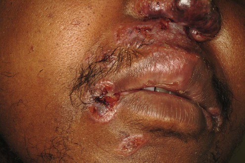
Sarcoidosis is a multisystem disease of unknown etiology characterized histologically by non-caseating granulomas. It is considered to be immune mediated, with a Th1-predominant cytokine profile. Skin manifestations are observed in approximately 25% of cases. Sarcoidosis has been reported to develop following exposure to inorganic particles in the environment. The use of polymerase chain reaction (PCR) techniques has also led to the identification of mycobacterial and propionibacterial DNA and RNA in sarcoidal tissue. Sarcoidosis may be the end result of immune responses to those or other specific triggers. Therapy for erythema nodosum associated with sarcoidosis is addressed in the chapter on erythema nodosum.
The treatment of cutaneous sarcoidosis depends on the type and extent of lesions present and is directed at suppressing the formation of granulomas. Guidelines for treating extracutaneous involvement can be found elsewhere, but it should be recognized that therapy for internal involvement may take precedence over skin disease and that response to treatment may be variable, depending on the type of tissue involved.
In small papular or extremely localized sarcoidosis, treatment with potent topical corticosteroids or intralesional triamcinolone acetonide (3.3–10 mg/mL) is reasonable. If this is ineffective or involvement is more diffuse, oral chloroquine (up to 3.5 mg/kg/day) or hydroxychloroquine (up to 6.5 mg/kg/day) may be effective in addition to methotrexate or tetracyclines. If no response is seen, or disfiguring lesions are present, oral prednisone can be used at a dose of 1 mg/kg daily (maximum 60 mg) for up to 3 months, and then tapered if improvement or a stable level is reached, to a maintenance dose of 5–10 mg on alternate days for several months. Periodic escalations in dose are necessary with flares of the disease.
Methotrexate may be used as a corticosteroid-sparing agent or as monotherapy in those patients with lupus pernio, ulcerative sarcoidosis, or severe disease that has not responded to prednisone. Initial doses of 15–20 mg weekly are favored. Mycophenolate mofetil has also shown promise as a steroid-sparing agent. Thalidomide, azathioprine, chlorambucil, isotretinoin, or allopurinol could be considered if methotrexate or mycophenolate mofetil fail in this subset of patients. Azathioprine and chlorambucil are better studied in patients with pulmonary sarcoidosis. Thalidomide seems to be more effective than isotretinoin and allopurinol in cutaneous disease. Reported failures are described with etretinate and allopurinol.
Biologic agents that inhibit tumor necrosis factor (TNF)-α are therapeutic modalities that should be considered in patients with lupus pernio, cutaneous sarcoidosis recalcitrant to systemic steroids or steroid sparing agents. Infliximab may be more effective than adalimumab, but may be associated with a higher rate of infection and autoimmune disease. Etanercept does not appear to be useful for the treatment of sarcoidosis; indeed some studies have reported on its potential role in patients developing sarcoidosis, or in relapse of pre-existing sarcoidosis.
Leflunomide and apremilast have shown promise in the treatment of cutaneous sarcoidosis unresponsive to other therapies, but further studies are needed to establish long-term usefulness.
Localized disease that does not respond to topical or intralesional corticosteroids, or in those cases in which the use of systemic antimalarial drugs or corticosteroids is undesirable, may represent a niche for other modalities, such as excision, laser, photodynamic therapy, PUVA, surgery (lupus pernio) or intralesional chloroquine.
Special stains, cultures, and polarization of biopsy specimens
Electrolytes, blood urea nitrogen, creatinine, serum calcium, liver function tests, complete blood count, 24-hour urine calcium, and serum angiotensin-converting enzyme
Ophthalmologic evaluation, including slit-lamp examination
Chest radiograph, pulmonary function tests
Electrocardiogram
FDG PET (18-fluoro-deoxyglucose positron-emission tomography) is useful in identifying sites for diagnostic biopsy (for patients without apparent lung involvement)
MRI with gadolinium (neurological sarcoidosis may occur without any other internal organ involvement)
DEXA (dual X-ray absorptiometry) bone density scans in patients on long-term oral corticosteroids
Iannuzzi MC, Rybicki BA, Teirstein AS. N Engl J Med 2007; 357: 2153–65.
A thorough review of the literature that discusses advances in the diagnosis and treatment of sarcoidosis and the challenges that clinicians may encounter.
Haimovic BA, Sanchez M, Judson MA, Prytowsky S. J Am Acad Dermatol 2012; 66: 699.e1–18.
The authors present an extensive review of cutaneous sarcoid including epidemiology, pathogenesis including the Th1 pathway, morphologies and treatment. The discussion includes a proposed treatment algorithm.
Corradin MT, Forcione M, Fiorentino R, Bordignon M, Alaibac M, Belloni-Fortina A. Eur J Dermatol 2010; 20: 659–60.
A 16-year-old African boy presented with violaceous, verrucous nodules on his forehead, right pectoral region, right arm and left elbow. The patient was treated with topical mometasone furoate 0.1% cream daily with clearance of lesions in 3 weeks.
Potent topical corticosteroid use is reported to be of some value in anecdotal reports. Most authors believe that intralesional corticosteroids are more effective, yet this too is anecdotal. Small lesions are optimal candidates; however, lupus pernio has also been reported to respond.
Kim YJ, Kim YD. Korean J Ophthalmol 2006; 20: 238–40.
A 29-year-old man presented with a cutaneous sarcoid on his right eyelid. The patient had no other findings of systemic sarcoidosis. A total of 0.6 mL of intralesional triamicinolone 40 mg/mL was injected at 1 cm intervals along the eyelid. Dramatic improvement of the lesion had occurred when he returned for his 1-month follow-up: he received an additional 1 mL of triamcinolone at the same site, which resolved the lesion completely.
Anecdotal reports suggest small, papular sarcoid responds best. The strength of triamcinolone acetonide varies from 2–40 mg/mL, and frequency varies from weekly to monthly.
Baughman RP, Lower EE. Clin Dermatol 2007; 25: 334–40.
Whereas the recommended starting dose of prednisone for pulmonary sarcoidosis is suggested to be 20–40 mg daily, the dose and duration of treatment for cutaneous sarcoidosis has not been established. The authors suggest using this as a benchmark and tapering the steroid dose after 1 or 2 months to one that controls the disease while avoiding toxicity. The authors report that the need for long-term systemic corticosteroids for treatment of chronic sarcoidosis occurs in about a quarter of patients.
Another suggested regimen for prednisone in cutaneous sarcoidosis is 30 mg orally on alternate days until the granulomas fade. The dose is then tapered over several months to 15 mg orally on alternate days. Other protocols report a good response with prednisone 30–40 mg orally daily, with a gradual taper to 10–20 mg orally on alternate days for 1 year, or prednisone 1 mg/kg orally daily (maximum 60 mg) for 8 to 12 weeks, with a taper to 0.25 mg/kg daily continued for 6 months.
With long-term treatment, prevention of corticosteroid-induced osteoporosis with bisphosphonates and Pneumocystis pneumonia prophylaxis with trimethoprim–sulfamethoxazole should be considered.
Zic JA, Horowitz DH, Arzubiaga C, King LE. Arch Dermatol 1991; 127: 1034–40.
A review of the efficacy and safety of chloroquine in the treatment of cutaneous sarcoidosis. The article cites four studies (three open clinical trials and one case series) that support the use of chloroquine. The authors recommend an initial dose of 250 mg twice daily for 14 days, then 250 mg daily for long-term suppression, though most studies have used 500 mg daily for several months. Relapses after discontinuation of treatment are frequent.
Jones EJ, Callen JP. J Am Acad Dermatol 1990; 23: 487–9.
Seventeen patients were treated with 200 mg or 400 mg daily of hydroxychloroquine. Cutaneous lesions regressed in 12 patients within 4 to 12 weeks, and three patients had a partial response. Of the 12 patients with the best response, six had a recurrence after a dosage reduction or discontinuation.
Hydroxychloroquine may have a better safety profile than chloroquine; however, chloroquine has been better studied in sarcoidosis. Although retinal toxicity is rare, in order to prevent this complication doses of chloroquine should not exceed 3.5 mg/kg/day and hydroxychloroquine should not exceed 6.5 mg/kg/day.
Baughman RP, Winget DB, Lower EE. Sarcoidosis Vasc Diffuse Lung Dis 2000; 17: 60–6.
Twenty-four patients with new-onset symptomatic pulmonary disease were randomized to receive either methotrexate or placebo, in addition to their prednisone. Although only 15 patients received at least 6 months of therapy, the methotrexate group required less prednisone than the placebo group. There was no difference in toxicity between the methotrexate and the placebo groups.
A favorable response is usually expected after several months of therapy, and the dose can be tapered weekly after 4 to 6 months. Some studies indicate that methotrexate may be particularly useful for those with ulcerative sarcoidosis.
Lower EE, Baughman RP. Arch Intern Med 1995; 155: 846–51.
Fifty patients were treated with methotrexate, 10 mg weekly, for a minimum of 2 years. Most patients did not have cutaneous involvement, but in those who did a good response was seen. In many patients methotrexate was used in conjunction with prednisone, with favorable results.
Kouba DJ, Mimouni D, Rencic A, Nousari HC. Br J Dermatol 2003; 148: 147–8.
A 53 year-old African American with extensive mucocutaneous disease and pulmonary involvement was recalcitrant to standard first-line treatments. He was given oral treatments with prednisone 60 mg tapered to 5 mg every other day over 5 months, hydroxychloroquine 6 mg/kg daily and mycophenolate mofetil 45 mg/kg daily for 12 months. Significant improvement was noted at 6 weeks and remained at 18 months follow-up with significant reduction of systemic disease.
Stagaki E, Mountford WK, Lackland DT, Judson, MA. Chest 2009; 135: 468–76.
This retrospective study of 54 patients with cutaneous sarcoid undergoing 116 treatment regimens showed infliximab-containing regimens to have a high likelihood of complete or near complete resolution compared to those containing corticosteroids, methotrexate, and hydroxychloroquine. Additionally, when systemic steroids were combined with a second agent there was a significant reduction in prednisone dose requirement.
Infliximab has become a second-line agent, particularly in cases of lupus pernio and neurocutaneous lupus. Studies suggest that infliximab can be used as an induction agent with long-term methotrexate. Superiority to etanercept has been demonstrated.
Recently, a double-blind randomized trial for treatment of ocular sarcoidosis with etanercept showed no comparative improvement in the sarcoidal lesions. Also, sarcoidosis was reported to develop in a patient with ankylosing spondylitis after being treated with etanercept. Therefore, etanercept does not appear to be useful for the treatment of sarcoidosis.
Doty JD, Mazur JE, Judson MA. Chest 2005; 127: 1064–71.
Ten patients with sarcoidosis recalcitrant to previous therapies were treated with infliximab. Infliximab is administered via intravenous infusion in doses ranging from 3 to 10 mg/kg/dose at 0, 2, 6, and every 8 to 19 weeks subsequently. Nine of 10 patients reported subjective improvement of their lesions, and all 10 were found to have objective measures of improvement. All five patients with lupus pernio experienced significant improvement or clearance of the lesions.
Infliximab, although effective, has been shown to increase the risk of tuberculosis reactivation, of other granulomatous infections, lymphomas, and autoimmune disease. Therapy is expensive, costing up to thousands of dollars per infusion. One must be aware of the side effects and cost.
Bachelez H, Senet P, Cadranel J, Kaoukhov A, Dubertret L. Arch Dermatol 2001; 137: 69–73.
Twelve patients with cutaneous sarcoidosis, three of whom had systemic involvement, were treated with minocycline at a daily dose of 200 mg orally for a median duration of 12 months. Ten patients showed a response, eight complete and two partial; one patient’s symptoms remained stable and one patient’s disease progressed. Four patients experienced relapse after discontinuation of minocycline; doxycycline was then utilized, resulting in remission.
Due to the safety profile and moderate success, tetracyclines can be considered a second-line therapy in limited cutaneous disease.
Nguyen YT, Dupuy A, Cordoliani F, Vignon-Pennamen MD, Lebbé C, Morel P, et al. J Am Acad Dermatol 2004; 50: 235–41.
A retrospective evaluation of 12 patients with cutaneous sarcoidosis, two with systemic involvement, 10 of whom were treated successfully with a treatment duration of 2 to more than 16 months, with a daily dose of thalidomide ranging from 50 to 200 mg orally daily. Two patients received combined therapy with oral corticosteroids (dose ranging from 7.5 to 30 mg daily), one patient used potent topical corticosteroids, and one received combined therapy with methotrexate (dose 25 mg weekly). The average response time was 2 to 3 months. The main adverse effect noted in this series was deep venous thrombosis in one patient.
Antony F, Layton AM. Br J Dermatol 2000; 142: 1052–3.
A 38-year-old Afro-Caribbean man with painful sarcoidal nodules around the ends of his fingers (but no evidence of sarcoidal arthritis) had a partial response to oral and intralesional steroids with subsequent recurrence of his lesions. He had no improvement with hydroxychloroquine, methotrexate, or azathioprine. He was started on allopurinol 100 mg twice daily, which was increased to 300 mg daily after 3 weeks. This resulted in sustained objective clinical improvement.
A few case reports demonstrate resolution of truncal and extremity plaques of sarcoidosis after 12 weeks of allopurinol.
Georgiou S, Monastirli A, Pasmatzi E, Tsambaos D. Acta Derm Venereol 1998; 78: 457–9.
A 31-year-old woman with nodules and plaques on the trunk and extremities showed a complete remission with 8 months of isotretinoin at 1 mg/kg daily; 15-month follow-up revealed continuing remission.
Two other cases report improvement with a 30-week course (0.67–1.34 mg/kg daily) and a 6-month course (0.4–1 mg/kg daily), respectively, of isotretinoin.
Hof DG, Hof PC, Godfrey WA. Am J Respir Crit Care Med 1996; 153: 870A.
Of the 21 patients in this study, eight had ‘multisystem’ involvement and one had skin-only involvement. All patients with extrapulmonary disease achieved a complete remission with azathioprine and a tapering dose of prednisone.
Israel HL, McComb BL. Sarcoidosis 1991; 8: 35–41.
In this study, 31 patients received chlorambucil because complicating diseases prevented them from receiving corticosteroids, or they did not respond to them. Marked improvement was noted in 15 patients, moderate improvement in 13 patients, but relapses were very common upon discontinuation. No immune suppression-related side effects were noted.
Yesudian PD, Azurdia RM. Clin Exp Dermatol 2004; 29: 552–4.
A 50-year-old woman with disfiguring scar sarcoidosis of the lips responded poorly to steroids and was unable to tolerate hydroxychloroquine; she was started on mepacrine 100 mg daily with significant remission to practically normal lip margins. She has had no flares in 10 months of follow-up visits.
The yellow discoloration of the skin and sclera observed with quinacrine (one-third of patients) makes chloroquine and hydroxychloroquine better alternatives.
Nowack U, Gambichler T, Hanefeld C, Kastner U, Altmeyer P. BMC Dermatol 2002; 24: 215.
Report of three patients with cutaneous sarcoidosis treated with fumaric acid esters for 4 to 12 months, resulting in complete clearance of cutaneous lesions.
Vano-Galvan S, Fernandez-Guarino M, Carmona LP, Harto A, Carrillo R, Jaen P. Eur J Dermatol 2008; 18: 89–90.
A 33-year-old female with lichenoid type cutaneous sarcoidosis was treated with 0.1% tacrolimus ointment twice daily. Near complete resolution was noted at 2 months and the patient remained free of disease at 4 months.
Liedtka JE. Int J Dermatol 1996; 35: 682–3.
Multiple injections of intralesional chloroquine hydrochloride (50 mg/mL) were effective in treating five lesions in a single patient, with minimal side effects.
Goldin JH, Jawad SMA, Reis AP. J Laryngol Otol 1983; 97: 1053–6.
Split-thickness and full-thickness skin grafts, dermabrasion, and primary closure have been attempted, with mixed results. Surgery has been used in ulcerative and non-ulcerative sarcoidosis.
Several recent reports utilizing primary closure, a paramedian forehead flap and a full thickness graft continue to show mixed long-term results.
Young HS, Chalmers RJ, Griffiths CE, August PJ. J Cosmet Laser Ther 2002; 4: 87–90.
CO2 laser resurfacing was used in two patients with lupus pernio, with a favorable cosmetic result.
Flashlamp pulsed dye laser and Q-switched ruby laser were beneficial in one case report of lupus pernio. However, exacerbations of lupus pernio, namely generalized ulceration in treated and untreated lesions, have been reported following this therapy.
Holzmann RD, Astner S, Forschner T, Sterry G. Dermatol Surg 2008; 34: 393–6.
A 10-year-old boy with a 1.0 cm lesion of varicella zoster-induced scar sarcoidosis on the left cheek had not responded to systemic antibiotics or systemic steroids. Clinical remission occurred after three pulsed dye laser treatments at 6-week intervals which consisted of two to four pulses using a 595 nm wavelength and a 0.5 ms pulse duration. At 12 months’ follow-up there was no evidence of recurrence, although the varicella scar became more visible once the sarcoidosis disappeared.
Roos S, Raulin C, Ockenfels H, Karsai S. Dermatol Surg 2009; 35: 1139–40.
A 63-year-old Caucasian female with nodular sarcoid on her back was treated with flashlamp pumped pulsed dye laser at 6 J/cm2 (585 nm, 0.5 ms, 12 mm) with complete resolution in 4 weeks. Prednisone was utilized for iridocyclitis after the laser treatments were completed. She remained clear 13 months after discontinuation of systemic steroids.
Pignone AM, Rosso AD, Fiori G, Matucci-Cerinic M, Becucci A, Tempestini A, et al. J Pineal Res 2006; 41: 95–100.
Melatonin was given to 18 chronic sarcoid patients for 2 years at a dose of 20 mg/day during the first year and 10 mg/day during the second year. Skin lesions, present in three patients, completely disappeared after 24 months of treatment. No side effects were reported.
Melatonin may increase drowsiness, and has been associated with hypothermia, hypotension, and bradycardia.
Baughman RP, Judson MA, Ingledue R, Craft NL, Lower EE. Arch Dermatol 2012; 148: 262–4.
A study of 15 patients on systemic steroids treated with 20 mg twice daily (once daily if not tolerated) apremilast (a novel phosphodiesterase type 4 inhibitor). A significant reduction in SASI (Sarcoidosis Area and Severity Index) induration score was observed at 4 and 12 weeks. Relapse was noted in three patients at the 3-month follow-up. Pentoxifylline has been reported as an effective treatment for pulmonary sarcoidosis; however, efficacy in cutaneous sarcoidosis needs further investigation.
Baughman RP, Lower EE. Sarcoidosis Vasc Diffuse Lung Dis 2004; 21: 43–8.
In a case series study, 32 patients with sarcoidosis involving the eye, lung, and/or skin were treated with leflunomide. Remission (complete and partial) was seen in 12 of 17 patients treated with leflunomide, and 13 of 15 patients treated with leflunomide and methotrexate. An initial dose of leflunomide 100 mg/day for 3 days was followed by 20 mg daily after that.
Adverse events included gastrointestinal problems and hypersensitivity reactions, including erythema multiforme and Stevens–Johnson syndrome, have been reported.
Heffernan MP, Smith DI. Arch Dermatol 2006; 142: 17–19.
A 46-year-old black woman with cutaneous sarcoidosis in the form of papules on the nose and ulcerating nodules on the legs had failed local therapy with topical clobetasol and intralesional triamcinolone, hydroxycholoquine, and pentoxifylline. She did not tolerate minocycline. Adalimumab was administered at a dose of 40 mg subcutaneously once weekly. After 10 weeks the nodules on her legs were completely healed, and the lesions on her nose were significantly improved.
Another case report highlights the successful use of adalimumab in a 55-year-old woman with ulcerative cutaneous sarcoidosis.
Mahnke N, Medve-Koenigs K, Berneburg M, Ruzicka T, Neumann NJ. J Am Acad Dermatol 2004; 50: 978–9.
An 82-year-old woman with cutaneous sarcoidosis affecting 80% of her body surface was treated with medium-dose UVA1 radiation four times weekly. She received 20 J/cm2 for the first three treatment sessions, 40 J/cm2 for the following 12 sessions, and then 60 J/cm2 for the next 35 sessions. After 50 sessions, nearly all lesions had resolved.
Gleeson CM, Morar N, Staveley I, Bunker CB. Br J Dermatol 2011; 164: 892–4.
A series of six patients with recalcitrant cutaneous sarcoidosis were treated with topical gel psoralen and UVA twice weekly at 0.2 J/cm2. Patients were placed on oral prednisolone; five patients were tapered at the start of therapy and one remained on long-term oral prednisolone. In follow-up at 4 months to three years, 3 patients had complete resolution and three patients had greater than 50% improvement.
Penrose C, Mercer SE, Shim-Chang H. J Am Acad Dermatol 2011; 65: e12–14.
A 52-year-old African-American female with topical and systemic steroid-resistant cutaneous sarcoidosis (likely lupus pernio) had significant improvement after 10 sessions of aminolevulinic acidadnphotodynamic therapy at 2-week intervals.
This is the sixth reported patient to be treated with photodynamic therapy and the first with Fitzpatrick skin type V.
Reddy R, Vitiello m, Kerdel F. Int J Dermatol 2011; 50: 1132–4.
A 67-year-old Hispanic male with biopsy proven cutaneous sarcoidosis who failed treatment with topical and intralesional steroids, topical tacrolimus, and oral cyclosporine showed improvement after starting oral prednisone and 100 mg dapsone daily.
Sarcoidosis is a great masquerader of other dermatoses. This case affirms the need for both diagnostic suspicion and combination therapy for patients with the disease.
Treatment of Skin Disease Comprehensive Therapeutic Strategies 4e
WhatsApp us


 Topical corticosteroids
 Intralesional corticosteroids
 Oral corticosteroids
 Chloroquine
 Hydroxychloroquine
 Methotrexate
 Mycophenolate mofetil
 Infliximab
 Minocycline, doxycycline
 Thalidomide
 Allopurinol
 Isotretinoin
 Azathioprine
 Chlorambucil
 Quinacrine
 Topical tacrolimus
 Intralesional chloroquine
 Excision
 Laser
Laser Melatonin
 Pentoxifylline
 Apremilast
 Leflunomide
 Adalimumab
 Medium-dose UVA1
 PUVA
PUVA Photodynamic therapy
 Dapsone