[level-membership-for-dermatology-category]
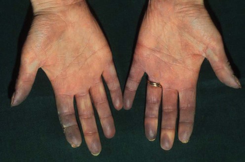
Raynaud disease and phenomenon
Sameh S. Zaghloul, Najat A.Y. Marraiki and Mark J.D. Goodfield
Specific investigations


First-line therapies
 Calcium channel blockers
 Glyceryl trinitrate
 Prostacyclin analogs
Second-line therapies
 Selective serotonin reuptake inhibitors (fluoxetine)
 Endothelin receptor antagonist (bosentan)
 Angiotensin II receptor type I antagonist (losartan)
 Serotonin antagonists (ketanserin)
 Phosphodiesterase inhibitors
 Oral vasodilators
 Hexopal
Third-line therapies
 Prostaglandin E1 (alprostadil)
 Dipyridamole and low-dose acetylsalicylic acid
 Calcitonin gene-related peptide
 L-Arginine
 H-O-U therapy
 Triiodothyronine
 Helicobacter pylori treatment
 Sympathectomy
 Low-level laser therapy
 Acupuncture
 Evening primrose oil supplementation
 Fish oil supplementation
 Biofeedback
 Botulinum toxin
 Spinal cord stimulation
[/level-membership-for-dermatology-category][not-level-membership-for-dermatology-category]
Raynaud disease and phenomenon
Sameh S. Zaghloul, Najat A.Y. Marraiki and Mark J.D. Goodfield
Specific investigations
First-line therapies
Buy Membership for Dermatology Category to continue reading. Learn more here
[/not-level-membership-for-dermatology-category]
