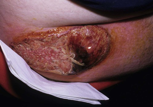
Pyoderma gangrenosum


First-line therapies
 Topical tacrolimus
 Topical corticoids
 Dapsone
 Intralesional corticoids
 Minocycline
 Nicotine
 Topical pimecrolimus
 Sodium cromoglycate
 Sulfasalazine
Second-line therapies
 Cyclosporine
 Systemic corticoids
Third-line therapies
 Infliximab
 Other TNFα-antagonists
 Alefacept
 Alkylating agents (cyclophosphamide, chlorambucil)
 Plasmapheresis (plasma exchange)
 Leukocytapheresis
 IVIG
IVIG Intralesional cyclosporine
 Tacrolimus (FK506)
 Azathioprine or mercaptopurine
 Colchicine
 Thalidomide
 Potassium iodide
 Topical nitrogen mustard (mechlorethamine)
 Mycophenolate mofetil
 GM-CSF
 Methotrexate
 Topical platelet-derived growth factor
 Recombinant human epidermal growth factor
 Clofazimine
 Hyperbaric oxygen
 Isotretinoin
 Anakinra
 Ustekinumab
 Imiquimod
 Visilizumab
 Topical phenytoin
 Surgical repair by graft or flap
Targeted treatment of pyoderma gangrenosum in PAPA (pyogenic arthritis, pyoderma gangrenosum and acne) syndrome with the recombinant human interleukin-1 receptor antagonist anakinra.
Brenner M, Ruzicka T, Plewig G, Thomas P, Herzer P. Br J Dermatol 2009; 161: 1199–201.
A single case of PG associated with an autosomal dominant genodermatosis.
A case of PG associated with inflammatory bowel disease has been reported not to respond.
