Published on 18/03/2015 by admin
Filed under Dermatology
Last modified 22/04/2025
This article have been viewed 4633 times
Cynthia O. Anyanwu, Preston W. Chadwick and Warren R. Heymann
Evidence Levels: A Double-blind study B Clinical trial ≥ 20 subjects C Clinical trial < 20 subjects D Series ≥ 5 subjects E Anecdotal case reports
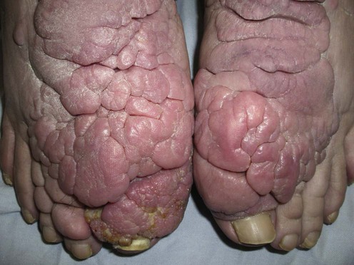
Pretibial myxedema, more accurately termed ‘thyroid dermopathy,’ is characterized by non-pitting edema and skin-colored to violaceous nodules or plaques. These are most commonly distributed pretibially, but can sometimes be seen over the arms, shoulders, head, and neck.
Pretibial myxedema is an autoimmune phenomenon which tends to occur following treatment of patients with Graves disease. The condition can, however, develop in hypothyroid and euthyroid patients. It is helpful to look for other clinical signs of thyroid disease, including thyroid acropachy and the presence of a goiter. Pretibial myxedema typically follows the onset of ophthalmopathy, often years after the diagnosis of hyperthyroidism. Goals of treatment include cosmesis and the prevention of long-term side effects such as elephantiasis, decreased range of motion, or foot drop from neural entrapment. Resolution may occur without treatment.
Patients with significant thyroid dermopathy should be started on a trial of high-potency topical corticosteroids, alone or under occlusion, for at least 2 months. If symptoms persist, intralesional corticosteroids may be effective. A combination of the above in conjunction with compression bandages can be beneficial when monotherapy proves inadequate. Both oral and intravenous corticosteroids have also been shown to improve lesions in several patients. However, their use is limited by systemic side effects.
Pentoxifylline, an analog of methylxanthine theobromine, has been shown to reduce the extent of lesions and can also be used in conjunction with topical and/or intralesional corticosteroids. Although there are conflicting data, the use of intravenous immuneglobulin (IVIG) may improve lesions of pretibial myxedema. Subcutaneous or intralesional octreotide, a somatostatin analog, yields conflicting results. Plasmapheresis has been reported to be beneficial in improving severe cases and has been successful when used in combination with rituximab.
Temporary improvement with cytotoxic agents has been observed. Pretibial myxedema is not a life-threatening condition, and so the use of such agents should be limited to severe, debilitating cases. Surgical excision has been shown to be effective in a minority of cases. The high risk of recurrence makes surgical intervention an infrequently used modality; however, post-operative intralesional steroids can minimize this risk. Complete decongestive physiotherapy has shown some success in treating the elephantiasic form of pretibial myxedema.
Pretibial ultrasonography, with or without digital infrared thermal imaging, to measure skin thickness may be useful in assessing treatment response. Measuring serum hyaluronic acid levels to follow therapeutic response may also be of value.
Thyroid function tests
Antithyroglobulin and antithyroid peroxidase antibodies
Anti-thyroid-stimulating hormone receptor antibodies
Pretibial ultrasound and/or digital infrared thermal imaging
Serum hyaluronic acid
Fatourechi V. In: Heymann WR (ed), Thyroid Disorders with Cutaneous Manifestations. London: Springer Verlag, 2008; 103–19.
Georgala S, Katoulis AC, Georgala C, Katoulis EC, Hatziolou E, Stavrianeas NG. J Eur Acad Dermatol Venereol 2002; 16: 380–3.
A 28-year-old Greek woman presented initially with asymptomatic pretibial myxedema, which ultimately led to a diagnosis of Graves disease. This patient had elevated anti-TSH-receptor antibodies.
An assessment of thyroid function is warranted because most patients with pretibial myxedema have clinical or laboratory evidence of autoimmune thyroid disease.
Shih SR, Lin MS, Li HY, Yang HY, Hsiao YL, Chang MT, et al. Eur J Endocrinol 2011; 164: 605–11.
Digital infrared thermal imaging (DITI) detects surface temperature, and sonography reflects composition changes in soft tissue. Lower leg temperatures of normal volunteers decreased gradually from proximal to distal parts. In all patients with pretibial myxedema, DITI showed abnormally low focal temperatures over the lesions. In Graves disease patients with mild diffuse non-pitting edema and Graves disease patients with normal appearance of lower legs, DITI showed abnormally low focal temperature in 90.9% and 65.2% of the patients respectively. Areas of clinically visible pretibial myxedema and low focal temperature detected by DITI were sonographically characterized with increased skin thickness, hypoechoic substance deposition in the cutaneous tissue, and blurred boundary lines between dermis and subcutaneous tissue.
The use of digital infrared thermal imaging and high-resolution ultrasonography to analyze pretibial skin of patients with Graves disease allows for early detection of pretibial myxedema in patients with and without visible dermopathy.
Fatourechi V, Pajouhi M, Fransway AF. Medicine 1994; 73: 1–7.
Treatment with topical 0.05–0.1% triamcinolone acetonide cream under occlusion for 2 to 10 weeks led to partial remission in 29 of 76 patients in this retrospective study; 1% had complete remission.
Takasu N, Higa H, Kinjou Y. Intern Med 2010; 49: 665–9.
The authors detail their experience of six patients with pretibial myxedema treated with triamcinolone 0.1% under occlusion. Only two patients responded to therapy; these patients had their treatments initiated within months of the appearance of pretibial myxedema.
A delay in treatment of 5 to 10 years or advanced Graves disease manifest by exophthalmos may be associated with refractory disease when treated with topical steroids under occlusion.
Lang PG, Sisson JC, Lynch PJ. Arch Dermatol 1975; 111: 197–202.
Seven of nine patients treated with monthly injections of 8 mL or less of intralesional triamcinolone acetonide solution (5 mg/mL, 1 mL per injection site) had complete remission of pretibial myxedema after a total of three to seven visits. The other two patients, despite withdrawing from the study prematurely for non-medical reasons, showed a partial improvement.
Frisch DR, Roth I. J Am Podiatr Med Assoc 1985; 75: 147–52.
A 29-year-old woman with pretibial myxedema was treated with rest, elevation, and topical 0.05% fluocinonide cream under occlusion. Outpatient therapy included weekly intralesional Celestone Soluspan injections followed by topical 0.05% fluocinonide cream under occlusion. Compression dressings with an Unna boot were applied weekly. After 2 months the lesions were greatly improved.
Compression stockings may also be beneficial. Unless contraindicated, compression should be used in conjunction with any therapeutic approach for this disorder.
Br J Dermatol 2012; 166: 457–9.
A patient with pretibial myxedema presenting as verruciform plaques failed initial treatment with monthly intralesional injections of 1 mL betamethasone, 0.2 mL at each of five points, but responded when the injections were increased to every 2 weeks. A second patient with tumorous lesions responded to surgical removal of tumors with post-operative intralesional triamcinolone acetonide 10 mg at five points every 3 days as prophylaxis to prevent recurrence.
Chang CC, Chang TC, Kao SC, Kuo YF, Chien LF. Acta Endocrinol 1993; 129: 322–7.
Pentoxifylline caused an in vitro dose-dependent decrease in fibroblast proliferation and glycosaminoglycan synthesis in fibroblast cultures taken from pretibial sites. A preliminary trial with a dose of 400 mg intravenously and 800 mg orally daily of pentoxifylline reduced the size of pretibial myxedema lesions within 1 week.
Engin B, Gumusel M, Ozdemir M, Cakir M. Dermatol Online J 2007; 13: 16.
A 32-year-old man achieved partial remission with clobetasol under occlusion combined with pentoxifylline 400 mg three times daily and intralesional triamcinolone (5 mg/mL).
Antonelli A, Navarranne A, Palla R, Alberti B, Saracino A, Mestre C, et al. Thyroid 1994; 4: 399–408.
Improvement of pretibial myxedema began after a few weeks in six patients treated with 400 mg/kg daily of high-dose IVIG given over 3 to 4 hours on five consecutive days. The cycle was repeated three times every 21 days. Maintenance therapy of 400 mg/kg for 1 day was then administered for seven to 15 more cycles every 21 days. Total treatment ranged from 7 to 12 months, with maximum response occurring after an average of 6 months.
Terheydem P, Kahaly GJ, Zillikens D, Brocker EB. Clin Exp Dermatol 2003; 28: 224–6.
IVIG did not significantly improve the lesions of a patient with elephantiasic pretibial myxedema.
Benoit FL, Greenspan FS. Ann Intern Med 1967; 66: 711–20.
Oral prednisolone, begun at 60 mg then tapered, and methylprednisolone starting at 40 mg cleared the pretibial lesions of four patients and improved the lesions of two others. Of the various corticosteroid treatments studied, the best results were obtained with high-dose systemic corticosteroids for 2 weeks.
Shinohara M, Hamasaki Y, Katayana I. Br J Dermatol 2000; 143: 1083–6.
Intralesional octreotide 200 µg daily improved the lesions of pretibial myxedema in a male patient with Graves disease after 4 weeks of therapy.
Octreotide inhibits insulin-like growth factor-1-induced hyaluronic acid secretion by lesional fibroblasts, which may play a role in the pathogenesis of pretibial myxedema.
Chang TC, Kao SC, Huang KM. Br Med J 1992; 304: 158.
Three patients with pretibial myxedema were successfully treated with 100 µg of octreotide three times daily.
The authors do not comment on the route of administration of the octreotide acetate. According to the Physicians Desk Reference, the drug may be administered either by subcutaneous injection or intravenously.
Rotman-Pikielny P, Brucker-Davis F, Turner ML, Sarlis NJ, Skarulis MC. Thyroid 2003; 13: 465–70.
Three women did not show a statistically significant benefit from octreotide 300 µg.
The conflicting results from these small studies regarding the use of octreotide for thyroid dermopathy mandates that only larger, controlled studies will verify whether it is a useful modality for this condition.
Kuzuya N, DeGroot LJ. J Endocrinol Invest 1982; 5: 373–8.
Two patients, one with elephantiasis-like lesions, were treated with 16 exchanges over 4 to 5 months with 1–2 L of the patient’s plasma removed and replaced with 1300 mL of purified protein fraction and 700 mL 0.9% saline. Immunoglobulin G fraction was separated out, thereby reducing total thyrotropin-binding inhibitory immunoglobulin (TBII) activity per unit of serum. The pretibial myxedema was partially and temporarily improved with plasmapheresis, and abnormal antibodies were reduced.
Noppen M, Velkeniers B, Steenssens L, Vanhaelst L. Acta Clin Belg 1988; 43: 381–3.
A patient with pretibial myxedema unresponsive to topical corticosteroids was cured after 5 days of plasmapheresis followed by 100 mg of azathioprine twice daily for 3 months. Azathioprine was tapered to 50 mg twice daily and continued for a year, at which time no recurrence was noted.
Hanke CW, Bergfeld WF, Guirguis MN, Lewis LJ. Cleve Clin Q 1983; 50: 183–8.
Fibroblasts from pretibial myxedema sites of a 44-year-old man showed reduced DNA content in vitro with the use of cytotoxic agents. Melphalan, which reduced hyaluronic acid levels to the greatest extent, was given orally (8 mg daily) for 4 days, and repeated monthly for 6 months. This regimen provided transient improvement, but the patient’s condition then worsened.
Felton J, Derrick EK, Price ML. Br J Dermatol 2003; 148: 825–6.
A 56-year-old man with pretibial myxedema was treated with surgical shave removal followed by daily subcutaneous octreotide injections for 6 months. His lesions did not recur over a 9-year follow-up period.
Matsuoka LY, Wortsman J, Dietrich JG, Pearson R. Arch Dermatol 1981; 117: 250–1.
A 47-year-old woman with hypothyroidism had recurrence of pretibial myxedema in a split-thickness skin graft 3 years after placement.
Heyes C, Nolan R, Leahy M, Gebauer K. Australas J Dermatol 2012; 53: e1–4.
Significant improvement in multi-treatment refractory elephantiasic thyroid dermopathy was achieved in a 55-year-old woman who underwent plasmapheresis once every 6 days and received rituximab infusions once per week for 1 to 7 weeks on seven occasions for a total of 29 doses of rituximab and 241 episodes of plasmapheresis over 3.5 years. She remained clear 6 months after completion of treatment.
Rituximab has been utilized successfully for an ever-expanding list of autoimmune disorders. Further study is warranted to determine if rituximab, without the concomitant use of plasmapheresis, would be of value for patients with thyroid dermopathy.
Susser WS, Heermans AG, Chapman MS, Baughman RD. J Am Acad Dermatol 2002; 46: 723–6.
A 67-year-old woman with elephantiasic pretibial myxedema had a 47% reduction of leg edema after 6 weeks of intensive complete decongestive physiotherapy. This response was sustained for 2 years after treatment.
Complete decongestive physiotherapy consists of manual massage of the lower extremities to promote lymphatic drainage, followed by compressive bandages, exercise, and skin care.
Treatment of Skin Disease Comprehensive Therapeutic Strategies 4e
WhatsApp us


 Topical corticosteroids with or without occlusion
 Intralesional corticosteroids
 Compression
 Pentoxifylline
 Pentoxifylline with topical and/or intralesional steroid
 Intravenous immunoglobulin
 Systemic corticosteroids (including pulse corticosteroid therapy)
 Octreotide
 Plasmapheresis
 Plasmapheresis with rituximab
 Cytotoxic therapy
 Surgery
 Complete decongestive physiotherapy