Published on 18/03/2015 by admin
Filed under Dermatology
Last modified 18/03/2015
This article have been viewed 1599 times
Alex Milligan and Graham A. Johnston
Evidence Levels: A Double-blind study B Clinical trial ≥ 20 subjects C Clinical trial < 20 subjects D Series ≥ 5 subjects E Anecdotal case reports
Pityriasis lichenoides et varioliformis acuta (PLEVA) is an eruption of small, erythematous papules which become vesicular and hemorrhagic. Some ulcerate and necrose, leaving pitted scars. The name refers to the morphology not the duration of the condition, because a significant proportion of cases regress with or without treatment, only to recur. Patients should be warned that relapse is common and that recurrent courses of therapy may be required. Febrile ulceronecrotic Mucha–Habermann disease is a rare and severe form of PLEVA characterized by an abrupt onset of an ulceronecrotic eruption associated with a high fever and systemic symptoms.
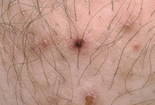
There are only a handful of controlled trials for this condition and large series are rare. In many therapeutic trials PLEVA is often grouped together with pityriasis lichenoides chronica and management strategies are therefore often similar or interchangeable.
Although a ‘wait and see’ approach is justifiable in infants, children should be given a 6-week course of high-dose erythromycin. Tetracycline should not be given because of its effects on dentition.
Second-line therapy in children, and possibly first-line in adults, is either UV light or psoralen plus UVA (PUVA) because the only comparative study has shown this to be more effective. Topical corticosteroids are only reported anecdotally in textbooks rather than in studies. They are used with antihistamines to reduce pruritus, but have no reported effect on disease course.
In more extensive or symptomatic disease low-dose methotrexate is useful, and systemic corticosteroids or cyclosporine have also been used.
Some authors have suggested that combination therapy (e.g., erythromycin and PUVA or methotrexate and PUVA) is effective, especially in the rare, febrile ulceronecrotic variant of Mucha–Habermann disease.
Consider skin biopsy
A diagnostic skin biopsy is unnecessary in clinically obvious cases, but may be useful to exclude lymphomatoid papulosis or before commencing aggressive systemic therapy.
An infective etiology for PLEVA is suggested by reports of clustering of cases, resolution following tonsillectomy, and occurrence in five members of a family. Case reports exist associating PLEVA with parvovirus, adenovirus in the urine, staphylococci from throat cultures, Epstein–Barr virus, toxoplasmosis, and HIV. No organism has been cultured from lesional skin and, unless there are clinical signs of infection, routine investigation for an infective agent does not appear to be useful.
Tomasini D, Tomasini CF, Cerri A, Sangalli G, Palmedo G, Hantschke M, et al. J Cutan Pathol 2004; 31: 531–8.
This study suggests that pityriasis lichenoides is mediated by a cytotoxic T-cell effector population.
The identification of parvovirus B19 DNA in nine cases may be interpreted ambiguously.
English JC III, Collins M, Bryant-Bruce C. Int J Dermatol 1995; 34: 642–4.
Biopsy-proven PLEVA in association with carriage of Gram-positive cocci cleared with ciprofloxacin in a 35-year-old woman. An identical eruption in her husband, who was found to have group A β-hemolytic streptococcus from a skin swab, cleared with erythromycin.
Rongioletti F, Delmonte S, Rebora A. Int J Dermatol 1999; 38: 367–76.
Biopsy-proven PLEVA in a patient with serology indicating acute toxoplasmosis failed to respond to azithromycin, but cleared with spiramycin followed by trimethoprim–sulfamethoxazole.
Treatment of Skin Disease Comprehensive Therapeutic Strategies 4e
WhatsApp us