[level-membership-for-dermatology-category]
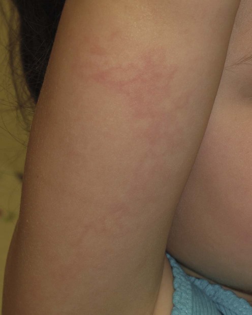
Parvovirus infection


 Reassurance
 Antipyretics (e.g., acetaminophen [paracetamol], ibuprofen)
Second-line therapies
 NSAIDs
 Systemic corticosteroids
 Blood transfusion
Third-line therapies
 Immunoglobulin infusion Immunoglobulin infusion |
C |
 Bone marrow/stem cell transplantation Bone marrow/stem cell transplantation |
D |
 Intrauterine transfusion Intrauterine transfusion |
C |
 HAART HAART |
E |
[/level-membership-for-dermatology-category][not-level-membership-for-dermatology-category]
Parvovirus infection
