Nephrogenic systemic fibrosis

Published on 18/03/2015 by admin

Filed under Dermatology

Last modified 22/04/2025

Print this page

rate 1 star rate 2 star rate 3 star rate 4 star rate 5 star
Your rating: none, Average: 0 (0 votes)

This article have been viewed 1555 times

Nephrogenic systemic fibrosis

Anjela Galan and Shawn E. Cowper

Evidence Levels:  A Double-blind study  B Clinical trial ≥ 20 subjects  C Clinical trial < 20 subjects  D Series ≥ 5 subjects  E Anecdotal case reports

image

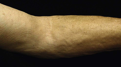
(From Nazarian, R.M., et al., 2011. Quantitative assessment of dermal cellularity in nephrogenic systemic fibrosis: a diagnostic aid. J Am Acad Dermatol 64(4), 741–7.)

Nephrogenic systemic fibrosis (NSF) affects patients with renal impairment who have been exposed to gadolinium-containing contrast agents (GCCA) for MRI studies. Onset is marked by cutaneous erythema, edema, hyperpigmentation, and woody induration. The clinical course commonly results in joint contractures and sometimes fibrosis of internal organs.

Management strategy

Prevention is paramount. Three GCCA (Magnevist, Omniscan, and Optimark) are now contraindicated in patients with renal dysfunction (acute kidney injury or chronic kidney disease [estimated GFR of <30 mL/minute/1.73 m2]). Patients should be screened prior to the use of GCCA to identify those at risk. Avoid using GCCA in patients with suspected or known impairment of drug elimination unless the need for the diagnostic information is essential and not available with non-contrasted MRI or other alternative imaging modalities. If exposure is unavoidable, optimal dosing and follow-up should involve the managing radiologist and nephrologist. Although not proven to prevent NSF, immediate hemodialysis following exposure to GCCA is recommended.

Specific investigations

There are no serologic tests for NSF. Diagnosis is based upon clinical and histopathological correlation in a patient with renal disease. Gadolinium identification is not required to establish a diagnosis.

First-line therapies

image Re-establishment of renal function  
 – Transplantation D
 – Dialysis D
image Physical therapy E
image Pain management E

Transplantation, particularly if performed soon after NSF onset, is often the best therapeutic recourse. Although the benefit is limited (approximately 50%), complete resolution may be achieved. Significant clinical improvement may not be observed for months or years following resumption of renal function, and in some cases may not occur at all; however, improvement of symptoms has been reported. Treatment with dialysis has mixed results. In one study half of patients on dialysis had clinical improvement, although complete resolution was not seen.

Rigorous physical therapy is mandatory to improve motion and maintain joint function.

Second-line therapies

imageExtracorporeal photopheresis E
imagePentoxifylline E
imageImatinib mesylate E
(prospective trials are pending)

Antifibrotic effect after low dose imatinib mesylate treatment in patients with nephrogenic systemic fibrosis: an open-label non-randomized, uncontrolled clinical trial.

Elmholdt TR, Buus NH, Ramsing M, Olesen AB. J Eur Acad Dermatol Venereol 2011. doi:10.1111/j.1468-3083.2011.04398.x [Epub ahead of print].

Anecdotal experience in two patients treated with 400 mg of imatinib daily showed significant improvement in symptoms at 15 weeks. Clinical effects on skin, but not on joint mobility, have been reported in three patients treated with low-dose imatinib mesylate. Additional rare case reports with similar findings are found in the literature. Two additional patients showed improvement in joint mobility and tethering that reversed upon discontinuation of therapy. The optimal period of treatment has not been determined.

Third-line therapies

imageSodium thiosulfate E
imageUVA1 E
imagePUVA with retinoids E
imagePhotodynamic therapy with methyl aminolevulinate E
imagePlasmapheresis E
imageIVIG E
imageCorticosteroids (topical, intralesional, and systemic) E
imageMethotrexate (systemic) E
imageAzathioprine E
imageCalcipotriene E
imageAlefacept E
imageRapamycin E