Monitoring integrity of the neuromuscular junction

Published on 07/02/2015 by admin

Filed under Anesthesiology

Last modified 22/04/2025

Print this page

rate 1 star rate 2 star rate 3 star rate 4 star rate 5 star
Your rating: none, Average: 0 (0 votes)

This article have been viewed 2243 times

Monitoring integrity of the neuromuscular junction

Jeffrey J. Lunn, MD

Neuromuscular transmissions

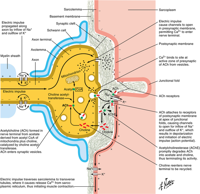
When a nerve impulse arrives at the neuromuscular junction, voltage-gated ion channels open, leading to an influx of calcium within the terminal that causes several hundreds of vesicles of acetylcholine to fuse with the nerve membrane (Figure 19-1). The acetylcholine within these vesicles is released into the synaptic cleft, combining with and activating nicotinic receptors on the motor endplate, the activation of which opens ion channels on the muscle membrane and depolarizes the membrane. The release of calcium from intracellular stores stimulates an interaction between actin and myosin, resulting in muscle contraction.

To facilitate tracheal intubation and many surgical procedures, neuromuscular blocking agents (NMBAs) are often administered to inhibit the nicotinic receptor. A multitude of factors affect patients’ responses to NMBAs, therefore making it essential to monitor the extent of blockade of the nicotinic receptor.

Neuromuscular monitoring

Generally, the degree of neuromuscular blockade induced by an NMBA is evaluated by the response induced by a supramaximal electrical stimulus delivered to a peripheral nerve with measurement of a mechanical response of the muscle to the stimulus. When a peripheral nerve is stimulated with a supramaximal stimulus, each muscle fiber enervated by that nerve responds in an all-or-none fashion, and the aggregate response of the whole muscle is dependent upon the number of individual fibers that respond. Muscle fibers with nicotinic receptors still inhibited by NMBAs do not respond to a stimulus.

Nerve stimulators

To test the degree of neuromuscular blockade—either to ensure muscle paralysis for tracheal intubation or a surgical procedure or to assess the adequacy of muscle strength at the end of an anesthetic—it is important to deliver a supramaximal stimulus, one that is at least 10% to 20% greater than the current necessary to produce a maximum response and 200% to 300% greater than the threshold stimulus. The ability to generate a supramaximal response requires that at least 50 to 80 mA of current be applied to the peripheral nerve. So that a constant current is applied throughout the entire impulse, the stimulus must be monophasic and delivered as a rectangular square wave with a duration of 0.2 to 0.3 ms. Pulses exceeding 0.5 ms may stimulate muscle directly or cause repetitive firing of nerves.

The resistance to current flow increases as skin temperature decreases; conversely, removing hair or degreasing the skin decreases resistance. Modern nerve stimulators deliver a constant current, despite the resistance, by varying voltage to ensure that the amount of current selected is equal to the amount of current delivered.

Nerve stimulators should be able to deliver several different types of stimuli, such as a single twitch, train of four (TOF), continuous (tetanic) stimulation, posttetanic stimulation, and double-burst stimulation (DBS). Nerve stimulators should also have polarity indicators, and most have a display indicating the amount of current applied.

Nerve stimulators have two cables, one with a positive and one with a negative polarity, that are attached to pregelled silver or silver chloride electrodes, each having a minimum of about an 8-mm diameter conducting area, that attach to the skin with a self-contained adhesive. Subcutaneous needle electrodes are rarely, if ever, used for routine neuromuscular monitoring.

Sites for monitoring

Stimulation of the ulnar nerve at the wrist—with measurement of the response in the adductor pollicis muscle—remains the most popular site for monitoring the degree of neuromuscular blockade. If the arm is not available, the facial nerve or the posterior tibial or the common peroneal nerve is stimulated, with monitoring of their respective muscle groups. The facial nerve can be stimulated with the electrodes placed on the mandible-maxilla; overlying the facial nerve. Placement of the electrodes behind and in front of the ear provides very good sensitivity and specificity, with little chance of direct nerve stimulation.

There are differences when these sites are stimulated because the muscles themselves have different sensitivity to NMBAs. The diaphragm itself is the most resistant muscle, requiring up to two times the dose of NMBAs required to block the adductor pollicis. The muscles of the face enervated by the facial nerve are less resistant to blockade than is the diaphragm but are more resistant than the adductor pollicis.

Modes of stimulation

Single twitch

Single twitches are supramaximal stimuli delivered at between 0.1 and 1 Hz. The response to the stimulus is measured by visual or tactile means and will remain static until approximately 75% of nicotinic receptors are blocked. A linear decrease occurs in response until no twitch can be observed, which occurs at about 95% of nicotinic receptor blockade.

Train of four

TOF comprises four supramaximal stimuli at 2 Hz that are delivered and may be repeated at a minimum of every 10 sec. Each stimulus can produce a motor response; the degree of receptor blockade is based upon the fade of the individual responses, the presence of subsequent responses, or both. All four responses are identical when no neuromuscular blockade is present. When the fourth stimulus produces no response (3-4), approximately 75% to 80% of receptors are blocked. When the third response is lost (2-4), approximately 85% of receptors are blocked. When the second response is lost (1-4), approximately 90% of receptors are blocked. If there are no responses to any stimuli (0-4), more than 95% of receptors are inhibited.

If there are four responses, the ratio of the strength of the fourth to the first twitch also can be used to assess the degree of neuromuscular function. At the end of a procedure, a T4/T1 of 0.9 is the gold standard, indicating that a sufficient degree of neuromuscular strength has returned so that clinicians can assure themselves that patients will be able to maintain adequate ventilation and can protect their airways. The advantage of a TOF is that no baseline control twitch is required. When measuring the ratio of T4/T1, an objective measure of the mechanical response is performed using a device that objectively quantifies the strength of the muscle contraction.

Though it is uncommon to use TOF monitoring in patients who have received a depolarizing NMBA, such as succinylcholine, for those patients who have prolonged weakness associated with depolarizing blockade, the TOF may be helpful. Normally, depolarizing blockade decreases the twitch height of all four responses equally. If fade occurs, and the first twitch is greater than subsequent twitches, a phase II block should be considered.

Techniques to monitor muscular response

As mentioned previously, response to stimulation is most frequently measured visually; however, tactile evaluation of the twitches is recommended. For research and for assessing degrees of neuromuscular blockade at either end of the spectrum, an objective measurement of response is desired.