Published on 19/03/2015 by admin
Filed under Dermatology
Last modified 22/04/2025
This article have been viewed 1644 times
E. Eugene Bain, III and Nathalie Zeitouni
Evidence Levels: A Double-blind study B Clinical trial ≥ 20 subjects C Clinical trial < 20 subjects D Series ≥ 5 subjects E Anecdotal case reports
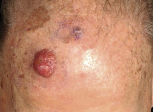
Merkel cell carcinoma (MCC) is a rare cutaneous neuroendocrine carcinoma with aggressive clinical behavior, initially described by Toker in 1972. MCC typically affects older individuals, with the median patient age being 69 years. Immunosuppressive conditions such as chronic lymphocytic leukemia, human immunodeficiency virus and solid organ transplantation have been linked to increased risk of developing MCC. In terms of the pathogenesis of MCC, there is evidence for both infectious and environmental factors. Support for viral promoted oncogenesis came with the discovery in 2008 of the Merkel cell polyomavirus. Ultraviolet (UV) radiation has also been described as a risk factor for the development of MCC, as most of these tumors develop at sites of sun damage with UV-associated cutaneous malignancies. The most common location is the head and neck region followed by the trunk.
Given the rarity of this malignancy, randomized controlled trials for therapies are lacking. As such, much of the data regarding the success of treatment modalities such as surgery, radiation and chemotherapy stems from case series or retrospective analyses of patient data. An algorithmic approach to treatment has been put forth by the National Comprehensive Cancer Network (NCCN).
Imaging is employed for initial work-up and staging (as clinically indicated) to detect regional or distant metastases. This may be done with either computed tomography (CT), magnetic resonance imaging (MRI) or FDG positron emission tomography-CT (PET-CT), though studies have not shown a convincing advantage for functional imaging modalities.
Surgical removal of the primary tumor is an important component of any treatment strategy, either with standard wide local excision or Mohs micrographic surgery. In addition to excision of the primary tumor, sentinel lymph node biopsy (SLNB) is often recommended, if feasible, given the tendency of the tumor to metastasize. Evidence of clinically positive lymphadenopathy at the time of presentation will often prompt lymphadenectomy.
Radiation may be used adjunctively for disease control, to the primary site as well as the draining lymph node basin. Not all studies have shown a statistically significant benefit with radiation therapy. Moreover, recurrences may respond less well to radiation therapy. In addition to traditional applications of radiation therapy, newer methods have been reported including surface-mold computer-optimized high-dose-rate brachytherapy. Chemotherapy is sometimes employed, though less evidence supports its use. Isolated limb perfusion or infusion has been used successfully for in-transit metastases.
Diagnostic imaging
Sentinal lymph node biopsy
Maury G, Dereure O, Du-Thanh A, Mariano-Goulart D, Guillot B. J Eur Acad Dermatol Venereol 2011; 25: 1420–7.
This retrospective study of 15 patients compared clinical staging with (18)FDG PET-CT as well as conventional CT. Investigators found that FDG PET-CT imaging did not change disease staging and/or management based on a combination of clinical examination, SLNB and conventional CT. Conventional CT and FDG PET-CT demonstrated identical sensitivity and specificity in this small patient cohort.
These results support data of other studies which have shown similar sensitivities when comparing functional and anatomic imaging modalities.
Fields RC, Busam KJ, Chou JF, Panageas KS, Pulitzer MP, Kraus DH, et al. Ann Surg Oncol 2011; 18: 2529–37.
SLNB was positive in 29% of patients with clinically negative nodes in this retrospective review of 153 patients. Primary tumor size and the presence of lymphovascular invasion were associated with sentinel lymph node positivity. The status of the sentinel lymph node was not associated with disease-specific survival or disease recurrence.
Gupta SG, Wang LC, Peñas PF, Gellenthin M, Lee SJ, Nghiem P. Arch Dermatol 2006; 142: 685–90.
This study presented a combination of a single institution retrospective review (30 patients) and literature based meta-analysis (92 patients) for total of 122 patients, and it found that SLNBs can detect clinically and radiographically occult disease. CT scans of lymph node basins have a low sensitivity (20%) for detecting both regional and distant metastases. Patients with positive SLNBs were more likely to receive adjuvant therapy and more likely to benefit from such therapy (improved relapse-free survival). The median follow-up period of this study of 12 to 15 months did not allow investigators to report disease-specific survival. Instead, only relapse-free survival data was reported.
O’Connor WJ, Roenigk RK, Brodland DG. Dermatol Surg 1997; 23: 929–33.
Examining 86 patients with MCC in their retrospective review, standard wide local excision was noted to be associated with a high rate of local persistence and regional metastasis (32% and 49%, respectively) as compared to Mohs micrographic surgical excision (8.3% for local persistence and 33.3% for regional metastasis). The mean follow-up for patients treated with standard surgical excision was 60 months, while the follow-up for those treated with Mohs excision was 36 months. Adjuvant radiation therapy reduces local persistence of tumor and regional metastases in Mohs treated patients.
This review only included 13 patients who received Mohs excision. In addition, the extended follow-up period for patients treated with standard surgical excision as compared to Mohs excision selects for more recurrences and metastases in the former group.
Fields RC, Busam KJ, Chou JF, Panageas KS, Pulitzer MP, Allen PJ, et al. Ann Surg 2011; 254: 465–73; discussion 473–475.
Retrospective review of a database of 412 patients found that sentinel lymph node status in those with clinically localized disease did not correlate with survival. The presence of clinically (but not microscopically) positive lymph nodes and lymphovascular invasion of the primary tumor was associated with disease specific mortality. In this review where patient data was available for a median of 3 years, overall and disease-specific survival were 56% and 30% respectively at 5 years.
Howle JR, Hughes TM, Gebski V, Veness MJ. J Am Acad Dermatol 2012; 67: 33–40.
In this retrospective study of 136 patients, adjuvant radiation therapy was found to be associated with improved disease-free survival, but not overall survival. The most frequent site of initial relapse was the regional lymph nodes. Nodal relapse rates were similar when comparing those who had received elective nodal treatment (elective lymphadenectomy, elective lymphadenectomy with radiation therapy or SLNB) as compared with those who had not. Increasing numbers of metastatic nodes were associated with decreased overall survival.
In addition to being a retrospective study, the extended time in accruing patient data (28 years) was a limitation of this study.
Mojica P, Smith D, Ellenhorn JD. J Clin Oncol 2007; 25: 1043–7.
This was a retrospective study of 1487 patients who received some form of cancer-directed surgery as part of their treatment. Those who also received adjuvant radiation therapy showed improved median survival as compared to those who did not undergo radiation.
Although this study included a large number of patients, the methods and regimens of radiation therapy delivered varied, with some patients receiving external beam radiation therapy and others receiving radioisotopes, implants or some combination of these.
Medina-Franco H, Urist MM, Fiveash J, Heslin MJ, Bland KI, Beenken SW. Ann Surg Oncol 2001; 8: 204–8.
This study was a combination of an institutional case series and literature review with a combined 1024 patients and demonstrated that radiation improves local control of MCC. Eleven of the case series reviewed by the authors (comprising a total 441 patients) reported data on local recurrence with and without adjuvant radiation therapy. The use of adjuvant radiation was associated with a reduced rate of local recurrence. Stage at presentation also strongly correlated with overall survival, with the median survival for stage I and II patients found to be 97 and 15 months, respectively.
The specifics of the radiation regimens employed by the studies included in this review were not detailed.
Eng TY, Naguib M, Fuller CD, Jones WE 3rd, Herman TS. Am J Clin Oncol 2004; 27: 576–83.
This retrospective analysis of 46 patients with recurrent or persistent MCC showed that patients with local or nodal recurrence treated with combination therapy (chemotherapy, radiation ± surgery) had improved survival as compared to persons treated with a single treatment modality (36.5 vs 17.5 months). The mean survival following local, nodal or distant recurrence was 32 months, 44 months, and 12 months, respectively. In this study, the overall survival following recurrence was 37%.
Zeitouni NC, Giordano CN, Kane JM. Dermatol Surg 2011; 37: 357–64.
In this series of 12 patients (two from the authors’ institution and 10 from the literature), isolated limb infusion was found to be less invasive than isolated limb perfusion. Two patients from the authors’ institution were treated with isolated limb infusion, while those reported from review of the literature had been treated with isolated limb perfusion. All patients were able to avoid limb amputation. Complete response was seen in 11/12 patients. One patient had a partial response. Mean duration of response was 21.8 months. The mean follow-up period was 25.3 months.
While both techniques appear to be reasonable options for the treatment of in-transit disease, the inclusion of only two cases of isolated limb infusion as compared to 10 cases of isolated limb perfusion makes comparison of their respective efficacies difficult. Although both isolated limb infusion and isolated limb perfusion allow for the regional treatment of in-transit or advanced local disease with high dose chemotherapy while minimizing systemic effects, isolated limb infusion is less invasive (percutaneous catheter placement) than isolated limb perfusion. In addition, isolated limb infusion may be performed in areas of prior lymphadenectomy with scarred vessels permitting repeat treatments as needed. Moreover, isolated limb infusion utilizes a low flow, hypoxic system with subsequently shorter treatment times.
Treatment of Skin Disease Comprehensive Therapeutic Strategies 4e
WhatsApp us


 Wide surgical excision
 Mohs micrographic surgery
 Radiation
 Chemotherapy and isolated limb perfusion/infusion