[level-membership-for-dermatology-category]
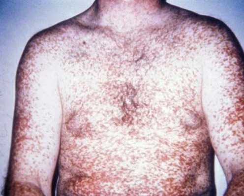
Mastocytoses
Specific investigations


First-line therapies
 H1 antihistamines
 H2 antihistamines
 Disodium cromoglycate
Second-line therapies
 Topical glucocorticoid with plastic-film occlusion
 Oral glucocorticoids
 Intralesional glucocorticoids
 NB-UVB phototherapy
 Oral PUVA photochemotherapy
 Psoralen baths plus UVA photochemotherapy
 UVA1 phototherapy
Third-line therapies
 Interferon-α
 Topical calcineurin inhibitors
 Leukotriene receptor inhibitor
 Cyclosporine
 Nifedipine
 Thalidomide
 Surgical excision
 Flashlamp-pumped pulsed dye and Nd : YAG lasers
 Electron beam radiation
 Imatinib mesylate
 Cladribine
 Omalizumab
[/level-membership-for-dermatology-category][not-level-membership-for-dermatology-category]
Mastocytoses
[level-membership-for-dermatology-category]
[/level-membership-for-dermatology-category][not-level-membership-for-dermatology-category]
