Management of acute spinal cord injury

Published on 07/02/2015 by admin

Filed under Anesthesiology

Last modified 22/04/2025

Print this page

rate 1 star rate 2 star rate 3 star rate 4 star rate 5 star
Your rating: none, Average: 0 (0 votes)

This article have been viewed 1264 times

Management of acute spinal cord injury

Eric L. Bloomfield, MD, MS, MMI, FCCM

Respiratory considerations

A lesion at a vertebra above T7 may alter respiratory function. Vital capacity, expiratory reserve volume, and forced expiratory volume typically are decreased. The resulting respiratory physiologic characteristics depend on three factors: intercostal muscle function, the diaphragmatic function, and the use of accessory muscles of respiration.

Neurons exiting the cord at C3, C4, and C5 provide innervation of the diaphragm, so a spinal cord injury at C3 results in paralysis of the diaphragm; if the injury is not recognized and treated immediately, patients with such lesions asphyxiate. With a C5 lesion, a patient may have signs of partial diaphragmatic paralysis. By comparison, a C6 lesion enables the patient to maintain ventilation because innervation of the diaphragm is intact. Even so, the patient will have some compromised respiratory function, with sternal retraction and paradoxical breathing. This problem of respiratory compromise is due to intercostal paralysis; the patient can still have a compromised cough and an inability to clear secretions.

Spinal cord lesions are not always static. They may be complete or incomplete, such as a Brown-Séquard syndrome. A spinal cord lesion may involve a spreading hematoma that then leads to increased edema and ischemia of the neural tissue. Ondine curse, “central” sleep apnea, can be caused by a lesion involving the anterolateral portion of C2 through C4 spinal cord segments. Patients who have traumatic injury to the spinal cord with associated neurologic deficit have an increased risk of developing deep venous thrombosis and pulmonary embolic events due to thrombus, or from a fat embolus, the latter associated with other bone-related injuries. Other pulmonary disorders associated with spinal cord injury are neurogenic pulmonary edema, aspiration pneumonia, and acute respiratory distress syndrome.

Cardiovascular considerations

After spinal cord injury, blood pressure and heart rate increase, sometimes dramatically, because of the associated sympathetic storm. Over time, parasympathetic activity increases, manifested by bradycardia, sinus node pauses, sick sinus syndrome, supraventricular arrhythmias, ventricular ectopy, and possibly ST-segment changes, based on the individual’s coronary anatomy.

Depending on the level and severity of injury, spinal shock may occur and last up to 6 weeks as a result of a loss of vascular tone and of the vasopressor reflex. Injury to the spinal cord from the level of the T1 to L2 damages the sympathetic nervous system (see Chapter 40) and may result in orthostatic hypotension and, potentially, bradycardia. The bradycardia results from the loss of cardiac acceleration fibers. Prompt treatment of spinal shock with intravascular fluids and vasoactive agents (see Chapter 88) to maintain a mean arterial pressure of 65 mm Hg to 75 mm Hg can potentially improve neurologic outcome.

Metabolic considerations

On initial presentation, patients with spinal cord injury often require emergency airway management. Succinylcholine is preferred by many in the emergency department or operating room to manage patients with acute spinal cord injury. When these same patients subsequently return to the operating room for additional surgical procedures, the use of succinylcholine can lead to a tremendous potassium release, which may cause ventricular fibrillation and cardiac arrest. With denervation of muscle, there is an increase in the number of nicotinic receptors: some from the muscle membrane where, previously, there were no nictonic receptors and also from the appearance of new isoforms of these nicotinic receptors. All of these receptors respond to succinylcholine with release of potassium from a larger area of muscle membrane than would normally occur, and hence, hyperkalemia results. The change in the number and type of receptors occurs as early as 3 days after injury and may be ongoing for up to 6 months. The marked hyperkalemic response has been reported to occur as early as 5 days after injury, but the hyperkalemia also occurs in patients who have an associated ongoing infectious process.

Patients with an injury to the spinal cord at between T1 and L2, with sufficient damage to the sympathetic nervous system, lose the ability to thermoregulate. Hypothermia can lead to vasoconstriction, metabolic acidosis from peripheral vasoconstriction and myocardial ischemia from coronary artery vasoconstriction. Conversely, patients with spinal cord lesions above C7 have an inability to sweat, which can be manifested by hyperthermia.

In patients with longstanding paralysis, bone reabsorption can lead to hypercalcemia. Patients with impaired ventilatory drive can present with respiratory acidosis, with or without a compensating alkalosis.

Anesthetic management

Preoperative management

Airway management mandates stabilization of the neck while intubating the trachea in an expeditious manner for the reasons mentioned previously. Chest physiotherapy, deep vein thrombosis prophylaxis (beginning 2-3 days after the injury to avoid hemorrhage at the site of injury), decompression of the stomach, administration of stress-related ulcer prophylaxis, and monitoring gas exchange are all considerations.

Airway management

All patients with cervical spine fractures are considered to have a difficult airway. The main goal of their care is to maintain cervical stability and, at the same time, oxygenate, ventilate and protect the airway by placing a tracheal tube in a timely manner. If the patient is in immediate need of an airway, inline stabilization with direct laryngoscopy may be the best choice. If timing allows, awake intubation may be the best choice to avoid manipulation of the cervical spine. Nasal intubation should be avoided in patients with basilar skull fractures, raccoon eyes, Battle sign, Le Fort fractures, or any evidence of cerebrospinal fluid leak.

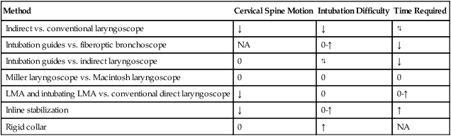
Intubation maneuvers (Table 140-1) can be gauged in accordance with the time needed and the amount of cervical neck manipulation associated with the use of a laryngoscope versus a fiberoptic bronchoscope.

Table 140-1

Comparison of Options for Anesthesia Management of Spinal Injury Procedures

Method Cervical Spine Motion Intubation Difficulty Time Required
Indirect vs. conventional laryngoscope ↓ ↓
Intubation guides vs. fiberoptic bronchoscope NA 0-↑
Intubation guides vs. indirect laryngoscope 0
Miller laryngoscope vs. Macintosh laryngoscope 0 0 0
LMA and intubating LMA vs. conventional direct laryngoscope 0 0-↑
Inline stabilization 0-↑
Rigid collar 0 NA

image

LMA, Laryngeal mask airway; NA, not applicable.

Cardiovascular considerations

Lesions located above T4 are associated with neurogenic shock. Restoration of an adequate perfusion pressure is paramount to preventing extension of the neurologic deficit. During surgery for spinal cord stabilization, monitoring of motor-evoked or sensory-evoked potentials, or both, is often used.

The use of corticosteroids, such as methylprednisolone (30 mg/kg bolus followed by 5.4 mg·kg−1·h−1 for 24-48 h), is associated with a small but statistically significant improvement in outcome—assuming that the cord is not completely transected—if the therapy is started within 8 h of injury.

The patient’s hemodynamic instability from shock will stabilize after 10 to 14 days. However, patients with lesions at T4 or above are at risk of developing autonomic hyperreflexia. Despite an injured patient’s lack of sensation below the site of injury, anesthesia may be necessary if the patient returns to the operating room, and precautions must be taken to attenuate if not avoid the complication.

In summary, the goals in the treatment of a patient with spine cord injury are the following: