Published on 18/03/2015 by admin
Filed under Dermatology
Last modified 22/04/2025
This article have been viewed 2204 times
Patrick O.M. Emanuel and Ben Tallon
Evidence Levels: A Double-blind study B Clinical trial ≥ 20 subjects C Clinical trial < 20 subjects D Series ≥ 5 subjects E Anecdotal case reports
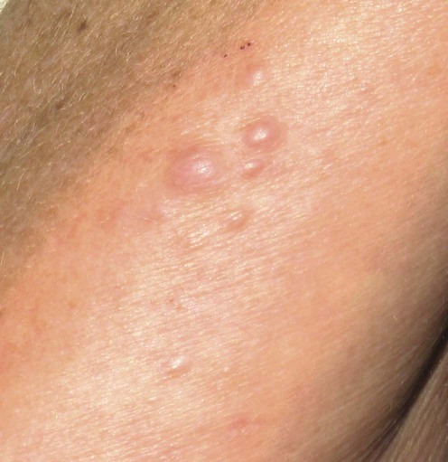
Lymphangioma circumscriptum is an uncommon lymphatic malformation. It presents on the skin surface as grapelike groups of thin-walled translucent lymph-filled vesicles, often compared to frog spawn. Hemorrhage within the lesions can create a deep red or brown appearance.
More commonly congenital, they are typically noted at birth or appear during childhood. They are most commonly found around the shoulder girdle and proximal limbs. There is a morphologically identical acquired variant related to lymphatic obstruction as a consequence of surgery, radiation, or malignancy.
Although observation is an appropriate option for many cases, cosmetic concern is the typical indication for treatment. Other indications may include persistent leakage of lymphatic fluid or blood, and recurrent infection. The risk of developing angiosarcoma and squamous cell carcinoma is trivial and should not be used to rationalize full surgical excision.
Treatment is challenging and often thwarted by local recurrences owing to the persistence of deep lymphatic cisterns which may delve deep into the subcuticular adipose tissue, skeletal muscle, and nerves. Every treatment option has associated recurrence rates/complication profiles. Consequently, there is disagreement in the literature as to which treatment option is the most effective. Although complete surgical excision has the lowest recurrence rates, it has the highest rate of complications; more extensive lesions may be deemed inoperable.
Sclerotherapy using a variety of sclerosants has been advocated as a less invasive and effective treatment modality which is either a first-line alternative or an adjunct to surgery.
Other authorities suggest that after definitive diagnosis and radiologic mapping, surgical excision and postoperative histologic assessment of excision margins provides the most effective treatment option.
Resurfacing of the lesions can be attempted and achieved even if proper surgical excision is not possible and/or sclerotherapy fails, but recurrence rates are usually higher than with other therapies. The high-energy, short-pulse CO2 laser has been found to yield functionally and cosmetically acceptable results. This seals communicating channels to the deeper cisterns by vaporizing the superficial lymphatics, and is said to have fewer complications than more aggressive treatment alternatives. Other laser methods, particularly the pulsed dye laser, have also been shown to be effective in selected superficial cases.
Biopsy
Imaging studies
MRI
Lymphangiography
Ultrasound
In the majority of cases the clinical diagnosis is straightforward. In some, the differential diagnosis may be broad: genital lesions are often associated with verrucous changes, which give them a warty appearance and are often confused with viral warts or squamous cell carcinoma; discoloration of the vesicles can lead to confusion with hemangiomas and even malignant melanoma; herpetic infection and dermatitis herpetiformis are less frequent differential diagnoses. Biopsy is diagnostic for clinically unusual cases and exhibits numerous thin-walled, dilated lymphatic channels encroaching onto the epidermis (which may become hyperkeratotic), expanding the papillary dermis and extending deep into the dermis and subcutis. Immunostaining with VEGFR3 and D2-40 decorates the vessels and confirms their lymphatic origin.
MRI can define the entire anatomy of a lesion and, when used preoperatively, can help prevent unnecessarily extensive or incomplete surgical resection. CT, ultrasound, and lymphoscintigraphy have also been useful in determining the extent of a lesion.
Secondary lesions may be investigated for an underlying cause if the cause is not clinically obvious. Lymphangiomas may be associated with rare disorders such as Proteus, Cobb and Klippel–Trenaunay syndromes, and so appropriate investigations and consultations should be sought if these conditions are suspected.
Intralesional sclerotherapy with group A Streptococcus pyogenes of human origin (OK-432) has emerged as an effective sclerosing agent.
Ogita S, Tsuto T, Nakamura K, Deguchi E, Iwai N. J Pediatr Surg 1994; 29: 784–5.
Ahn SJ, Chang SE, Choi JH, Moon KC, Koh JK, Kim DY. J Am Acad Dermatol 2006; 55: S106–7.
Okazaki T, Iwatani S, Yanai T, Kobayashi H, Kato Y, Marusasa T, et al. J Pediatr Surg 2007; 42: 386–9.
The main advantage of OK-432 over other sclerosing agents is the absence of perilesional fibrosis. OK-432 is an effective agent for cases of a single or limited numbers of vesicles. In larger lesions the technique is useful as a pretreatment adjunct to surgical excision.
Al Ghamdi KM, Mubki TF. J Cosmet Dermatol 2011; 10: 156–8.
Bikowski JB, Dumont AM. J Am Acad Dermatol 2005; 53: 442–4.
Hypertonic saline has been reported to be effective for the management of lymphangioma circuscriptum to the shoulder, although wider use of this agent in comparison with other treatments for lymphangioma circumscriptum has not been reported.
Molitch HI, Unger EC, Witte CL, van Sonnenberg E. Radiology 1995; 194: 343–7.
Five patients with unresectable lymphangiomas of the pelvis (n=2), neck (n=1), abdomen (n=1), or leg (n=1) were treated at two medical centers with sclerotherapy, using doxycycline as the sclerosant.
Park CO, Lee MJ, Chung KY. Dermatol Surg 2005; 31: 1451–3.
Two cases of lymphangioma circumscriptum were treated with a sclerosant, sodium tetradecyl sulfate, and were almost cleared with several treatments of sclerotherapy.
Primary surgical excision was significantly more successful than sclerotherapy, but the results are often unsatisfactory because of complications, including damage to surrounding structures, particularly nerves and blood vessels, scarring, and recurrence owing to incomplete excision. Ultrasound and magnetic resonance imaging demonstrates deep cisterns, thereby ensuring deeper excision of these structures and less risk of recurrence.
Browse NL, Whimster I, Stewart G, Helm CW, Wood JJ. Br J Surg 1986; 73: 585–9.
Confirmation of the completeness of excision can be obtained using frozen section analysis of the lateral and deep margins.
Sah SP, Yadav R, Rani S. J Obstet Gynaecol Res 2001; 27: 293–6.
A case report of vulval lymphangioma circumscriptum, clinically diagnosed as a genital wart. Following biopsy, the patient required extensive vulval surgery and there was no recurrence after 16 months.
Latifoglu O, Yavuzer R, Demir Y, Ayhan S, Yenidünya S, Atabay K. Plast Reconstruct Surg 1999; 103: 175–8.
An extensive lymphangioma circumscriptum of the penis and scrotum that was treated by wide excision in single-stage surgery. At the 14th postoperative month the patient was free of recurrence.
Bond J, Basheer MH, Gordon D. Dermatol Surg 2008; 34: 271–5.
Two cases of surgical excision with no recurrence at 1 and 2 years. Imaging of deep communicating structures and histologic assessment of excision margins provide the greatest chance of non-recurrence.
Bailin PL, Kantor GR, Wheeland RG. J Am Acad Dermatol 1986; 14: 257–62.
CO2 laser in a vaporization mode successfully ablated superficial cutaneous lesions in seven patients with lymphangioma circumscriptum.
Eliezri YD, Sklar JA. J Dermatol Surg Oncol 1988; 14: 357–64.
Three patients were treated with CO2 laser vaporization, with good to excellent cosmetic results and complete resolution of their symptoms.
Huilgol SC, Neill S, Barlow RJ. Dermatol Surg 2002; 28: 575–7.
Focal recurrence and an area of localized persistence were noted in both patients with lymphangioma circumscriptum treated with CO2 laser therapy.
Haas AF, Narurkar VA. Dermatol Surg 1998; 24: 893–5.
A difficult case of lymphangioma circumscriptum was treated successfully with a high-energy, short-pulse CO2 laser following two failed surgical excisions.
Treharne LJ, Murison MS. Lymphat Res Biol 2006; 4: 101–3.
Widespread disease of the scrotum had excellent symptomatic relief following treatment with the CO2 laser.
Thissen CA, Sommer A. Int J Dermatol 2007; 46: 16–18.
An intense pulsed light source led to good cosmetic results.
Lapidoth M, Ackerman L, Amitai DB, Raveh E, Kalish E, David M. Dermatol Surg 2006; 32: 790–4.
Treatment was rated as ‘excellent’ in four patients and ‘good’ in two. Swelling, erythema, and pain were present in all patients, and ulcers and scarring in two.
Lai CH, Hanson SG, Mallory SB. Pediatr Dermatol 2001; 18: 509–10.
A child with a symptomatic lymphangioma circumscriptum was treated with pulsed dye laser, with good results.
Yildiz F, Atahan IL, Ozhar E, Karcaaltincaba M, Cengiz M, Ozyigit G, et al. Int J Gynecol Cancer 2008; 18: 556–9.
A patient was treated successfully with a course of external radiotherapy following failed attempts with sclerosing agents and surgery.
Denton AS, Baker-Hines R, Spittle MF. Clin Oncol (R Coll Radiol) 1996; 8: 400–1.
Localized radiotherapy has been successfully used.
Treatment of Skin Disease Comprehensive Therapeutic Strategies 4e
WhatsApp us


 Conservative/observation
 Antibiotics
 Sclerotherapy
 OK-432
 Hyperosmolar saline
 Sodium tetradecyl sulfate
 Surgery
 CO2 laser
 Pulsed dye laser
 Radiotherapy
 Cryotherapy
 Argon laser
 Suction-assisted lipectomy