Lung Cancer: Etiologic and Pathologic Aspects

Published on 12/06/2015 by admin

Filed under Pulmolory and Respiratory

Last modified 22/04/2025

Print this page

rate 1 star rate 2 star rate 3 star rate 4 star rate 5 star
Your rating: none, Average: 0 (0 votes)

This article have been viewed 1244 times

20

Lung Cancer

Etiologic and Pathologic Aspects

Carcinoma of the lung is a public health problem of immense proportions. It has been a source of great frustration to individual physicians and the medical profession in general. Half a century ago, the primary cause of carcinoma of the lung—cigarette smoking—was identified beyond a shadow of a doubt. Fortunately, the prevalence of smoking in developed countries has been falling gradually after peaking in the mid-1970s. Unfortunately, any optimism is tempered by the following concerns: (1) about 20% of all American adults still smoke; (2) although smoking prevalence declined substantially in the years before 1990, it has changed only slowly since then; and (3) tobacco use is continuing to increase in developing countries, where the tobacco industry is focusing its marketing efforts.

A few statistics put the magnitude of the problem of lung cancer into perspective. Worldwide in 2008, there were an estimated 1,600,000 new cases of lung cancer and an estimated 1,380,000 deaths. In the United States, nearly 220,000 new cases of lung cancer are diagnosed annually, and approximately 160,000 individuals each year die as a result of the disease. For many years, carcinoma of the lung has been the leading cause of cancer deaths among men, and in 1985 lung cancer surpassed breast cancer as the leading cause of cancer deaths among women. Lung cancer is responsible for 25% to 30% of all deaths attributable to cancer and approximately 5% of all deaths from any cause, killing more people than cancers of breast, prostate, colon, liver, kidney, and melanoma combined. It is sobering to realize that during the last 5 years, more Americans died from lung cancer than were killed in all of the wars in U.S. history.

The number of cases and the number of deaths related to lung cancer have increased dramatically over the last several decades. For no other form of cancer has the increase approached that of lung cancer. For men, the death rate appears to have reached a peak in 1990 and fortunately has been decreasing since then. In women the death rate increased fivefold in the 30 years from 1960 to 1990 but more recently appears to be reaching a plateau. Despite the magnitude of the problem, our ability to treat carcinoma of the lung has improved only minimally. Five-year survival has increased from roughly 7% to 14% during the last several decades, making the prognosis of this disease still dismal in the vast majority of cases.

The discussion of carcinoma of the lung is presented in two parts. This chapter considers what is known about the etiology and pathogenesis of lung cancer, followed by a description of the pathologic aspects and classification of the different types of tumors. Chapter 21 continues with a discussion of the clinical aspects of the disease, including diagnostic and therapeutic considerations. Chapter 21 concludes with a brief discussion of two additional types of neoplastic disease affecting the respiratory system, bronchial carcinoid tumor (bronchial adenoma) and mesothelioma, along with a consideration of the common problem of the patient with a solitary pulmonary nodule.

Etiology and Pathogenesis

For no other common cancer affecting humans have the causative factors been identified as well as for lung cancer. Cigarette smoking clearly is responsible for the vast majority of cases (>85%, according to some estimates), and additional risk factors associated with occupational exposure have been identified. This chapter begins with a discussion of the two major risk factors—cigarette smoking and occupational exposure—and considers genetic factors as a potential contributor to lung cancer risk. Next is a brief description of the importance of previous scarring within the pulmonary parenchyma, which has been implicated in the development of “scar carcinomas,” and several miscellaneous proposed risk factors are mentioned. Finally, the role of oncogenes and tumor suppressor genes in the pathogenesis of lung cancer is discussed.

Smoking

Cigarette smoking is the single most important risk factor for the development of carcinoma of the lung. As might be expected, the duration of smoking history, number of cigarettes smoked each day, depth of inhalation, and amount of each cigarette smoked all correlate with the risk for developing lung cancer. As a rough but easy way to quantify prior cigarette exposure, the number of years of smoking can be multiplied by the average number of packs smoked per day, giving the number of “pack years.”

Although the evidence linking smoking with lung cancer is incontrovertible, the responsible component of cigarette smoke has not been identified with certainty. Cigarette smoke consists of a gaseous phase and a particulate phase, and potential carcinogens have been found in both phases, ranging from nitrosamines to benzo[a]-pyrene and other polycyclic hydrocarbons. Filters appear to decrease but certainly do not eliminate the potential carcinogenic effects of cigarettes. A substantially lower risk for lung cancer is associated with cigar and pipe smoking, presumably related to the fact that cigar and pipe smoke is generally not inhaled deeply into the lungs in the same manner as cigarette smoke. Marijuana and cocaine smoking are also associated with the precancerous histologic changes observed among cigarette smokers, and both are believed to be risk factors for lung cancer.

Development of lung cancer due to smoking requires many years of exposure. However, histologic abnormalities before the development of a frank carcinoma are well documented in the bronchial epithelium of smokers with lesser degrees of exposure. These changes, including loss of bronchial cilia, hyperplasia of bronchial epithelial cells, and nuclear abnormalities, may be the pathologic forerunners of a true carcinoma. If a person stops smoking, many of these precancerous changes appear to be reversible. Epidemiologic studies have suggested that the risk for developing lung cancer decreases progressively after cessation of smoking but probably never fully returns to the level in nonsmokers, even after more than 10 to 15 years. In many cases, the initial cellular changes leading to or predisposing to malignant transformation have already developed by the time the patient stops smoking, and it is merely a matter of time before the carcinoma develops or becomes clinically apparent.

Data indicate that the risk of lung cancer is increased for nonsmoking spouses because of their exposure to sidestream or “secondhand” smoke. Although the risk attributable to “passive smoking” is relatively small compared with the risk of active smoking, involuntary exposure to cigarette smoke is likely responsible for some cases of lung cancer occurring in nonsmokers. The comparably small but real risk of lung cancer from passive smoking has been a major justification for legislation prohibiting smoking in shared spaces such as commercial aircraft, restaurants, and offices.

Occupational Factors

A number of potential environmental risk factors have been identified, most of which occur with occupational exposure. Perhaps the most widely studied of the environmental or occupationally related carcinogens is asbestos, a fibrous silicate formerly in wide use because of its properties of fire resistance and thermal insulation. Shipbuilders, construction workers, and those who worked with insulation and brake linings are among those who may have been exposed to asbestos.

Carcinoma of the lung is the most likely malignancy to result from occupational asbestos exposure, although other tumors, especially mesothelioma (see Chapter 21), are also strongly associated with prior asbestos contact. (Low-level nonoccupational exposures to asbestos in schools or among residents living near asbestos mines or processing facilities are of much lesser significance.) The risk for development of lung cancer is particularly high among smokers exposed to asbestos, in which case these two risk factors probably have a multiplicative rather than a simple additive effect. Specifically, asbestos alone appears to confer a twofold to fivefold increased risk for lung cancer, whereas smoking alone is associated with an approximately 10-fold increased risk. Together, the two risk factors make the person who smokes and has an asbestos exposure 20 to 50 times more likely to have carcinoma of the lung than a nonsmoking, nonexposed counterpart. Like other forms of asbestos-related disease, a long time elapses before complications develop. In the case of lung cancer, the tumor generally becomes apparent more than 2 decades after exposure.

A number of other types of occupational exposure have been implicated. Examples include exposure to arsenic (in workers making pesticides, glass, pigments, and paints), ionizing radiation (especially in uranium miners), halo ethers (bis[chloromethyl] ether and chloromethyl methyl ether in chemical industry workers), and polycyclic aromatic hydrocarbons (in petroleum, coal tar, and foundry workers). As is the case with asbestos, there is generally a long latent period of at least 2 decades from the time of exposure until presentation of the tumor.

Genetic Factors

Why lung cancer develops in some heavy smokers and not in others is a question of great importance but with no definite answer at present. The assumption is that genetic factors must place some individuals at higher risk for lung cancer after exposure to carcinogens. The finding of an increased risk of lung cancer among first-degree relatives of lung cancer patients—even after confounding factors have been taken into account—supports this hypothesis.

Candidate genetic factors have primarily included specific enzymes of the cytochrome P450 system. These enzymes may have a role in metabolizing products of cigarette smoke to potent carcinogens, and genetically determined increased activity or expression of the enzymes may be associated with a greater risk of developing lung cancer following exposure to cigarette smoke. One example is the enzyme aryl hydrocarbon hydroxylase, which can convert hydrocarbons to carcinogenic metabolites. This enzyme is induced by smoking, and genetically determined inducibility of this enzyme by smoking may correlate with the risk for lung cancer. Another enzyme of the cytochrome P450 system can be identified by its ability to metabolize the antihypertensive drug debrisoquine. Some data suggest an association between extensive metabolism of debrisoquine and development of lung cancer. Presumably, the action of this enzyme on a potentially carcinogenic substrate from cigarette smoke affects an individual’s risk for developing lung cancer. However, the available data for both of these cytochrome P450 enzymes are inconsistent, and a role for these enzymes is not universally accepted.

Other as yet unidentified genetic factors potentially affect susceptibility to environmental carcinogens and may include the activity of tumor suppressor genes. If such factors eventually are recognized, then preventing susceptible individuals from being exposed to the known environmental carcinogens or targeting more aggressive screening techniques toward the populations at greatest risk may be possible.

Parenchymal Scarring

Scar tissue within the lung can be a locus for the subsequent occurrence of lung cancer, called a scar carcinoma. The scarring may be either localized (e.g., resulting from an old focus of tuberculosis or another infection) or diffuse (e.g., from pulmonary fibrosis, whether idiopathic or associated with a specific cause). Most frequently, scar carcinoma of the lung is an adenocarcinoma and often a specific subtype that has traditionally been called bronchioloalveolar carcinoma. These cell types are discussed in the section on adenocarcinoma.

Although it is easy to consider carcinomas occurring within or adjacent to scar tissue to be scar carcinomas, adenocarcinomas of the lung also may develop fibrotic areas within the tumor. Therefore, in some cases it may be impossible to know whether the scar preceded or followed development of the carcinoma.

Miscellaneous Factors

The lay press has expressed a great deal of interest and publicized the risk of lung cancer from exposure to radon, a gas that is a decay product of radium-226 (itself a decay product of uranium-238). Exposure to this known carcinogen may occur indoors in homes built on soil that has a high radium content and is releasing radon into the surrounding environment. Although the finding of unacceptably high levels of radon in some home environments has sparked concern about the risk of lung cancer and interest in widespread testing of houses, uncertainty remains about the overall risk posed by exposure to radon. At the extreme, it has been suggested that radon is the second most important factor contributing to lung cancer and potentially is responsible for 20,000 lung cancer deaths per year in the United States. However, this magnitude of risk is not universally accepted.

Some evidence suggests that dietary factors may affect the risk of lung cancer. Some studies have reported an association between low intake and serum levels of β-carotene, the provitamin form of vitamin A, with an increased risk of lung cancer. However, the data relating to this issue are controversial. An increased risk associated with low dietary intake of β-carotene, if it exists, is relatively minor compared with the risk posed by cigarette smoking. Three large randomized trials have failed to demonstrate a protective effect of β-carotene, α-tocopherol, or retinoid supplementation on lung cancer risk. The issue is further complicated by data suggesting an increase in the incidence of lung cancer in some trials of individuals given supplements.

Human immunodeficiency virus (HIV) infection appears to increase the risk of lung cancer, and this association may become more important as advances in antiretroviral treatment decrease mortality from infectious causes in this population. Patients who have received radiation therapy to the thorax (e.g., as treatment for breast cancer or Hodgkin lymphoma) are at increased risk for lung cancer. Finally, in developing countries, chronic exposure to wood smoke is believed responsible for a sizable fraction of lung cancers, particularly among women.

Concepts for Lung Cancer Pathogenesis

There has been a great deal of interest in identifying the cell(s) of origin (i.e., histogenesis) of the various types of lung cancer and elucidating the genetic changes involved in malignant transformation of these cells. For many years it was assumed that the different histopathologic types of lung cancer (described in the section on pathology) were each associated with a different cell of origin. It was thought that previously well-differentiated normal cells underwent a process of dedifferentiation and unrestricted growth when exposed to a carcinogenic stimulus. However, based in part on the common finding of cellular heterogeneity (i.e., more than one cell type within a single tumor), it is currently believed that many if not all types of lung cancer arise from an undifferentiated precursor or stem cell. During this cell’s malignant transformation, it differentiates along one or more particular pathways that determine its ultimate histologic appearance—that is, its cell type(s).

Alterations in genes that code for proteins controlling or regulating cell growth have been found in a high proportion of patients with lung cancer. These molecular changes may play a central role in lung cancer pathogenesis. Two types of oncogenes have been identified: proto-oncogenes (which code for growth-promoting factors) and tumor suppressor genes (which code for factors having a negative regulatory effect on cell proliferation). A mutation in one of the paired alleles of a proto-oncogene can result in production of a protein with a growth-promoting effect such that a “dominant” behavior or effect would be observed. In contrast, both alleles of a tumor suppressor gene must be altered before the absence of the gene product would be clinically manifest as increased cell growth or malignant transformation. This requirement produces a “recessive” pattern of clinical expression.

Specific common alterations in proto-oncogenes that have been identified in lung cancer include mutations in the ras, EGFR, HER2, and BCL2 families of dominant oncogenes. A variety of mutations in recessive tumor suppressor genes also have been identified, including the retinoblastoma (rb) and p53 genes. In addition, deletion of genetic material from chromosome 3p (the short arm of chromosome 3) has been recognized in lung cancer, and it is thought that deletion may involve loss of one or more tumor suppressor genes. Particularly interesting experimental data link a carcinogenic metabolite of benzo[a]pyrene, which is found in cigarette smoke, to those mutations of the p53 gene that are most commonly seen in lung cancer.

Pathology

The term bronchogenic carcinoma is often used interchangeably with the term lung cancer, implying that lung cancers arise from bronchi or bronchial structures. Many if not most lung cancers do originate within airways, but other tumors arise in the periphery of the lung and may not necessarily originate in an airway. This section focuses on the currently accepted classification of lung cancer and summarizes what is known about the behavior patterns of the various types of tumors. Of note, whereas older classification schemes of lung cancer relied exclusively upon microscopic examination of stained specimens, immunohistochemistry and genetic analysis now play an increasing role in characterizing lung malignancies.

Most lung cancers fall within one of four histologic categories: (1) squamous cell carcinoma, (2) small cell carcinoma, (3) adenocarcinoma, and (4) large cell carcinoma. Within each category are several subcategories that, for our purposes, are less important. Of note, nearly 24% of lung cancers are considered “other non–small cell” tumors, a category that includes malignancies too anaplastic to permit further subtyping, as well as rarer variants that will not be discussed here.

One of the most important distinctions to make is between small cell carcinoma and all the other cell types, which are grouped together as non–small cell carcinoma. The importance of this distinction relates to the propensity of small cell carcinoma for early clinical and subclinical metastasis, which affects the approaches to staging and treatment of this tumor compared with those of all the other cell types. Recent increased emphasis on differentiating adenocarcinomas and squamous cell cancers from other non–small cell carcinomas has arisen because of differences in responsiveness to certain drugs such as epidermal growth factor receptor tyrosine kinase inhibitors.

Each of the four major categories of lung cancer is associated with cigarette smoking, but the statistical association between smoking and the individual cell types is strongest for squamous cell carcinoma and small cell carcinomas, seen almost exclusively in smokers. Even though smoking also increases the risk for adenocarcinoma and large cell carcinoma, these cell types also occur with some regularity in nonsmokers.

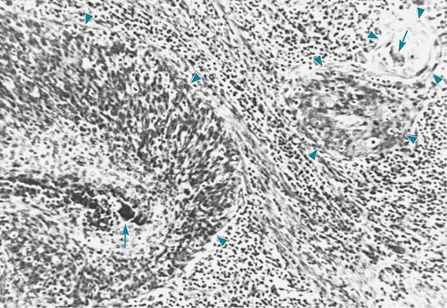
Squamous Cell Carcinoma

Formerly the most common histopathologic type encountered, squamous cell tumors currently account for only about 20% of all bronchogenic carcinomas. These malignancies originate within the epithelial layer of the bronchial wall, in which a series of progressive histologic abnormalities result from chronic or repetitive cigarette smoke–induced injury.

Initially there is metaplasia of normal bronchial columnar epithelial cells, which are replaced by squamous epithelial cells. Over time these squamous cells become more and more atypical in appearance until there is development of a well-localized carcinoma (i.e., carcinoma in situ). Eventually the carcinoma extends beyond the bronchial mucosa and becomes frankly invasive. After the tumor reaches this stage, it generally comes to eventual clinical attention by producing either symptoms or radiographic changes. In some cases, detection of the carcinoma is made at the earlier in situ stage, usually by recognition of the malignant cells in a specimen of sputum obtained for cytologic examination or by biopsy of grossly abnormal-appearing bronchial mucosa during bronchoscopic evaluation undertaken for other reasons.

Specific histologic features of squamous cell carcinoma allow the pathologist to establish this diagnosis. These tumors are characterized by the visible presence of keratin, “squamous pearls,” and intercellular desmosomes or bridges (Fig. 20-1).

Squamous cell carcinomas tend to be located in relatively large or proximal airways, most commonly at the subsegmental, segmental, or lobar level. With growth of the tumor into the bronchial lumen, the airway may become obstructed. The lung distal to the obstruction frequently collapses (becomes atelectatic), and a postobstructive pneumonia may develop. Sometimes a cavity develops within the tumor mass; this finding of cavitation is much more common with squamous cell than with other types of bronchogenic carcinoma.

Spread of squamous cell carcinoma beyond the airway usually involves (1) direct extension to the pulmonary parenchyma or other neighboring structures or (2) invasion of lymphatic vessels, with spread to local lymph nodes in the hilum or mediastinum. These tumors have a general tendency to remain within the thorax and cause problems by intrathoracic complications rather than by distant metastasis. The overall prognosis in terms of 5-year survival is better for patients with squamous cell carcinoma than for patients with any of the other cell types.

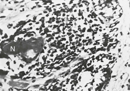
Small Cell Carcinoma

Small cell carcinoma, constituting 14% of all lung cancers, was previously considered to have several subtypes, of which oat cell carcinoma was the most important. Since 1999, classifications of lung cancer no longer include oat cell carcinoma as a separate subtype. Like squamous cell carcinoma, small cell carcinomas generally originate within the bronchial wall, most commonly at a proximal level. The cell of origin of small cell carcinoma is disputed. An older theory proposed that these tumors arise from a neurosecretory type of epithelial cell termed the Kulchitsky cell or K cell. These cells have the capacity for polypeptide production and are considered a type of APUD cell (i.e., capable of amine precursor uptake and decarboxylation). A more recent theory suggests that small cell carcinomas, like other lung cancers, have their origin from a pluripotent stem cell. The eventual cell type then depends on the pattern and degree of differentiation from this precursor cell. Molecular and chromosomal studies have shown that more than 90% of small cell carcinomas demonstrate deletions on the short arm of chromosome 3 (3p).

In small cell carcinoma, the malignant cells appear as small, darkly stained cells with sparse cytoplasm (Fig. 20-2). Local growth of the tumor often follows a submucosal pattern, but the tumor quickly invades lymphatics and submucosal blood vessels. Hilar and mediastinal nodes are involved and enlarged early in the course of the disease and frequently are the most prominent aspect of the radiographic presentation.
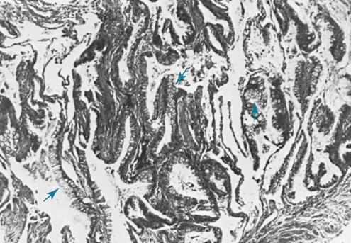
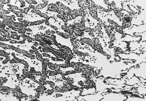
Rapid dissemination of small cell carcinoma makes metastatic spread to distant sites a common early complication. Distant disease, which may be clinically occult at the time of presentation, often affects the brain, liver, bone (and bone marrow), and adrenal glands. It is this propensity for early metastatic involvement that gives small cell carcinoma the worst prognosis among the four major categories of bronchogenic carcinoma.

Adenocarcinoma

Adenocarcinoma has surpassed squamous cell carcinoma as the most frequent cell type, accounting for about 39% of all lung tumors. Because the majority of adenocarcinomas occur in the lung periphery, it is much harder to relate their origin to the bronchial wall. At present, these tumors are believed to arise at the level of bronchioles or alveolar walls. Adenocarcinomas sometimes appear at a site of parenchymal scarring that is either localized or part of a diffuse fibrotic process.

Adenocarcinoma is the most common type of lung cancer to develop among nonsmokers. Nearly 18% of lung adenocarcinomas are diagnosed in nonsmokers, versus less than 2% of squamous cell carcinomas and less than 1% of small cell carcinomas. A link between human papillomavirus and adenocarcinoma of the lung has been hypothesized but is not definitively established.

The characteristic appearance defining adenocarcinoma is the tendency to form glands and in many cases produce mucus (Fig. 20-3). When the malignant cells seem to grow and spread along the preexisting alveolar walls, almost as though they were using the alveolar wall as scaffolding for their growth, the tumors have been subcategorized as bronchioloalveolar carcinomas (Fig. 20-4). Of note, a 2011 joint statement from the International Association for the Study of Lung Cancer, the American Thoracic Society, and the European Respiratory Society recommended elimination of this specific term.

The usual presenting pattern of adenocarcinoma is a peripheral lung nodule or mass. Occasionally the tumors arise within a relatively large bronchus and therefore may become apparent clinically because of complications of localized bronchial obstruction, as seen with squamous cell carcinoma. The bronchioloalveolar subcategory can manifest in several ways: as a nodule or mass lesion, as a localized infiltrate simulating a pneumonia, or as widespread parenchymal disease.

Although adenocarcinoma may spread locally to adjacent regions of lung or to pleura, it also has a propensity for hilar and mediastinal lymph node involvement and distant metastatic spread. Like small cell carcinoma, it spreads to liver, bone, central nervous system, and adrenal glands. In comparison with small cell carcinoma, however, adenocarcinoma is more likely to be localized at the time of presentation, particularly when it manifests as a solitary peripheral lung nodule. The overall prognosis for adenocarcinoma is not surprising given this behavior; its natural history and survival rates are intermediate between those of squamous cell and small cell carcinomas.

Large Cell Carcinoma

Large cell carcinoma accounts for approximately 3% of all lung cancers. It is the most difficult carcinoma to describe microscopically because the tumors often are defined by the characteristics they lack—that is, the specific features that would otherwise classify them as one of the other three cell types. Microscopically they appear as collections of large polygonal cells with prominent nucleoli and a moderate amount of cytoplasm.

The behavior of these tumors is relatively similar to that of adenocarcinoma. They often appear in the periphery of the lung as mass lesions, although they tend to be somewhat larger than adenocarcinomas. Their natural history is also similar to that of adenocarcinoma in terms of both propensity for spread and overall prognosis.

Table 21-1 (see Chapter 21) summarizes the distinguishing features of each cell type and reiterates many of the points discussed here.

References

Etiology and Pathogenesis

Alberg, AJ, Ford, JG, Samet, JM. Epidemiology of lung cancer. ACCP evidence-based clinical practice guidelines, 2nd ed. Chest. 2007;132:29S–55S.

Brennan, P, Hainaut, P, Boffetta, P. Genetics of lung cancer susceptibility. Lancet Oncol. 2011;12:399–408.

Dacic, S. Pulmonary preneoplasia. Arch Pathol Lab Med. 2008;132:1073–1078.

Darby, S, Hill, D, Auvinen, A, et al. Radon in homes and risk of lung cancer: collaborative analysis of individual data from 13 European case-control studies. BMJ. 2005;330:223–229.

Denissenko, MF, Pao, A, Tang, M, Pfeifer, GP. Preferential formation of benzo[a]pyrene adducts at lung cancer mutational hotspots in p53. Science. 1996;274:430–432.

Dhala, A, Pinsker, K, Prezant, DJ. Respiratory health consequences of environmental tobacco smoke. Med Clin North Am. 2004;88:1535–1552.

Doll, R, Peto, R, Boreham, J, et al. Mortality in relation to smoking: 50 years’ observations on male British doctors. BMJ. 2004;328:1519–1527.

Field, RW, Krewski, D, Lubin, JH, et al. An overview of the North American residential radon and lung cancer case-control studies. J Toxicol Environ Health A. 2006;69:599–631.

Goodman, GE. Prevention of lung cancer. Thorax. 2002;57:994–999.

Herbst, RS, Heymach, JV, Lippman, SM. Lung cancer. N Engl J Med. 2008;359:1367–1380.

Kratz, JR, Yagui-Beltrán, A, Jablons, DM. Cancer stem cells in lung tumorigenesis. Ann Thorac Surg. 2010;89:S2090–S2095.

Miller, YE. Pathogenesis of lung cancer. 100 year report. Am J Respir Cell Mol Biol. 2005;33:216–223.

Rezazadeh, A, Laber, DA, Ghim, SJ, et al. The role of human papilloma virus in lung cancer: a review of the evidence. Am J Med Sci. 2009;338:64–67.

Rom, WN, Hay, JG, Lee, TC, et al. Molecular and genetic aspects of lung cancer. Am J Respir Crit Care Med. 2000;161:1355–1367.

Sato, M, Shames, DS, Gazdar, AF, et al. A translational view of the molecular pathogenesis of lung cancer. J Thorac Oncol. 2007;2:327–343.

Spiro, SG, Silvestri, GA. One hundred years of lung cancer. Am J Respir Crit Care Med. 2005;172:523–529.

Subramanian, J, Govindan, R. Lung cancer in never smokers: a review. J Clin Oncol. 2007;25:561–570.

Tanvetyanon, T, Bepler, G. Beta-carotene in multivitamins and the possible risk of lung cancer among smokers versus former smokers: a meta-analysis and evaluation of national brands. Cancer. 2008;113:150–157.

Whitesell, PL, Drage, CW. Occupational lung cancer. Mayo Clin Proc. 1993;68:183–188.

Pathology

Dacic, S. Molecular diagnostics of lung carcinomas. Arch Pathol Lab Med. 2011;135:622–629.

Franklin, WA. Diagnosis of lung cancer: pathology of invasive and preinvasive neoplasia. Chest. 2000;117(Suppl):80S–89S.

Müller, K-M. Lung cancer: morphology. Eur Respir Mon. 2001;17:34–47.

Schwartz, AM, Henson, DE. Diagnostic surgical pathology in lung cancer. ACCP evidence-based clinical practice guidelines, 2nd ed. Chest. 2007;132:78S–93S.

Travis, WD. Pathology of lung cancer. Clin Chest Med. 2002;23:65–81.

Travis WD, Brambilla E, Muller-Hermlink HK, et al, eds. World Health Organization classification of tumours. Pathology and genetics of tumours of the lung, pleura, thymus and heart. Lyon: IARC Press, 2004.

Travis, WD, Brambilla, E, Noguchi, M, et al. International Association for the Study of Lung Cancer/American Thoracic Society/European Respiratory Society international multidisciplinary classification of lung adenocarcinoma. J Thorac Oncol. 2011;6:244–285.

Verbeken, EK, Brambilla, E. WHO classification of lung and pleural tumours. The WHO/IASLC 1999 revision. Eur Respir Rev. 2002;12(review 84):172–176.