Lower extremity block: Psoas compartment block

Published on 07/02/2015 by admin

Filed under Anesthesiology

Last modified 22/04/2025

Print this page

rate 1 star rate 2 star rate 3 star rate 4 star rate 5 star
Your rating: none, Average: 0 (0 votes)

This article have been viewed 1295 times

Lower extremity block: Psoas compartment block

Sandra L. Kopp, MD

The psoas compartment block (PCB) is a lumbar plexus block utilizing a posterior approach. Introduced in the 1970s in response to criticism that the femoral “3-in-1” block did not reliably block the femoral, obturator, and lateral femoral cutaneous nerves, the PCB did not gain immediate popularity owing to the unreliability of the loss-of-resistance technique. Not until the introduction of nerve stimulation techniques in 1989 did practitioners begin to commonly perform this peripheral nerve block.

Relevant anatomy

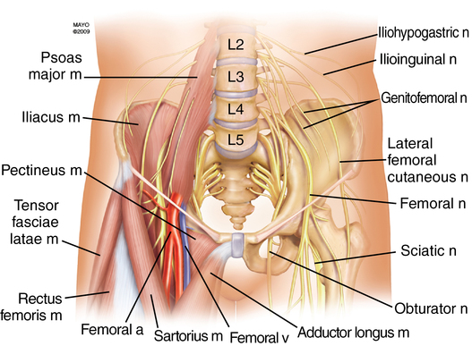
The lumbar plexus is most commonly formed from the ventral rami of L1 through L4, although frequently a branch of T12 and, occasionally, a branch from L5 are included. The plexus lies anterior to the transverse processes of the lumbar vertebrae and descends vertically with the psoas muscle. The branches of the lumbar plexus emerging from the psoas muscle are the femoral nerve (L2-L4), obturator nerve (L2-L4), lateral femoral cutaneous nerve (L2-L3), iliohypogastric nerve (L1), ilioinguinal nerve (L1), and genitofemoral nerve (L1-L2) (Figure 128-1). It provides sensory innervation to the anterior thigh and to the medial portion of the lower leg via the saphenous nerve (distal branch of the femoral nerve), as well as the majority of the femur, ischium, and ilium.

The surface anatomy consists of three main landmarks: (1) the intercristal line, the line connecting the iliac crests; (2) the midline, identified by a line connecting the spinous processes; and (3) the posterior superior iliac spine (PSIS), a projection at the posterior aspect of the ilium.

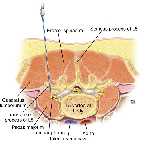
As the needle passes from posterior to anterior at the level of L4 through L5, the following structures are encountered: skin, subcutaneous adipose tissue, posterior lumbar fascia, paraspinous muscles, anterior lumbar fascia, quadratus lumborum, and the psoas muscle (Figure 128-2). The distance from skin to lumbar plexus varies greatly with sex and body mass index, whereas the distance from the transverse process of L4 to the lumbar plexus consistently ranges from 1.5 to 2.0 cm in both sexes.

Technique

Patient position

The patient is positioned laterally with the hips flexed and perpendicular to the horizontal plane (similar to the position utilized for an intrathecal injection) with the operative leg uppermost.

Needle insertion site

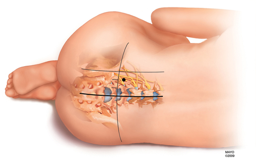
One of several needle insertion sites can be used, although the landmarks described by Capdevila and colleagues use localization of the L4 transverse process, thereby reducing the likelihood of excessive needle depth. The intercristal line is identified and drawn. A horizontal line is drawn identifying the midline. A line originating at the PSIS is drawn parallel to midline. The distance between the PSIS and midline is dissected into thirds. The needle insertion site is 1 cm cephalad to the intercristal line at the junction of the lateral one-third line and the medial two-thirds line (Figure 128-3).

With the use of a nerve stimulator, the needle is advanced perpendicular to the skin at the entry site until contact is made with the transverse process of L4. The needle is then withdrawn and “walked off” the transverse process in a caudad direction until a motor response of the lumbar plexus is elicited. A motor response of the quadriceps femoris muscle is ideal, although any motor response of the lumbar plexus may be utilized. Once the desired motor response is obtained, the local anesthetic solution is slowly administered with frequent aspiration for blood or cerebrospinal fluid. For a continuous-catheter technique, a 20G catheter is threaded through an 18G insulated needle approximately 4 to 5 cm into the psoas compartment.

Needle redirection cues

If contact with the transverse process is not made on the first pass, the needle is redirected, first caudad, then cephalad, searching for the transverse process. If the transverse process has not been contacted and the desired motor response has not been elicited, the needle is redirected slightly medial, and the preceding steps are repeated until a lumbar plexus motor response is obtained. Owing to an increased incidence of complications, extreme medial redirection of the needle should be avoided. Dural sleeves surround the roots of the lumbar plexus at this level; therefore, stimulation at currents less than 0.5 mA could indicate needle placement within the dural sleeve. Injection of a local anesthetic agent within a dural sleeve could cause significant epidural or subarachnoid spread. If a motor response of the hamstring muscles is obtained, the needle was inserted too caudally. The needle should be withdrawn and reinserted in a more cephalad direction. In some patients, the normal kidney may extend down to the level of the L3 vertebrae; therefore, it is important to avoid extreme cephalad redirection when the needle insertion site is at the level of L4.

Side effects and complications

Unlike the relatively minor complications associated with the use of other lower extremity nerve blocks, the risks associated with a PCB can be quite severe. Because of the proximity of the neuraxis, intrathecal or epidural injection of a local anesthetic agent or catheter placement is a potential complication. Epidural spread of local anesthetic agent is the most common complication, with an incidence of approximately 1.8% to 16%. The factors that may contribute to epidural spread are a medially directed needle, injection of large volumes of local anesthetic agent, and the presence of a spinal deformity (scoliosis). Less commonly, intrathecal or subarachnoid injection or catheter placement have been reported, leading to a high spinal anesthetic.

Because the PCB results in injection of a local anesthetic agent into or in close proximity to large, richly vascularized muscles (psoas, quadratus lumborum), severe retroperitoneal or renal capsular hematomas are possible. Most patients who have developed these hematomas had undergone a PCB while they were anticoagulated, or they received anticoagulation medication shortly after a PCB was placed or in the presence of a continuous psoas catheter. Although larger studies are needed, the American Society of Regional Anesthesia conservatively recommends that patients having a PCB be managed in much the same way as those patients undergoing neuraxial blockade when thromboprophylaxis is ordered.

Although nerve injury is uncommon, lumbar plexus injury has occurred following continuous psoas compartment blockade. Nerve injury during PCB may be caused by direct needle trauma to the nerve roots. Local anesthetic agents should not be injected if the patient complains of pain or paresthesia; therefore, in adults, this block should not be performed in patients who have received general anesthesia, although, because of the depth of the plexus from the skin, sedation is usually required. Hypotension is rare due to a unilateral sympathectomy, although if epidural spread or an intrathecal injection occurs, significant hypotension may be seen.