Published on 18/03/2015 by admin
Filed under Dermatology
Last modified 22/04/2025
This article have been viewed 4189 times
Lacy L. Sommer, Christian R. Millett and Donald J. Baker
Evidence Levels: A Double-blind study B Clinical trial ≥ 20 subjects C Clinical trial < 20 subjects D Series ≥ 5 subjects E Anecdotal case reports
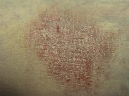
Lichen simplex chronicus (LSC, or neurodermatitis circumscripta) is characterized by pruritic, lichenified plaques that most often occur on the neck, anterior tibias, ankles, wrists, and anogenital region in response to chronic localized scratching or rubbing. Primary LSC evolves on apparently normal skin from pruritus of unclear etiology, whereas secondary LSC is superimposed upon pre-existing dermatoses, especially atopic dermatitis, psoriasis, or dermatophytosis. Psychogenic factors can play an important contributory role, and psychopathology has been found at higher rates in LSC groups as compared to control groups. Neurological abnormalities may sometimes be a factor in the etiology of LSC, as an association between long-standing LSC on the limbs and chronic cervical or lumbar radiculopathy has been reported.
The objective of treatment is to remove environmental trigger factors, break the itch–scratch cycle, and treat any underlying cutaneous or systemic disease. Patients’ understanding of their role in the itch–scratch cycle is essential if their cooperation in avoiding scratching is to be enlisted, thereby facilitating a more complete and permanent recovery. Recurrences are frequent and complete resolution often requires multiple approaches to therapy. Environmental trigger factors such as harsh skin care products or bathing regimens, friction, and excessive moisture or dryness should be minimized or eliminated. High-potency topical corticosteroids, such as clobetasol, diflorasone, and betamethasone, as creams or ointments, are the initial treatments of choice. The potency and/or frequency of application of topical corticosteroids should be decreased as the lesion resolves to avoid atrophy with their long-term use. Adjunctive therapies such as doxepin cream may be introduced if topical corticosteroids are not easily tapered. Occlusion has been found to be a successful aid to therapy because it provides a physical barrier to prevent scratching and permits enhanced and prolonged application of topical medications. Occlusive plastic film or hydrocolloid dressings have been used alone or over mid-potency corticosteroids. Flurandrenolide tape is very effective as both an occlusive and anti-inflammatory measure and is usually changed once daily, although a short occlusion-free period each day will help minimize the side effects of occlusion therapy. In chronic, difficult cases on the lower leg, an Unna boot (a gauze roll impregnated with zinc oxide) may be applied for up to 1 week, provided there is no concomitant infection of the occluded area. Calcineurin inhibitors such as tacrolimus and pimecrolimus also have been successfully used as monotherapy to treat LSC, and offer a good alternative for treatment in steroid sensitive areas such as the genitalia.
Intralesional injections of triamcinolone at monthly intervals can rapidly induce involution. Although highly effective, repeated injections may cause depigmentation or thinning of the epidermis. Therefore, other therapies should be used if several treatments with intralesional corticosteroids do not clear LSC. Infected areas should not be injected with corticosteroids because of the risk of abscess formation. Secondary infections should be treated with appropriate topical or systemic antibiotics. Intralesional botulinum toxin has been reported to offer lasting relief in patients with recalcitrant LSC.
A variety of other therapies have been reported to be effective in the management of LSC. Doxepin cream, capsaicin cream, or aspirin/dichloromethane solution are occasionally of value alone, but are probably best used as adjunctive therapy when LSC does not quickly clear with topical or intralesional corticosteroids. Oral antihistamines may be useful for their sedative effect on patients who scratch during their sleep. Cryosurgery and surgical excision have been reported to help some patients with nodular neurodermatitis. Non-invasive transcutaneous electrical nerve stimulation (TENS) has emerged as a possible effective treatment for pruritic dermatoses such as LSC. Pruritus and neuralgia of varying etiologies have responded to the anticonvulsant gabapentin, which may explain its usefulness in treating LSC. In more severe or recalcitrant conditions, psychotherapy and/or the use of psychopharmacologic agents may be needed for sustained improvement. Benzodiazepines, amitriptyline, pimozide, and doxepin have been used to treat neurotic excoriations and severe neurodermatitis. Neurodermatitis has improved with habit-reversal behavioral therapy, biofeedback, and hypnotherapy in certain individuals. Acupuncture and electroacupuncture are labor intensive, but have been effective in treating some cases of LSC.
Skin biopsy with periodic acid–Schiff stain
LSC that is atypical in appearance or poorly responsive to therapy should be biopsied and cultured to look for pre-existing dermatoses and underlying cutaneous malignancy or infection. The presence of lice or scabies infestation should be excluded. Patch testing may be useful, especially in patients whose biopsy is suggestive of allergic contact dermatitis. In particularly refractory or unusual cases, systemic disease and malignancy should be ruled out.
Datz B, Yawalkar S. J Am Acad Dermatol 1991; 25: 1157–60.
In 127 patients with chronic, localized atopic dermatitis or LSC, healing was reported in 65.1% of those treated with halobetasol propionate ointment (a superpotent group I topical corticosteroid) compared with 54.7% of those treated with clobetasol propionate (a weaker group I topical corticosteroid). Success rates, early onset of therapeutic effect, and adverse effects were similar in the two treatment groups.
Guzzo CA, Weiss JS, Mogavero HS, et al. J Am Acad Dermatol 1991; 25: 1179–83.
Two vehicle-controlled, double-blind studies were performed: a paired comparison study in 124 patients and a parallel group study in 100 patients. In both studies, treatments were applied twice daily for 2 weeks. Severity scores and patient ratings favored halobetasol propionate over the vehicle-treated group. Global assessments showed complete resolution or marked improvement for 83% of patients using halobetasol propionate versus 28% using vehicle. The study concluded that 0.05% halobetasol propionate is highly effective and well tolerated with rapid action and a high degree of clearing.
Group I topical corticosteroids should not be used for more than 2 weeks. They are therefore best combined with adjuvant therapies such as topical doxepin or pimecrolimus cream.
Bard JW. J Ky Med Assoc 1969; 67: 668–70.
Of the 18 patients in the study, 10 used flurandrenolone tape and eight used a topical corticosteroid preparation without occlusion. Lasting remissions were seen in 70% of those using the tape versus 25% of those using topical corticosteroids without occlusion. Duration of therapy is not mentioned.
The use of occlusion with topical corticosteroids is considered the treatment of choice for LSC despite the lack of adequate clinical trials.
Richards RN. J Cutan Med Surg 2010; 14: 19–23 [Review].
In the absence of any formal clinical studies since the 1960s, Richards compiled a review of peer-reviewed literature, six standard dermatology textbooks, and questionnaires from dermatologists to summarize the available information on the use of intralesional steroids in localized dermatoses. Until more definitive and controlled studies are performed, we can be guided by the pooled clinical experience presented in this review, which suggests that triamcinolone acetonide 2.5 mg/mL administered in total doses of 7.5–20 mg intralesionally every 3 to 4 weeks has proven to be a safe, economical, and highly effective treatment for localized dermatoses such as LSC.
Corticosteroid injections are considered first-line therapy despite the lack of adequate controlled clinical trials.
Doxepin Study Group. Drake LA, Millikan LE. Arch Dermatol 1995; 131: 1403–8.
A multicenter double-blind trial conducted to evaluate the safety and antipruritic efficacy of 5% doxepin cream in patients with LSC (n=136), nummular eczema (n=87), or contact dermatitis (n=86). Patients treated with doxepin versus vehicle had significantly greater pruritus relief. Of doxepin-treated patients, 60% experienced relief from pruritus within 24 hours with a response rate of 84% by the end of the study.
Kelekci HK, Uncu HG, Yilmaz B, Ozdemir O, Sut N, Kelekci S. J Dermatolog Treat 2008; 19: 274–8.
Twelve post-menopausal diabetic women with vulvar LSC were treated with pimecrolimus 1% cream twice daily for 3 months. All patients experienced a statistically significant reduction in pruritus when evaluated at 4 weeks and 12 weeks of applying pimecrolimus. After 3 months of treatment, 10 women (83.3%) had a complete cure and the pruritus was improved in the two remaining women.
Tupker RA, Coenraads PJ, van der Meer JB. Acta Derm Venereol 1992; 72: 463.
In this small, open study, two patients with corticosteroid-unresponsive neurodermatitis circumscripta were treated with 0.25% capsaicin, applied five times daily, resulting in flatter lesions and marked relief of itching.
McDow RA, Wester MM. J Derm Surg Oncol 1989; 15: 621–3.
This case report presents a 42-year-old male patient with an 8-month history of a persistent nodular, pruritic lesion, diagnosed clinically as neurodermatitis. Cryosurgery with Refrigerant 12 yielded successful clinical and aesthetic results.
Aschoff R, Wozel G. J Dermatolog Treat 2007; 18: 115–17.
This case report describes a 13-year-old boy with LSC on the face who was treated with tacrolimus 0.1% ointment for 9 months. His lesions healed completely during that time, and the patient was symptom-free 3 years after treatment cessation.
Yüksek J, Sezer E, Aksu M, Erkokmaz U. J Dermatol 2011; 38: 546–52.
Eight patients diagnosed with lichen simplex (LS) by clinical examination and eight patients with macular amyloidosis (MA) underwent TENS with a pulsed electric current to the pruritic area self-identified with the most intense itching. The 30-minute treatments were given thrice weekly for 4 weeks for a total of 12 sessions. A visual analog scale was used to assess the severity of itching at week 0, 2, and 4. The Dermatology Life Quality Index (DLQI) was used to assess the impact of skin disease at week 0, 2, and 4. Six (75%) of the patients with LS and all MA patients reported relief of their pruritus with TENS therapy. By week 2, there was a statistically significant difference from baseline in median VAS scores (p = 0.007) and DLQI total scores (p = 0.006) in the LS group.
Kikindjanin V, Vukaviic T, Stevanocvic V. Dermatol Monatsschr 1990; 176: 741–4.
Seventeen children with neurodermatitis were treated with ketotifen, a mast cell stabilizer, at a dosage of 1 twice daily. Alleviation of the itching occurred within 2 weeks and the patients became itch-free after 20 days on average. Skin lesions cleared between 7 and 9 months of treatment.
Yang Q. J Tradit Chin Med 1997; 17: 57–8.
Acupuncture was used to treat 96 patients with localized neurodermatitis and 43 patients with generalized neurodermatitis. A course of treatment was 10 days, and 3- to 5-day rest periods were given in between multiple courses of therapy. An 81% cure rate and 14% improvement rate were reported, but the number of courses of therapy and long-term follow-up were not specified.
Acupuncture and electroacupuncture (where acupuncture needles are stimulated with low-voltage, high-frequency stimulation) may be used to reduce the proinflammatory neuropeptide state in pruritic and inflamed skin, and thereby promote a more normal state of neuropeptide homeostasis.
Heckmann M, Heyer G, Brunner B, Plewig G. J Am Acad Dermatol 2002; 46: 617–19.
Four patients received 20 units of botulinum toxin type A (100 U/mL) per 2 cm × 2 cm area of an LSC plaque. 1 week after the injection, there was a noticeable reduction in pruritus levels. Three patients were free of itching and one had over 50% reduction in itching. After 12 weeks, three patients remained asymptomatic.
Yosipovitch G, Sugeng M, Chan YH, Goon A, Ngim S, Goh CL. J Am Acad Dermatol 2001; 45: 910–13.
In this double-blind, crossover, placebo-controlled study, 29 patients with LSC were randomized to receive aspirin/dichloromethane solution treatment followed by placebo or vice versa. In the aspirin/dichloromethane treatment group 46% achieved a significant response; 12% of the placebo group achieved a comparable improvement.
Gencoglan G, Inanir I, Gunduz K. Dermatol Ther 2010; 23: 194–8.
Five patients with LSC and four with prurigo nodularis refractory to antihistamines, corticosteroids, phototherapy, and antidepressants were treated with gabapentin. Gabapentin therapy was initiated at 300 mg/day and titrated up by 300 mg/day every 3 days to a final dose of 900 mg/day. Doses were subsequently decreased during a total treatment period ranging from 4 to 10 months. After 2 months, some reduction in pruritus was noted in all patients. Clinical improvement was maintained during the 3-month follow-up period after patients discontinued gabapentin. All patients had improvement in their pruritus, with residual itching responding to topical lubricants. Side effects were limited to tolerable sedation in some patients.
Rosenbaum MS, Ayllon T. Behav Res Ther 1981; 19: 313–8.
Four patients with neurodermatitis received a single treatment session in which they learned to substitute a competing response for their urges to scratch. There was a rapid reduction in scratching in all patients. At 6 months, scratching had been eliminated in one patient and markedly reduced in three patients.
Lehman RE. Am J Clin Hypn 1978; 21: 48–51.
One patient with extensive neurodermatitis was treated with eight sessions of hypnotherapy. She was clear within 2 weeks after her last session, and remained clear at 4-year follow-up.
Harris BA, Sherertz EF, Flowers FP. Int J Dermatol 1987; 26: 541–3.
Two patients with chronic neurotic excoriations had improvement in symptoms and in clinical signs of their skin condition within several weeks of receiving oral doxepin (30–75 mg daily).
Porter WM, Bewley A, Dinneen M, Walker CC, Fallowfield M, Francis N, et al. Br J Dermatol 2001; 144: 343–6.
Two patients with nodular LSC of the scrotum were treated by surgical excision of LSC plaques with remission lasting over 12 months.
Treatment of Skin Disease Comprehensive Therapeutic Strategies 4e
WhatsApp us

 Topical corticosteroids
 Occlusion – flurandrenolide tape
 Intralesional corticosteroids
 Doxepin cream
 Pimecrolimus cream
 Capsaicin cream
 Cryosurgery
 Tacrolimus ointment
 Transcutaneous electrical stimulation
 Ketotifen
 Acupuncture and electroacupuncture
 Botulinum toxin
 Aspirin
 Gabapentin
 Psychotherapy
 Hypnosis
 Psychopharmacotherapy
 Surgical excision