Leprosy (including reactions)

Published on 18/03/2015 by admin

Filed under Dermatology

Last modified 18/03/2015

Print this page

rate 1 star rate 2 star rate 3 star rate 4 star rate 5 star
Your rating: none, Average: 0 (0 votes)

This article have been viewed 1257 times

Leprosy (including reactions)

Anne E. Burdick and Ivan D. Camacho

Evidence Levels:  A Double-blind study  B Clinical trial ≥ 20 subjects  C Clinical trial < 20 subjects  D Series ≥ 5 subjects  E Anecdotal case reports

image

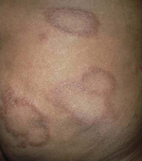
Leprosy or Hansen disease is an infection caused by Mycobacterium leprae (M. leprae), affecting the skin, respiratory mucosa, peripheral nerves, and other organs. The cardinal signs are anesthetic skin lesions, enlarged peripheral nerves, peripheral neuropathy with acid-fast bacilli in tissue biopsies or skin slit smears. A patient’s cell-mediated immunity against M. leprae determines his or her presentation of leprosy in the Ridley–Jopling spectrum: tuberculoid (TT), borderline tuberculoid (BT), mid-borderline (BB), borderline lepromatous (BL) and lepromatous (LL). A patient with TT leprosy has high cell-mediated immunity to M. leprae and few well-demarcated lesions located on cooler parts of the body. A patient with LL leprosy has low cell-mediated immunity to M. leprae and numerous disseminated infiltrated erythematous papules, plaques, and nodules, often presenting with peripheral neuropathy. BB leprosy patients have multiple annular ‘dimorphous’ plaques (see photo). On biopsy, TT lesions reveal rare acid-fast bacilli (AFB) and LL lesions have innumerable AFB. Indeterminate (I) leprosy presents as a single or few hypopigmented patches that tend to resolve spontaneously but may progress to leprosy anywhere on the spectrum. Pure neural leprosy is limited to peripheral nerve involvement without skin lesions.

Leprosy reactions occur in 25–50% of patients before, during or after treatment. BT, BB, and BL patients may develop a delayed hypersensitivity reaction, a type 1 ‘reversal’ reaction, with red edematous lesions and an acute peripheral neuritis. HIV patients may develop BT lesions and a type 1 reversal reaction as an immune reconstitution inflammatory syndrome within 6 months of starting highly active antiretroviral therapy if they also have a subclinical leprosy infection. Patients on infliximab and adalimumab may also manifest leprosy and a type 1 reversal reaction if they have concomitant subclinical leprosy.

Erythema nodosum leprosum, a type 2 reaction, is an immune complex reaction that develops in BL and LL cases. In a type 2 reaction, crops of new red indurated tender papules, plaques or nodules develop usually with fever and occasionally with iritis or orchitis. Lucio’s phenomenon, a rare vasculitis reaction, presents as tender purpuric and necrotic lesions.

Management strategy

Leprosy treatment consists of multidrug therapy with rifampin and dapsone, with or without clofazimine. For treatment purposes, the World Health Organization (WHO) classifies I, TT, and BT cases as paucibacillary and recommends monthly rifampin 600 mg and daily dapsone 100 mg for 6 months. WHO classifies BB, BL and LL cases as multibacillary and recommends monthly rifampin 600 mg, daily dapsone 100 mg and clofazimine 300 mg monthly and 50 mg daily for 12 months.

The US National Hansen’s Disease Program guidelines are different. For paucibacillary cases, daily rifampin 600 mg and daily dapsone 100 mg are recommended for 12 months. For multibacillary cases, daily rifampin 600 mg, daily dapsone 100 mg and daily clofazimine 50 mg are recommended for 24 months. In the US, clofazimine is only available through the National Hansen’s Disease Program as an investigational drug.

If clofazimine is not available or declined due to skin hyperpigmentation, or if a patient cannot take rifampin or dapsone, alternative antibiotics are minocycline 100 mg, ofloxacin 400 mg, levaquin 500 mg, clarithromycin 500 mg or moxifloxacin 400 mg. Rifampin is prescribed monthly for patients on prednisone, warfarin, oral contraceptives and other drugs with metabolism affected by rifampin. If multidrug therapy is interrupted for any reason, it should be resumed when possible for the remaining months required to complete the recommended duration of multidrug therapy.

Multidrug therapy should be continued during reactions. Mild type 1 reversal reactions and type 2 reactions are treated with non-steroidal anti-inflammatory drugs. For a severe type 1 reversal reaction with acute neuritis, particularly involving the face, aggressive therapy with systemic steroids are started to prevent potentially irreversible nerve damage. Prednisone 0.5–1 mg/kg daily should be started and tapered slowly, 5–10 mg every 2 to 4 weeks, for 3 to 6 months or longer depending on the patient’s response. Since high doses of steroids are often required to control a type 1 reversal reaction, the addition of a steroid sparing drug should be considered. Cyclosporine and methotrexate are effective for a type 1 reversal reaction, permitting a decreased dose or discontinuation of prednisone. In one case report, topical tacrolimus improved a severe type 1 reversal reaction.

Buy Membership for Dermatology Category to continue reading. Learn more here