Published on 18/03/2015 by admin
Filed under Dermatology
Last modified 22/04/2025
This article have been viewed 2321 times
Darrell S. Rigel, Ellen S. Marmur and John A. Carucci
Evidence Levels: A Double-blind study B Clinical trial ≥ 20 subjects C Clinical trial < 20 subjects D Series ≥ 5 subjects E Anecdotal case reports
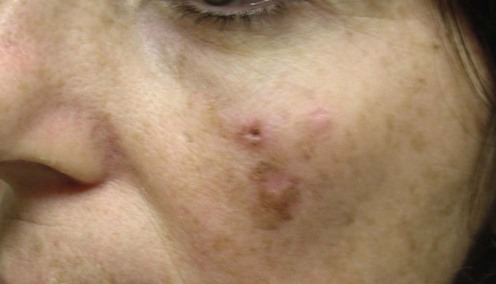
Lentigo maligna (LM) is a type of melanoma in situ that classically presents as irregular pigmented patches on sun-exposed areas of the face and neck in older individuals. If left untreated, the lifetime risk of LM progressing to invasive melanoma (lentigo maligna melanoma, LMM) varies from 4.7% to 2.2%. LM and LMM are the most common forms of melanoma on the head and neck, are commonly found on the cheek, often present as a cosmetic concern, and their incidence is increasing. Treatment has been difficult owing to high recurrence rates, which are attributed to subclinical extension and the difficulty of determining tumor margins.
The first treatment of choice for LM is wide local excision with a margin of at least 5 mm, which leads to clearance rates of 24–70% and recurrence rates of 7–20%. Staged surgical excision is associated with recurrence rates of 0% to 9.7%. In some patients, surgical treatment will inevitably lead to large skin defects and complex surgical reconstructions. Combination therapy with both surgical and non-surgical modalities, and surgery with wider margins are at the forefront of research in the field of LM/LMM.
Successful management of LM depends on early diagnosis and definitive removal. The differential diagnosis includes lentigo, macular seborrheic keratosis, pigmented actinic keratosis, pigmented squamous cell carcinoma in situ, and pigmented superficial basal cell carcinoma. Confirmatory biopsy is necessary prior to definitive treatment. The use of a Wood’s lamp may help to determine the perimeter of the lesion. Dermoscopy may also be helpful. Biopsy of the entire lesion is ideal in order to ascertain its maximum depth. However, the lesion tends to be large (>1 cm) owing to its propensity for extensive radial growth prior to vertical growth into the dermis. Therefore, biopsy of part of the lesion is often performed. Treatment is primarily surgical, although eradication by other methods may be considered. Patients with a history of LM should have periodic full-body skin examinations by a dermatologist to allow for early detection of recurrence, progression, or a second primary skin cancer. Patients with LM also require a consultation about sun protection behaviors.
Skin biopsy and patient evaluation
NIH Consensus Statement, 1992; 10: 1–26.
Biopsy of sufficient depth is critical for diagnosis and management of pigmented lesions. Punch, saucerization, excision, or incisional biopsy may be acceptable. On microscopic examination LM is characterized by increased numbers of atypical melanocytes, which may be solitary or arranged in nests, but do not invade the dermis. Evaluation should include a personal and family history, complete skin examination, and palpation of regional lymph nodes. Blood tests or imaging studies are not indicated.
NIH Consensus Statement 1992; 10: 1–26.
Current recommendations are based on the NIH consensus for melanoma in situ, which suggests excision of the lesion or biopsy site with a margin of 0.5 cm of clinically normal skin and layer of subcutaneous tissue. In general, margins of 0.5–1.0 cm are suggested for LM where feasible. Difficulties may arise in determination of clinical margins due to diffuse background sun damage. A Wood’s lamp may be useful in defining subclinical extension. Accurate determination of margins is key, because LM is likely to recur after inadequate excision.
Johnson TM, Headington JT, Baker SR, Lowe L. J Am Acad Dermatol 1997; 37: 758–64.
With this technique, a margin of 0.5–1.0 cm is outlined with angled corners to facilitate processing. A peripheral strip of tissue 2–4 cm wide is excised and processed for evaluation of permanent sections. Residual tumor is subsequently excised in directed fashion based on mapping. There were no recurrences in 35 patients at 2 years.
Cohen LM, McCall MW, Zax RH. Dermatol Surg 1998; 24: 673–7.
A report of successful treatment at 29.2 months in 45 patients with LM and LMM with Mohs micrographic surgery (MMS) aided by rush permanent sections. There was one recurrence at 50 months (97% cure rate).
Bene NI, Healy C, Coldiron BM. Dermatol Surg 2008; 34: 660–4.
Patients (n=116) with melanoma in situ (MIS) in sun-exposed skin of LMM type, were treated by MMS with subsequent evaluation of the final margin with paraffin-embedded sections that were cut en face, over a period of 12 years (mean follow-up, 50 months; median 48 months; 594.5 patient-years). The clearance rate by MMS technique using frozen sections was 94.1% for MIS LM type. The cure rate was 99.0% for MIS LM type.
MMS is a viable option for treatment of MIS that may increase cure rate and reduce the size of the defect especially in cosmetically and functionally sensitive areas.
Zalla MJ, Lim KK, Dicaudo DJ, Gagnot MM. Dermatol Surg 2000; 26: 771–84.
This modification to Mohs technique was developed because difficulties may arise in distinguishing sun-damaged melanocytes from residual LM on frozen sections. Special histologic stains have been used to enhance sensitivity. Zalla et al. reported successful treatment of melanoma by MMS using Melan-A, HMB-45, Mel-5, and S100. There were no recurrences in 68 patients after a mean follow-up of 16 months.
Hilari H, Llorca D, Traves V, Villanueva A, Serra-Guillén C, Requena C, et al. Actas Dermosifiliogr 2012; 103: 614–23.
A review of the clinical records of patients with LM of the head treated definitively with conventional surgical excision or slow MMS at the dermatology department of Instituto Valenciano de Oncología between January 1993 and April 2011 was reported. Surgical margins larger than 0.5 cm were required in 69.2% of recurrent LM and 26.5% of primary LM. Factors associated with the need for wider margins were prior treatment that might have interfered with the clinical delineation of the border, lesions in the center of the face, and skin phototypes III to V. Surgical margins of 0.5 cm are inadequate for the treatment of a considerable number of LM lesions located on the head, particularly if these are recurrent. Slow MMS using paraffin-embedded sections appears to be the treatment of choice in such cases, particularly for recurrent lesions or lesions with poorly defined borders or possible subclinical extension.
A potential disadvantage of relying on ‘slow mohs’ is the amount of time added to each procedure. In addition, the technique depends on off-site tissue processing and interpretation, thereby magnifying the possibility of error.
Abdelmalek M, Loosemore MP, Hurt MA, Hruza G. Arch Dermatol 2012; 148: 599–604.
All patients with a diagnosis of LM and LMM treated by staged excision from 1999 to 2007. The rate of recurrence after geometric staged excision was 1.7% (four of 239), with a mean of 32.3 months of follow-up. The mean margin to clearance after excision was 6.6 mm for LM and 8.2 mm for LMM. A total of 11.7% of LMM was initially diagnosed as LM on biopsy, with the invasive component discovered during the excision.
Hazan C, Dusza SW, Delgado R, Busam KJ, Halpern AC, Nehal KS. J Am Acad Dermatol 2008; 58: 142–8.
The authors conducted a retrospective study of 117 LM and LMM cases treated with a staged margin-controlled excision technique with rush paraffin-embedded sections. The mean total surgical margin required for excision of LM was 0.71 cm and was 1.03 cm for LMM. The study concluded that the standard excision margins for LM and LMM are often inadequate, and occult invasive melanoma occurs in LM.
Hedblad MA, Mallbris L. J Am Acad Dermatol 2012; 67: 60–8.
Patients (n=593) were treated with Grenz rays (GR) (primary therapy, n=350; partial excision followed by GR, n=71; radical excision followed by GR as recurrence-prophylactic treatment, n=172) at the Department of Dermatology, Karolinska University Hospital, Stockholm, Sweden, between 1990 and 2009. The treatment was given twice a week over 3 consecutive weeks in total doses of 100–160 Gy. Dosage depended on the stage of LM and the depth of periadnexal atypical melanocytic extension in histologically examined materials before treatment. Four hundred and twenty-five patients have been followed up for at least 2 years; of these, 241 for 5 years. Overall, 520 of 593 patients (88%) showed complete clearance after one fractionated treatment. Residual lesions were seen in 15 patients, and 58 relapsed, 53 of whom (72%) within 2 years.
Schmid-Wendtner MH, Brunner B, Konz B, Kaudewitz P, Wendtner CM, Peter RU, et al. J Am Acad Dermatol 2000; 43: 477–82.
Sixty-four patients with LM (42) and LMM (22) were treated with fractionated radiotherapy. There was no recurrence in any of the patients with LM over a mean follow-up of 23 months (range 1–96 months).
One of the advantages of this soft X-ray or Miescher’s technique is exclusion of underlying bone and minimization of risk of bony necrosis. The potential disadvantage is inadequate depth of penetration.
McLeod M, Choudhary S, Giannakakis G, Nouri K. Dermatol Surg 2011; 37: 1210–28.
Twelve studies examining staged surgical excision (SSE), nine using MMS, six investigating cryosurgery, 22 investigating imiquimod, seven using lasers, nine investigating radiation therapy, and two investigating electrosurgery and curettage. SSE and MMS are associated with the lowest recurrence rates for LM. Cryotherapy and radiation therapy may be considered the options for treatment of LM in patients who cannot tolerate surgery.
Kauvar ANB, Geronemus R. Lasers Surg Med 1995; 48.
Tumor eradication was achieved in three of four patients during a 6–24-month follow-up. The fourth patient developed amelanotic LM at 1 month. Q-switched ruby laser may be prone to failure due to inadequate depth of penetration.
Orten SS, Waner M, Dinehart SM, Bardales RH, Flock ST. Otolaryngol Head Neck Surg 1999; 120: 296–302.
Eight patients with LM were treated with the Nd : YAG laser. Three patients were treated with both 532 and 1064 nm. Two of these had complete eradication without recurrence at 3.5 years. The other showed a partial response, but died of unrelated causes prior to completion of therapy.
The Nd : YAG laser emits energy at 532 nm and 1064 nm, which may be especially suited to LM. Melanin has greater absorption at 532 nm, whereas the longer wavelength, 1064 nm, may provide deeper penetration.
Lee H, Sowerby LJ, Temple CL, Yu E, Moore CC. Arch Facial Plast Surg 2011; 13: 398–403.
Seventy-three patients aged 39 to 93 years (mean age, 64.8 years) were included in the study. Twenty-seven patients were treated with surgical excision, 31 patients with radiation therapy, and 15 patients with CO2 laser ablation. The median follow-up times were 16.6 months for surgical excision, 46.3 months for radiation therapy, and 77.8 months for CO2 laser ablation (p<0.001). Recurrence rates by treatment modality were 4.2% (one of 27) for surgical excision, 29.0% (nine of 31) for radiation therapy, and 6.7% (one of 15) for CO2 laser ablation. A trend toward lower recurrence rates with surgical excision and CO2 laser ablation was identified, but the results were not statistically significant.
CO2 laser ablation may have a role as an alternative treatment for lentigo maligna among patients in whom standard treatments, such as surgical excision and radiation therapy, are declined or carry significant morbidity.
Cornejo P, Vanaclocha F, Polimon I, Del Rio R. Arch Dermatol 2000; 136: 428–30.
Interferon-α is a biological response modifier indicated for adjunctive treatment of high-risk melanoma. The mechanism involves both immunomodulation and direct antiproliferative effects. Successful treatment of 11 LM lesions in 10 patients at doses of 3 × 10⁶ to 6 × 10⁶ IU administered three times weekly is reported, with clearance in all patients after between 12 and 29 doses.
Carucci JA, Leffell DJ. Arch Dermatol 2000; 136: 1415–16.
In this case report, clinical and histologic clearance was achieved after a total dose of 39 million units. Toxic effects included fever, chills, and flu-like symptoms, and were transient when interferon-α was used intralesionally.
Wong JG, Toole JW, Demers AA, Musto G, Wiseman MC. J Cutan Med Surg 2012; 16: 245–9.
Twenty-seven patients were reviewed. There were 20 responders (74.1%) and seven failures. The mean tumor size (area of an ellipse) was 6.69 cm2, and the mean treatment duration was 17.7 weeks. Neither the size of the tumor (p=0.86) nor treatment duration (p=0.18) was related to resolution of the lesion. Imiquimod is an effective treatment for LM that provides patients with a cosmetically favorable outcome when standard surgery is not an option.
Van Meurs T, Van Doorn R, Kirtschig G. Dermatol Surg 2010; 36: 853–8.
Ten patients with LM were treated with imiquimod 5% cream between 2004 and 2007 with a median follow-up of 31 months (range 11–56 months). Histological clearance was assessed in all patients using post-treatment biopsies. Complete clinical clearance was achieved in nine of 10 patients after treatment with imiquimod. During follow-up, three clinical and histological recurrences were observed at 9, 10, and 27 months after treatment cessation. In a fourth patient, histological recurrence without clinical signs was demonstrated 17 months after treatment. Five of 10 patients were in sustained clinical remission.
Chimenti S, Carrozzo AM, Citarella L, De Felice C, Peris K. J Am Acad Dermatol 2004; 50: 101–3.
In this series, two elderly patients with facial LM were treated with daily application of tazarotene gel 0.1% for 6 to 8 months. No recurrence in either patient was observed after follow-up periods of 18 and 30 months, respectively. These results should be interpreted with caution, considering the relatively short follow-up.
Cotter MA, McKenna JK, Bowen GM. Dermatol Surg 2008; 34: 147–51.
Forty patients with biopsy-confirmed LM were treated five times a week for 3 months with 5% imiquimod cream before staged excision. Tazarotene 0.1% gel was added when no clinical signs of erythema developed with imiquimod alone after 1 month (10 patients). After the course of topical therapy, patients were assessed for clinical and complete histologic clearance after staged excision. Thirty-three of 40 patients had a complete clinical response as determined by the absence of remaining clinical lesion on physical examination. Upon histologic review, 30 of 40 patients had no evidence of LM, whereas 10 of 40 harbored residual disease. One patient was found to have histologic evidence of invasion after completing the topical protocol. After a mean follow-up of 18 months (range, 12–34 months) and after complete surgical excision of the treatment site, none of the imiquimod-treated patients had evidence of recurrence. Combination therapy can be an effective adjunctive treatment for LM but does not qualify as a replacement therapy for surgery.
Hyde MA, Hadley ML, Tristani-Firouzi P, Goldgar D, Bowen GM. Arch Dermatol 2012; 148: 592–6.
Ninety patients with 91 LMs were randomized into two groups. One group received imiquimod, 5%, cream 5 days a week for 3 months, while the other group also received tazarotene, 0.1%, gel 2 days a week for 3 months. Following topical therapy, all patients underwent staged excisions and frozen section analysis with melan-A immunostaining to confirm negative margins. Forty-six patients with 47 LMs were randomized to receive monotherapy: Forty-two of 47 LMs reached the intended treatment duration, with 27 complete responses (64%). Forty-four patients with 44 LMs were randomized to receive combined therapy. Thirty-seven of 44 LMs reached the intended treatment duration, with 29 complete responses (78%). This difference did not reach statistical significance (p = 0.17). There have been no recurrences to date, with a mean follow-up period of 42 months.
Pretreating LM with imiquimod, 5%, cream may decrease surgical defect sizes; however, total reliance on topical imiquimod as an alternative to surgery may put the patient at increased risk of a local recurrence.
Treatment of Skin Disease Comprehensive Therapeutic Strategies 4e
WhatsApp us


 Excision
 Mohs micrographic surgery
 Modified Mohs surgery
 Staged excision
 Radiation therapy
 Cryotherapy
 Q-switched ruby laser
 Q-switched Nd : YAG laser
 CO2 laser
 Interferon-α
 Imiquimod
 Tazarotene
 Combination therapy