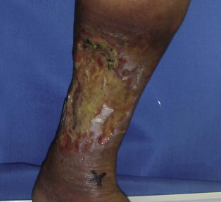
Leg ulcers

Published on 18/03/2015 by admin

Filed under Dermatology

Last modified 18/03/2015

Print this page

rate 1 star rate 2 star rate 3 star rate 4 star rate 5 star
Your rating: none, Average: 0 (0 votes)

This article have been viewed 1173 times

Leg ulcers

Eri Fukaya and David J. Margolis

Evidence Levels:  A Double-blind study  B Clinical trial ≥ 20 subjects  C Clinical trial < 20 subjects  D Series ≥ 5 subjects  E Anecdotal case reports

image

In general, leg ulcers are slow to heal. These wounds are on the most distal aspects of the circulatory, lymphatic, and nervous system supply and as a result there are routes for collateral vessel formation. The lower extremities are also the bearers of weight and gravity. Arterial and venous problems, infection, trauma and systemic problems such as diabetes, immunodeficiency, malnutrition or medications add to impairment. Leg ulcers secondary to other causes such as infection, basal or squamous cell cancer, or pyoderma gangrenosum can also occur but are discussed elsewhere.

Management strategy

Wound care

All chronic wounds are contaminated but not necessarily infected. Many feel that infection can be optimally treated by debridement, which is the removal of slough and necrotic tissue to prevent and/or contain infection as well as minimizing the presence of inflammatory materials. Wound infections prolong inflammation by maintaining high levels of proinflammatory cytokines and tissue proteases. These degrade granulation tissue and delay collagen deposition and healing. Debridement and aggressive cleaning can be done mechanically with curettage, with a scissor or with a scalpel. It can also be accomplished with pressurized liquids, ultrasound, biomechanically (i.e., maggots), and enzymatically.

Dressing

Wound dressings should be chosen to address exudate control, wound protection, and pain relief. The frequency of dressing changes is dependent on the dressing and the reason for its use. The goal is to provide a moist wound environment to promote epithelialization. Wound exudates contain vital proteins and cytokines which facilitate autolytic debridement and promote healing. Using hydrocolloids, hydrogels, alginates, transparent films, and other bandages provide coverage and may promote the function of these enzymes. However, excessive exudate can saturate the wound bed diminishing these beneficial properties as well as macerate surrounding tissue making it more prone to injury. Finally, choosing a dressing that does not adhere to the wound bed can be important to assure that it does not disrupt re-epithelialization when it is removed.

Accurately identifying the key cause of wounds is essential in the management of leg ulcers though many wounds are complex and may be of more than one etiology. Below are specific approaches to different leg ulcers.

Arterial ulcers

Buy Membership for Dermatology Category to continue reading. Learn more here