Inhalation anesthetic agents

Published on 07/02/2015 by admin

Filed under Anesthesiology

Last modified 22/04/2025

Print this page

rate 1 star rate 2 star rate 3 star rate 4 star rate 5 star
Your rating: none, Average: 0 (0 votes)

This article have been viewed 1623 times

Inhalation anesthetic agents

Bradley Anderson, MD and Michael J. Murray, MD, PhD

The inhaled anesthetic agents form a cornerstone for modern anesthetic delivery. Their roots run deep into medical history as they have helped pave the way toward modern-day surgical procedures. Today, four main inhaled anesthetic agents are in use in the United States: N2O, isoflurane, sevoflurane, and desflurane. Each possesses differing characteristics in their relative pharmacokinetic and pharmacodynamic profiles, making them uniquely suitable for use in different situations.

Originally used in nineteenth-century dentistry, N2O, a colorless gas at room temperature, is commonly referred to as laughing gas. Although N2O is not often used today, when it is used, it is in combination with other inhalation anesthetic agents rather than as a sole agent.

Desflurane, sevoflurane, and isoflurane are all fluorinated inhalation anesthetic agents. Compared with ether, inhalation anesthetic agents that have been halogenated with fluorine have reduced flammability as well as greater molecular stability. The use of desflurane and sevoflurane is gradually replacing that of older inhalation anesthetic agents because desflurane and sevoflurane result in the fastest and second-fastest respectively, induction and emergence times (because of their low blood solubility).

Uptake

The uptake of an anesthetic agent from the lung into the bloodstream is dependent on three main factors (excluding the concentration and second-gas effects) (Box 64-1). The first is the alveolar–mixed venous partial pressure difference P(Aimage). The next is the solubility of the anesthetic agent in the blood, defined as the blood-gas partition coefficient (λ), and the last is cardiac output (CO). Using these factors, a simple calculation can be used to help determine the uptake of any given inhaled anesthetic agent. From this equation, it is apparent that, if any of the three factors is increased, the result will be a larger uptake of the anesthetic agent:image

Minimum alveolar concentration

Minimum alveolar concentration (MAC), the concentration of an inhalation anesthetic agent that will prevent movement in response to a surgical stimulus in 50% of patients, is expressed as a percentage of the partial pressure of the anesthetic drug in relation to the barometric pressure. More simply stated, if the MAC of isoflurane is 1.14 at sea level, the partial pressure of isoflurane at steady state must be 0.0114 × 760 mm Hg, which is 8.66 mm Hg. For N2O with a MAC of 104, the partial pressure would be 790.4 mm Hg—a partial pressure that could only be obtained under hyperbaric conditions. Using this terminology, inhalation anesthetic agents can be compared using multiples of MAC (e.g., 0.5, 1.0, 1.2) to express their relevant effects at a given concentration. It is far easier to compare inhalation anesthetic agents in terms of their MAC than their partial pressures, which can vary greatly, depending on the agent, the altitude, and other factors.

Just as a MAC can be determined for the absence of a response to surgical stimulus, the MAC can also be determined for additional depths of anesthesia. For example, the MACs needed to prevent verbal as well as autonomic responses have been identified. Conveniently, the standard deviation of the MAC is approximately 10%; therefore, 1.2 MAC is roughly the concentration required to prevent response to a surgical stimulus in 97% of patients.

When fluorinated hydrocarbon agents are used in combination with N2O, their MACs are additive. For example, if a patient inhales 0.75 MAC of N2O, then only 0.25 MAC of a second inhalation anesthetic (e.g., isoflurane) is required to achieve a combined MAC of 1.0.

Blood-gas partition coefficient

The λ describes the relative solubility of an anesthetic agent in blood, compared with its solubility in a gas (Table 64-1). Simply put, it is the concentration of anesthetic agent in the blood divided by the concentration in gas when the two phases are in equilibrium with one another. Soluble anesthetic agents, or ones that have a high λ, have higher concentrations in the blood phase than in the gas phase. Therefore, for a soluble anesthetic agent to exert a partial pressure in the blood phase equal to that of the gas phase, a relatively large number of molecules must be absorbed into the blood, translating into a slower rate of rise of the PA.
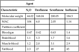
Table 64-1

Pharmacologic Characteristics of Inhalation Anesthetic Agents

  Agent
Characteristic N2O Desflurane Sevoflurane Isoflurane
Molecular weight 44.02 168.04 200.05 184.5
MAC 104 6.0 2.05 1.14
Partition coefficient        
Blood-gas 0.47 0.42 0.63 1.4
Brain-blood 1.1 1.3 1.7 1.6
Muscle-blood 1.2 2.0 3.1 2.9
Fat-blood 2.3 27 48 45

image

MAC, minimum alveolar concentration.

Just as the λ describes the solubility of anesthetic agents in blood compared with solubility in gas, the tissue-blood coefficient is used to describe the solubility of anesthetic agents in tissue, compared with their solubility in blood. Tissues with high tissue-blood coefficients (e.g., fat) require more molecules of anesthetic agent to be dissolved into them for equilibrium with the blood to be reached.

Concentration effect and second-gas effect

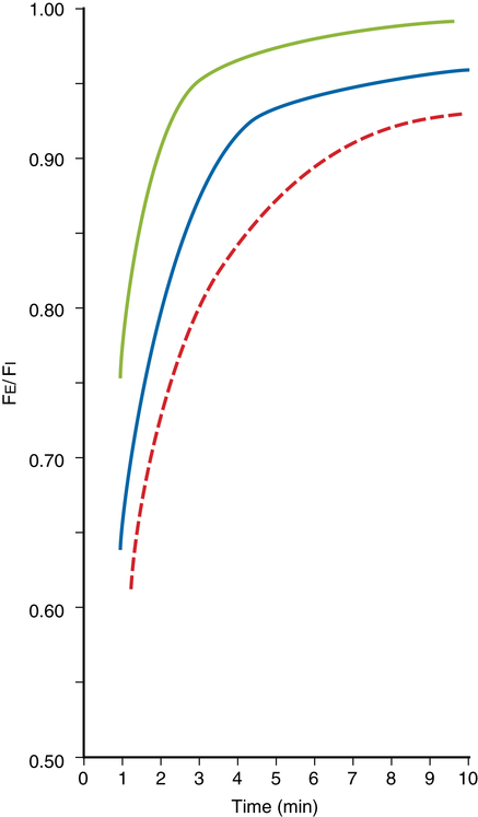
When anesthetic agents are combined, two phenomena—known as the concentration effect and second-gas effect—occur. When N2O is delivered in combination with other gases, the higher the inspired N2O concentration is, the faster the alveolar concentrations of N2O and the other gases will approach their respective inspired concentrations (PI concentration effect) (Figure 64-1). For example, patients receiving a PI of 80% N2O will experience a more rapid increase in their PA/PI ratio, as compared with patients receiving 60% N2O. As pulmonary capillary blood removes N2O from the alveoli, the gases in the anatomic dead space (e.g., bronchi) will be entrained into the alveoli, which results in an even faster rise in the alveolar concentration of the agent (second-gas effect).

Shunts

Right-to-left intracardiac shunts slow the rate of rise of the anesthetic PA during induction. This delay is due to the dilution of pulmonary blood entering the left side of the heart with venous blood that has not been exposed to the inhalation anesthetic agent within the lungs. Right-to-left intracardiac shunts therefore slow an inhalation induction.

Left-to-right intracardiac shunts deliver pulmonary blood containing inhalation anesthetic agents back to the pulmonary circulation for a second pass. As a result, a smaller amount of inhalation anesthetic agent diffuses from the alveoli to capillary blood. From a clinical standpoint, the induction rate is unchanged if there is only a left-to-right shunt. However, when combined right-to-left and left-to-right intracardiac shunts are present, depending on the anatomic location and size of the shunts, left-to-right shunts can affect induction times. The normally delayed induction experienced with a right-to-left shunt can be offset by a left-to-right shunt because unsaturated blood from the right side going into the left side has the opportunity to pass back to the right side, perfuse alveoli, and take up anesthetic drug.

Alveolar—mixed venous partial pressure difference

The relationship between the PA compared with the partial pressure of gases in the mixed venous blood returning to the lungs (Pimage) is known as the alveolar–mixed venous partial pressure difference: P(Aimage). During induction, the P(Aimage) is at its highest. Blood has not yet been exposed to anesthetic agents, and the high PA created by the inhaled gases leads to a large P(Aimage). Over time, more anesthetic agent in the alveoli equilibrates with pulmonary capillary blood until, eventually, blood returning to the lungs carries back some of the anesthetic agent, resulting in a smaller P(Aimage). As various tissues in the body become more saturated, the Pimage increases even further, and the amount of anesthetic agent taken up at the alveolar-capillary interface progressively declines because of the decrease in the P(Aimage). This decrease in anesthetic uptake necessitates a decrease in the amount of anesthetic agent administered to the patient over time.

The effect of CO on uptake and emergence

CO plays a major role in the uptake and induction time of inhaled anesthetic agents: uptake of the inhaled anesthetic agent is directly proportional to the CO. With a greater CO, more blood is delivered into the pulmonary capillary beds per unit of time; more blood absorbs more anesthetic agent, slowing the rate of rise of the agent and its pressure within the alveoli (Pa). Similarly, in patients with a high CO, even though more anesthetic agent is absorbed, it is dissolved in a larger volume of blood, leading to a lower Pa (and thus a lower PCNS ) of the inhaled agent. In patients with a low CO, the opposite occurs: blood spends more time in the pulmonary circulation, allowing for the anesthetic agent to equilibrate with the smaller volume of blood, and the anesthetic agent in the alveoli achieves steady state more quickly. Because the PA of the drug reaches equilibrium more quickly, so too does the Pa of the blood, and as this blood is delivered to the tissues, it translates into a more rapid rise in the PCNS.