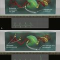
Figure 25-1 Solid tumors are systems within the human system The tumor system includes normal tissue/organ cells (pink), tumor cells usually containing a range of genetic alterations (yellow), and cancer stem cells (orange), along with immune cells (e.g., dendritic cells, granulocytes, macrophages, lymphocytes) and other stromal cells, such as fibroblasts and vasculature, that all play a part in tumor development, progression, metastasis, and response to therapy, based in part on their spatial relationships.
Table 25-1
Key Tumor System Processes and Biomarkers
| System Process | Example Biomarkers | References |
| Proliferation | Ki-67, Aurora A kinase | 294, 295 |
| Apoptosis | p53, Apo-1/Fas, FasL, TRAIL receptors, caspases, pAKT, Survivin, MCL-1, Bcl-2 | 296–303 |
| Cell cycle control | p53, p21, p27, p16, cyclins D1, E | 304–310 |
| Adhesion | E-cadherin, beta-catenin, CD44, CD24, Claudin-1 | 311–315 |
| Migration/motility | CXCR4, alpha6beta4 integrin, Net1, matrix metalloproteinases | 316–318 |
| Angiogenesis | VEGF, Flt-4, HIF-1alpha, pericyte markers | 319–323 |
| Immune responses | CD68, CD45RO, CD3zeta, CD4, CD8, PD-L1, FOXP3, CD1a, cytokines | 224, 324–327 |
| Inflammation | NF-kappaB, COX2, CSF-1R | 214, 328, 329 |
| Fibroblasts | Fibroblast activation protein-alpha, PDGF-beta | 330, 331 |
Adapted from Critchley-Thorne RJ, Miller SM, Taylor DL, et al. Applications of cellular systems biology in breast cancer patient stratification and diagnostics. Comb Chem High Throughput Screen. 2009;12:860-869.
Although the development of HCA has focused on the application of fluorescent probes, chromogenic probes continue to be used extensively for labeling tissue sections. Table 25-3 ∗∗ compares the advantages and disadvantages of fluorescent and chromogenic probes. Most HCA systems are optimized for the use of fluorescent probes, principally for their high sensitivity, high specificity, broad range of cellular functional readouts, broad range of wavelengths for multiplexing, and ability to engineer cells to express fluorescent proteins and biosensors. Because HCA makes use of automated imaging and quantitative image analysis, there is no need for direct viewing of the labeled specimen, and once the images are acquired, there is no further need for the specimen other than for institutional or clinical requirements. In traditional pathology, on the other hand, chromogenic probes have some advantages. The human brain is still the most sophisticated and reliable image processor for the interpretation of small numbers of images. 76 Readily available, low-cost chromogenic probes provide stable and dense labeling for visualization in a transmitted light microscope or by digital image pathology, while simultaneously viewing the contextual morphology of the cells. Although providing somewhat lower resolution and more limited multiplexing than fluorescent probes, chromogenic probes still provide a good labeling strategy where one to three biomarkers per slide can be useful.
Success in the Human Genome Project demanded tools to define the functions of the coding and noncoding portions of the genome, to define the dynamic interplay of cellular constituents within and between cells, and to characterize subpopulations, as well as to define the relationships between populations of cells in higher order biological systems. The field was named cellomics, and the platform technology was named HCA. HCA harnesses the ability to implement combinatorial treatments on large sample sizes by using microplates, patterned microarrays 77 and microfluidic devices 78 for cells, microplates for small organisms, and mounted sections/microarrays for tissues. These large sample sizes are required for statistical analyses and exploration by computational and systems biology. 16,21,63,79,80
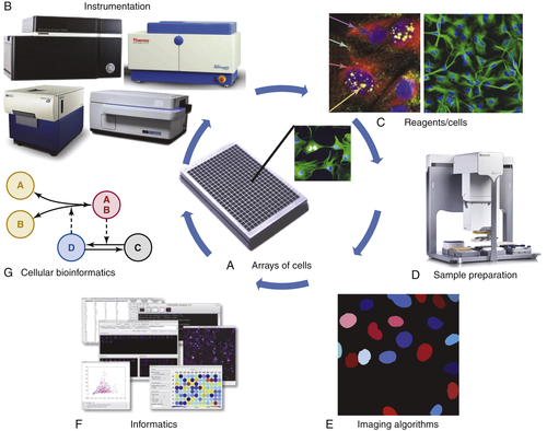
Figure 25-2 The components of high-content analysis (HCA) HCA is defined by the integration of (A) arrays of cells/tissues for high-throughput biology; (B) automated microscope systems available from multiple vendors; (C) a wide range of reagents and cell types; (D) automated sample preparation systems and protocols; (E) imaging algorithms typically designed to measure multiple features for each cell; (F) informatics to review and further process the data, for example, to fit dose-response curves; and (G) bioinformatics to relate multiparameter cellular features to biological functions.
Imaging Live Cells and Model Organisms with HCA
Imaging living cells and model organisms by HCA has the advantage of allowing the investigation of the dynamic, temporal-spatial interplay of cellular constituents that define normal and abnormal cell and tissue functions. Single time points and/or kinetic measurements can be generated and analyzed. 20,62 It is also possible to harness advanced, fluorescence-based probes and biosensors to measure physiological parameters not readily measured in fixed samples, such as cyclic protein translocations, pH, free Ca2+, membrane potentials, and a growing number of physiological biosensors by fluorescence microscopy. 63,69 A disadvantage of investigating living systems by HCA is that biological processes can change from the time of imaging the first well in a microplate to the last well and this issue is multiplied when going from 96 to higher-density well plates. Depending on the time course for the specific biological process, 81 including cyclic changes 82 and the protocol for the addition of experimental treatments, large-scale, living samples usually have to be profiled in smaller batch sizes.
All HCA profiling or screening studies start with living systems that receive some combinatorial application of small molecules or biologics, 83–86 RNAi for knockdowns, 87–89 and/or nucleic acids for transfections or transductions. Although more demanding to perform, a recent investigation studied the kinetics of response of individual cells to drug treatments demonstrating the variability of cellular responses in a population. 19 Measuring kinetic responses should increase as even more biosensors are developed and the complex and dynamic aspects of signaling processes are investigated.
Imaging Fixed Cells and Model Organisms with HCA
The main advantage of using fixed samples is that large-scale sample preparation and robotic screening of many microplates or slides is possible without changes in the biology during the readout. Therefore, many combinatorial treatments can be prepared at one time and the microplates/slides stacked in a robotic system for screening/profiling. There are many fluorescence-based reagents including antibodies, fluorescence in situ hybridization (FISH) probes, and fluorescent proteins that can be used to define single time-point localizations, relative concentrations, and activities. 63 In order to optimally interpret fixed samples, either the half-time of a process under investigation must be determined in live sample profiles, or multiple time points must be generated in distinct wells or plates.
Table 25-2
Classes of Fluorescence-Based Reagents, Readouts, and Online Resources for HCA
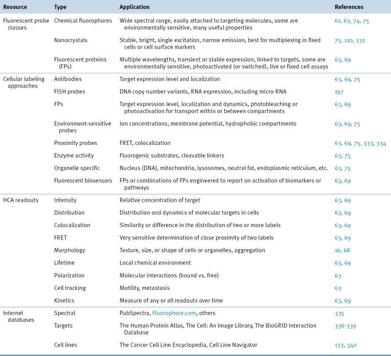
FISH, Fluorescence in situ hybridization; FRET, fluorescence resonance energy transfer; HCA, high-content analysis.
Table 25-3
Comparison of Fluorescent and Chromogenic Readouts 177,341–344
| Reporter Type | Advantages | Disadvantages |
| Fluorescent | Present standard in cell analysis High sensitivity and specificity Quantitative readout Multiplex targets that are colocalized and/or in close proximity Broad spectrum of wavelengths Higher resolution with confocal imaging |
Reagents are less stable for long-term storage More expensive fluorescence, more expensive imaging systems More expensive reagents |
| Chromogenic | Present standard in tissue analysis Long-term stability of labeling Brightfield microscopes Greater amplification |
Variable sensitivity and specificity Multiplexed targets must be spatially separated Precipitates cause fuzziness around target |
HCA has been extensively applied as a phenotypic approach to cancer drug discovery over the past few years, in both primary and secondary screens, either using live-cell 90–93 or fixed-cell 21,39,69,94 screening. Although specific molecular targets guide many of these screens, pathway modulations and phenotypic profiling are central to the approach. 39,71,95,96 Examples of cancer biologies explored include energy metabolism, 97 viral induction, 98 apoptosis, 99 cell cycle, 25,45,91 autophagy, 84 tumor invasion and metastasis, 50 pathway modulations, 47,100 a panel of biologies, 101 and phenotypic changes compared to mutants. 44,102 In many cases, HCA is also used in structure-activity relationship (SAR) to optimize lead compounds. 103,104 However, it is still important to ultimately identify the mechanism(s) of action of lead compounds. The role of HCA in cancer drug discovery and development has been further advanced with the application of more quantitative analyses of profiles using computational biology and systems biology approaches, 105–107 as explored in detail next.
Multiplexed to Hyperplexed Fluorescence-Based HCA
It has been the goal of imaging cytometry to increase the number of specific molecular parameters that can be measured in the same sample, so that complex interplays of components, pathway mapping, and heterogeneity of biological processes can be analyzed in increasing detail. We have defined multiplexed fluorescence in imaging applications as the combination in a sample of up to seven fluorescent probes that can be discriminated by spectral selection. Multiplexing has been accomplished in both live and fixed samples using a range of fluorescent probes. 63,108 Multiplexing by flow cytometry has reached the level of 15 to 18 distinct fluorescent probes, 109 but flow cytometry does not permit analyses of the temporal-spatial dynamics within or between cells. Hence, imaging technologies are being advanced to produce more parameters, especially in fixed samples. There have been a number of technical developments to increase the number of fluorescently labeled antibodies and FISH probes per sample, including new types of probes such as quantum dots, 110 new algorithms such as spectral unmixing, 111 and new protocols such as sequentially labeling, imaging, and quenching the fluorescence, and then repeating the process. 112–115 Recently, the GE Global Research Center has demonstrated that more than 60 fluorescence-based biomarkers can be applied to a single tissue sample using a sequential labeling approach. 111,116 This novel platform technology should have a great impact on basic cancer research, drug discovery, and diagnostics/prognostics. Generating the multiplexed to hyperplexed datasets creates a powerful platform that will enable the application of advanced computational methods to directly define pathways and modifications due to perturbations, as well as to characterize and understand heterogeneity. It is also possible to harness fluorescence lifetime imaging to gain some parameters, 117 as well as the application of mass spectroscopy 118 applied to single cells and tissues, but these latter approaches are not covered here.