Hepatic physiology and preoperative evaluation

Published on 13/02/2015 by admin

Filed under Anesthesiology

Last modified 22/04/2025

Print this page

rate 1 star rate 2 star rate 3 star rate 4 star rate 5 star
Your rating: none, Average: 0 (0 votes)

This article have been viewed 965 times

Hepatic physiology and preoperative evaluation

Frank D. Crowl, MD

Patients with preexisting severe hepatic dysfunction are known to be at significant risk for experiencing perioperative death. Patients with mild to moderate hepatic dysfunction are at increased risk for death.

Some patients develop unexpected hepatic dysfunction (jaundice) in the postoperative period. The reported incidence of postoperative hepatic dysfunction, as demonstrated by abnormalities in liver function tests, is between 1 in 239 and 1 in 1091 anesthetics delivered. Interestingly, some of these patients had preexisting hepatic dysfunction that was not clinically apparent. One study found that 1 in 700 healthy asymptomatic ASA class I and II patients admitted for elective operations had unexplained abnormalities on preoperative liver function tests. After cancellation of their operations, one third of these patients developed clinical jaundice.

Metabolic function

Glucose homeostasis

The liver maintains glucose homeostasis through a combination of mechanisms: the conversion of fats and proteins to glucose by gluconeogenesis, glycogenesis (glucose → glycogen, 75 g stored in liver ∼ 24-h supply), and the release of glucose from glycogen by glycogenolysis. Insulin stimulates glycogenesis and inhibits gluconeogenesis and the oxidation of fatty acids. Glucagon and epinephrine have the opposite effect by inhibiting glycogenesis and stimulating gluconeogenesis.

Hepatic blood flow

Total hepatic blood flow (HBF) is approximately 100 mL·100 g−1·min−1, 75% of which flows through the portal vein, which is rich in nutrients from the gut but is partially deoxygenated, and can therefore supply 50% to 55% of hepatic O2 requirements. The hepatic artery supplies 25% of HBF and 45% to 50% of hepatic O2 requirements.

Splanchnic vessels supplying the portal vein receive sympathetic innervation from T3 through T11. Hypoxemia, hypercarbia, and catecholamines produce hepatic artery and portal vein vasoconstriction and decrease HBF. β-Adrenergic blockade, positive end-expiratory pressure, positive-pressure ventilation (increased intrathoracic pressure increases hepatic vein pressure, which in turn decreases HBF), inhalation anesthetic agents, regional anesthesia with a sensory level above T5, and surgical stimulation (proximity of surgery to the liver determines the degree of HBF reduction) can all cause a reduction in HBF.

Preoperative hepatic assessment

Two indices are used to assess preoperative risk in patients with underlying advanced liver disease. The Child-Pugh score, the first scoring system used to stratify the severity of end-stage hepatic dysfunction, comprises five criteria: ascites, hepatic encephalopathy, INR (international normalized ratio), serum albumin, and bilirubin concentration. Patients are then stratified into three risk categories: A, minimal; B, moderate; C, severe.

The Model for End-stage Liver Disease (MELD) score, developed at the Mayo Clinic, uses only three laboratory values in its assessment of end-stage liver disease: INR, serum creatinine, and serum bilirubin concentration:

< ?xml:namespace prefix = "mml" />MELD = 3.78 [ln bilirubin] + 11.2 [ln INR] + 9.57 [ln creatinine] + 6.43

image

Patients with scores less than 10 are acceptable candidates for elective operations; in patients with scores of 10 to 20, operations are associated with increased risk; operations in patients with scores greater than 20 should be avoided unless other options have been exhausted. The MELD score is also used by the United Network for Organ Sharing (UNOS) to allocate cadaveric livers for transplantation.

Albumin

Serum albumin concentration is an indirect measure of the synthetic capacity of the liver and may have a predictive value for survival in hepatic disease. Because plasma half-life is 14 to 21 days, serum albumin concentrations change slowly with decreased synthesis. Albumin binds and transports neutral and acidic drugs, hormones, and bilirubin and helps maintain plasma oncotic pressure. Causes of decreased serum albumin concentration include liver disease, decreased synthesis (malnutrition), and increased losses (e.g., nephrotic syndrome, burns, ascites, and protein-losing enteropathies). Although malnutrition and hepatic dysfunction may decrease serum albumin concentration, the most common cause of decreased serum albumin concentration is severe illness. Decreased serum albumin may increase the free fraction of protein-bound drugs and decrease the amount of drug necessary to produce a desired effect. If the dose of a drug administered is not decreased in these circumstances, undesirable effects of the drug may be more frequent or pronounced. Total body albumin can be elevated in patients with cirrhosis who have a low serum albumin level but a large amount of albumin in ascitic fluid.

Bilirubin

Unconjugated (indirect) bilirubin is water insoluble, minimally excreted by the kidneys, and a neurotoxin. In the liver, unconjugated bilirubin is rapidly conjugated (direct bilirubin) and secreted in bile. Because it is water soluble, conjugated bilirubin in plasma can be cleared and secreted by the kidneys. Serum levels of conjugated bilirubin do not begin to rise until the liver has lost at least half of its excretory capacity.

Unconjugated bilirubin is produced by the breakdown of heme (hemoglobin, myoglobin, and cytochrome enzymes) and is then bound to albumin for transport to the liver, where it is conjugated via glucuronyltransferase and subsequently excreted into biliary canaliculi. Overt jaundice, which occurs with total bilirubin levels above 3 mg/dL, may be accompanied by pruritus, encephalopathy, and renal insufficiency. Hemolysis causes an increase in unconjugated bilirubin, with a decrease in hemoglobin and an increase in reticulocyte count. Gilbert syndrome is a genetic defect in the conjugation pathway of bilirubin, resulting in an increase in unconjugated bilirubin without a decrease in hemoglobin and a decrease in free haptoglobin values (haptoglobin binds unconjugated bilirubin, limiting its neurotoxicity) or an increase in reticulocyte count. Intrinsic hepatic disease is reflected by an increase in conjugated bilirubin. Frequently, elevated bilirubin postoperatively can be attributed to hemolysis.

Transaminases

Transaminases are sensitive, but not specific, indicators of hepatic dysfunction. Transaminases are released in response to acute hepatic injury. The magnitude of rise in serum concentration does not always correlate with the severity of the disease. They are helpful in testing for regression or progression of hepatic disease. Higher levels are present in acute hepatic cell death (acute viral hepatitis A and B, overdose of acetaminophen, and shock). Mild elevations are seen in fatty liver disease (alcohol, diabetes, obesity), hepatitis C, hemochromatosis, Wilson disease, α1-antitrypsin deficiency, autoimmune hepatitis, celiac sprue, Crohn disease, and ulcerative colitis. Transaminase levels may be normal or decreased in patients who undergo gastrointestinal bypass surgery and in patients with hemochromatosis, fatty liver of obesity, or end-stage hepatic disease. Skeletal muscle injury can produce marked increases in transaminase levels.

Summary

A liver biopsy remains the gold standard for the diagnosis, grading, and staging of liver disease. Liver function tests may be useful for the differential diagnosis of liver disease, as depicted in Table 49-1.

Table 49-1

Liver Function Tests and Differential Diagnosis

Hepatic Dysfunction Bilirubin Transaminase Enzymes Alkaline Phosphatase Causes
Prehepatic Increased unconjugated fraction Normal Normal Hemolysis
Hematoma resorption
Bilirubin overload from transfusion of red blood cells
Intrahepatic (hepatocellular) Increased conjugated fraction Markedly increased Normal to slightly increased Viral
Drugs
Sepsis
Hypoxemia
Cirrhosis
Posthepatic (cholestatic) Increased conjugated fraction Normal to slightly increased Markedly increased Stones
Sepsis

image

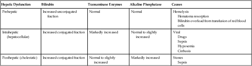
Reprinted with permission from Marschall KE. Diseases of the liver and biliary tract. In: Hines RA, Marschall KE, eds. Stoelting’s Anesthesia and Co-Existing Disease, 5th ed. Philadelphia: Churchill Livingstone; 2008:259-278.